



3'-Deoxycytidine

Based on 1 Customer Validation
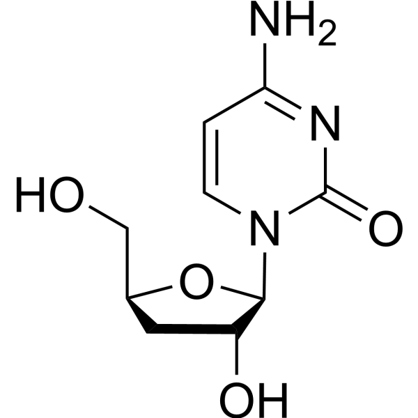
4-Amino-1-((2R,3R,5S)-3-hydroxy-5-(hydroxymethyl)tetrahydrofuran-2-yl)pyrimidin-2(1H)-one (3'-Deoxycytidine) is a purine nucleoside analogue. Purine nucleoside analogs have broad antitumor activity targeting indolent lymphoid malignancies. Anticancer mechanisms in this process rely on inhibition of DNA synthesis, induction of apoptosis, etc.
For research use only. We do not sell to patients.
- Purity : 98.0%
- CAS No.: 7057-33-2
- Formula: C9H13N3O4
- Molecular Weight:227.22
-
Storage:
4°C, protect from light
* In solvent : -80°C, 6 months; -20°C, 1 month (protect from light)

To place orders, for customer services and technical support, please contact: MedChemExpress USA
Tel: 609-228-6898 E-mail: [email protected] [email protected]


-
 Biological Activity
-
Chemical Information
-
Solvent & Solubility
- Protocols
- Purity & Documentation
- References
-
 Help & FAQs




Biological Activity
Description
Cellular Effect
|
Cell Line

|
Type | Value | Description | References |
|---|---|---|---|---|
| CCRF S-180 | ED50 |
15 μM
Compound: 22
|
Effective dose to inhibit the replication of S180 cell line in vitro.
Effective dose to inhibit the replication of S180 cell line in vitro.
|
[PMID: 1995894] |
| CCRF-CEM | ED50 |
25 μM
Compound: 22
|
Inhibit the replication of CCRF-CEM cell line in vitro.
Inhibit the replication of CCRF-CEM cell line in vitro.
|
[PMID: 1995894] |
| L1210 | ED50 |
5 μM
Compound: 22
|
Effective dose to inhibit the replication of L1210 cell line in vitro.
Effective dose to inhibit the replication of L1210 cell line in vitro.
|
[PMID: 1995894] |
| P388 | ED50 |
2.5 μM
Compound: 22
|
Effective dose to inhibit the replication of P388 cell line in vitro.
Effective dose to inhibit the replication of P388 cell line in vitro.
|
[PMID: 1995894] |


Chemical Information
-
CAS No. 7057-33-2
-
Appearance Solid
-
Molecular Weight 227.22
-
Formula C9H13N3O4
-
Color White to light yellow
-
SMILES
O=C1N([C@H]2[C@H](O)C[C@@H](CO)O2)C=CC(N)=N1
-
Shipping
Room temperature in continental US; may vary elsewhere.
-
Storage
4°C, protect from light
* In solvent : -80°C, 6 months; -20°C, 1 month (protect from light)


Solvent & Solubility
In Vitro:
DMSO : 100 mg/mL (440.10 mM; Need ultrasonic; Hygroscopic DMSO has a significant impact on the solubility of product, please use newly opened DMSO)
Please refer to the solubility information to select the appropriate solvent. Once prepared, please aliquot and store the solution to prevent product inactivation from repeated freeze-thaw cycles.
Storage method and period of stock solution: -80°C, 6 months; -20°C, 1 month (protect from light). When stored at -80°C, please use it within 6 months. When stored at -20°C, please use it within 1 month.
Please refer to the solubility information to select the appropriate solvent. Once prepared, please aliquot and store the solution to prevent product inactivation from repeated freeze-thaw cycles.
Storage method and period of stock solution: -80°C, 6 months; -20°C, 1 month (protect from light). When stored at -80°C, please use it within 6 months. When stored at -20°C, please use it within 1 month.
Concentration (start) × Volume (start) = Concentration (final) × Volume (final)
In Vivo:
Select the appropriate dissolution method based on your experimental animal and administration route.
- For the following dissolution methods, please ensure to first prepare a clear stock solution using an In Vitro approach and then sequentially add co-solvents:
- To ensure reliable experimental results, the clarified stock solution can be appropriately stored based on storage conditions. As for the working solution for In Vivo experiments, it is recommended to prepare freshly and use it on the same day.
- The percentages shown for the solvents indicate their volumetric ratio in the final prepared solution. If precipitation or phase separation occurs during preparation, heat and/or sonication can be used to aid dissolution.
Add each solvent one by one: 10% DMSO 40% PEG300 5% Tween-80 45% Saline
Solubility: ≥ 2.5 mg/mL (11.00 mM); Clear solution
This protocol yields a clear solution of ≥ 2.5 mg/mL (saturation unknown).
Taking 1 mL working solution as an example, add 100 μL DMSO stock solution (25.0 mg/mL) to 400 μL PEG300, and mix evenly; then add 50 μL Tween-80 and mix evenly; then add 450 μL Saline to adjust the volume to 1 mL.
Preparation of Saline: Dissolve 0.9 g sodium chloride in ddH₂O and dilute to 100 mL to obtain a clear Saline solution.
Add each solvent one by one: 10% DMSO 90% (20% SBE-β-CD in Saline)
Solubility: ≥ 2.5 mg/mL (11.00 mM); Clear solution
This protocol yields a clear solution of ≥ 2.5 mg/mL (saturation unknown).
Taking 1 mL working solution as an example, add 100 μL DMSO stock solution (25.0 mg/mL) to 900 μL 20% SBE-β-CD in Saline, and mix evenly.
Preparation of 20% SBE-β-CD in Saline (4°C, storage for one week): 2 g SBE-β-CD powder is dissolved in 10 mL Saline, completely dissolve until clear.
In Vivo Dissolution Calculator
Please enter the basic information of animal experiments:
-
-
-
-
Recommended: Prepare an additional quantity of animals to account for potential losses during experiments.
Please enter your animal formula composition:
-
%DMSO +
Recommended: Keep the proportion of DMSO in working solution below 2% if your animal is weak.
-
%+
-
+%Tween-80 + +
-
%Saline +
The co-solvents required include: DMSO, . All of co-solvents are available by MedChemExpress (MCE). , Tween 80. All of co-solvents are available by MedChemExpress (MCE).
Working solution concentration: 0.22 mg/mL
Method for preparing stock solution: mg drug dissolved in μL DMSO. Stock solution concentration: mg/mL. * In solvent : -80°C, 6 months; -20°C, 1 month (protect from light)
1. Take μL DMSO stock solution;
2. Add μL .
μL , mix evenly;
3. Then add μL Tween 80, mix evenly;
4. Then add μL
Please ensure that the stock solution in the first step is dissolved to a clear state, and add co-solvents in sequence. You can use ultrasonic heating (ultrasonic cleaner, recommended frequency 20-40 kHz), vortexing, etc. to assist dissolution.


Purity & Documentation
-
Data Sheet (274 KB)
-
SDS (393 KB)
- English - EN (393 KB)
- Français - FR (393 KB)
- Deutsch - DE (393 KB)
- Norwegian - NO (393 KB)
- Español - ES (393 KB)
- Swedish - SV (393 KB)
- Italian - IT (393 KB)
- Korean - KR (393 KB)
- Portuguese - PT (393 KB)
-
Handling Instructions (2659 KB)
References


Complete Stock Solution Preparation Table
Please refer to the solubility information to select the appropriate solvent. Once prepared, please aliquot and store the solution to prevent product inactivation from repeated freeze-thaw cycles.
Storage method and period of stock solution: -80°C, 6 months; -20°C, 1 month (protect from light). When stored at -80°C, please use it within 6 months. When stored at -20°C, please use it within 1 month.
| Optional Solvent | Concentration Solvent Mass | 1 mg | 5 mg | 10 mg | 25 mg |
|---|---|---|---|---|---|
| DMSO | 1 mM | 4.4010 mL | 22.0051 mL | 44.0102 mL | 110.0255 mL |
| 5 mM | 0.8802 mL | 4.4010 mL | 8.8020 mL | 22.0051 mL | |
| 10 mM | 0.4401 mL | 2.2005 mL | 4.4010 mL | 11.0026 mL | |
| 15 mM | 0.2934 mL | 1.4670 mL | 2.9340 mL | 7.3350 mL | |
| 20 mM | 0.2201 mL | 1.1003 mL | 2.2005 mL | 5.5013 mL | |
| 25 mM | 0.1760 mL | 0.8802 mL | 1.7604 mL | 4.4010 mL | |
| 30 mM | 0.1467 mL | 0.7335 mL | 1.4670 mL | 3.6675 mL | |
| 40 mM | 0.1100 mL | 0.5501 mL | 1.1003 mL | 2.7506 mL | |
| 50 mM | 0.0880 mL | 0.4401 mL | 0.8802 mL | 2.2005 mL | |
| 60 mM | 0.0734 mL | 0.3668 mL | 0.7335 mL | 1.8338 mL | |
| 80 mM | 0.0550 mL | 0.2751 mL | 0.5501 mL | 1.3753 mL | |
| 100 mM | 0.0440 mL | 0.2201 mL | 0.4401 mL | 1.1003 mL |


3'-Deoxycytidine Related Classifications
HY-W012293 Related Classifications
 Powered by Bioz