



AZD-7648
Based on 57 publication(s) in Google Scholar
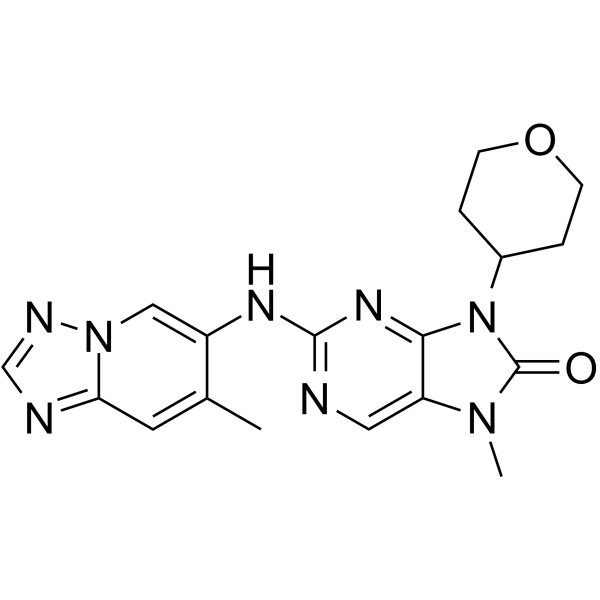
AZD-7648 is a potent, orally active, selective DNA-PK inhibitor with an IC50 of 0.6 nM. AZD-7648 induces apoptosis and shows antitumor activity.
For research use only. We do not sell to patients.
- Purity : 99.86%
- CAS No.: 2230820-11-6
- Formula: C18H20N8O2
- Molecular Weight:380.40
-
Storage:Powder -20°C, 3 years , 4°C, 2 years ; In solvent -80°C, 6 months , -20°C, 1 month

To place orders, for customer services and technical support, please contact: MedChemExpress USA
Tel: 609-228-6898 E-mail: [email protected] [email protected]


-
 Biological Activity
-
Chemical Information
-
Solvent & Solubility
- Protocols
- Purity & Documentation
- References
-
 Help & FAQs


-
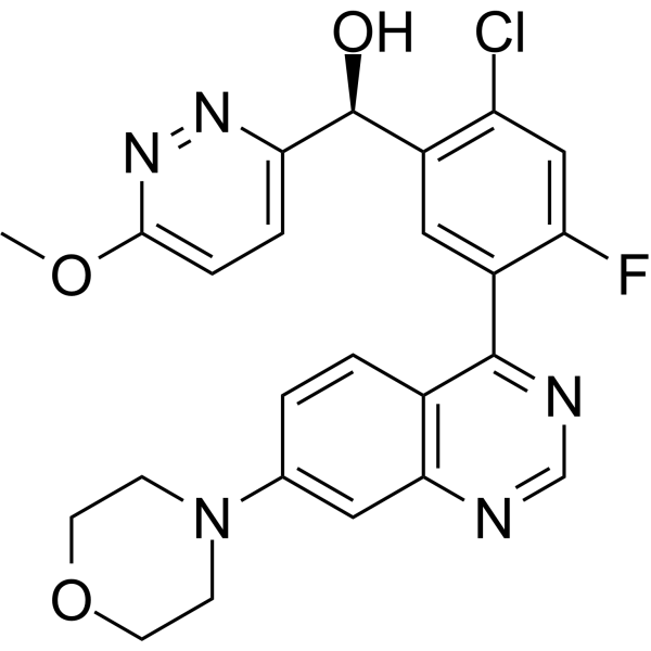
HY-101570
NedisertibDNA-PK Inhibitor -
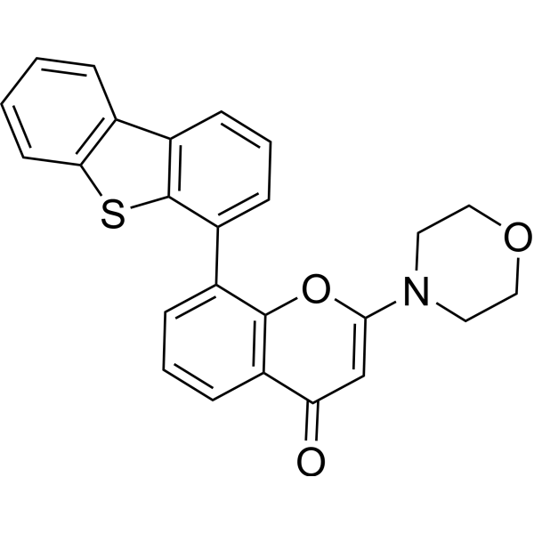
HY-11006
KU-57788DNA-PK Inhibitor -
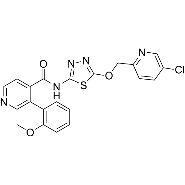
HY-150279
PolQi2Polθ Inhibitor -
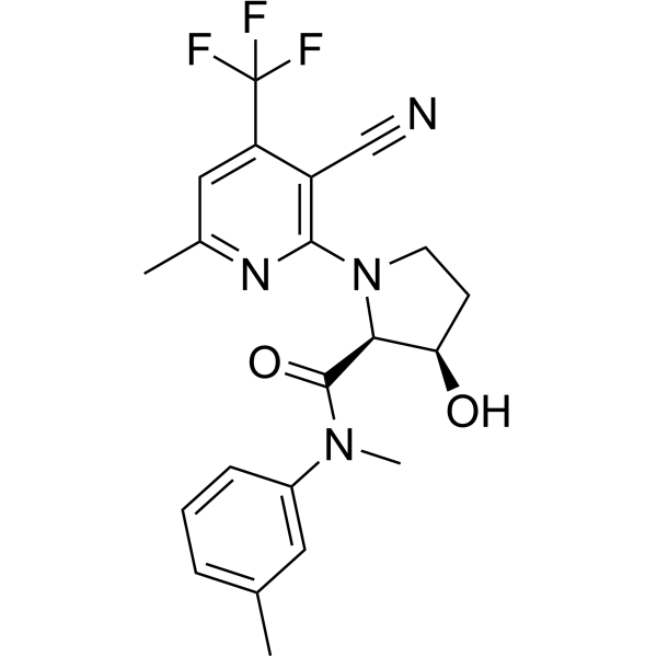
HY-141520
ART558Polθ Inhibitor -
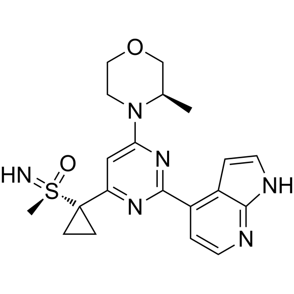
HY-19323
CeralasertibATR Inhibitor -
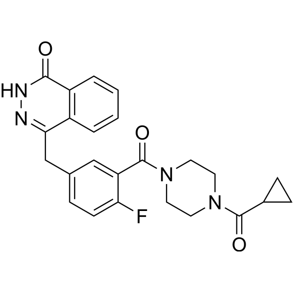
HY-10162
OlaparibPARP Inhibitor -
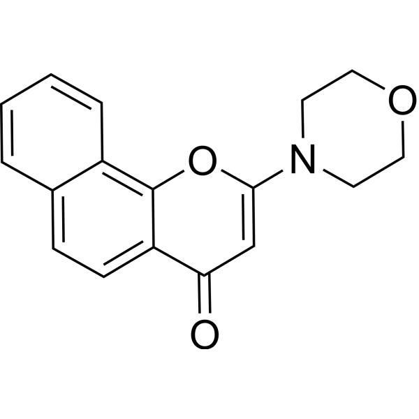
HY-15719
NU 7026DNA-PK Inhibitor -
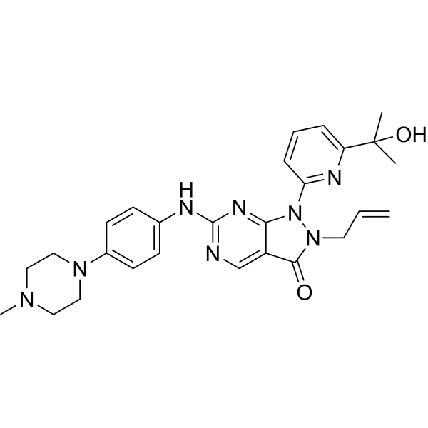
HY-10993
AdavosertibWee1 Inhibitor -

HY-13259
MG-132Proteasome Inhibitor -
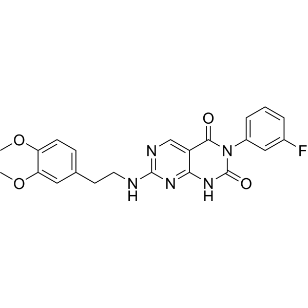
HY-122727
STL127705Ku 70/80 Heterodimer Protein Inhibitor -
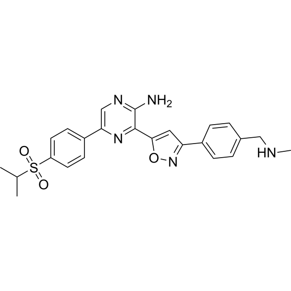
HY-13902
BerzosertibATR Inhibitor -
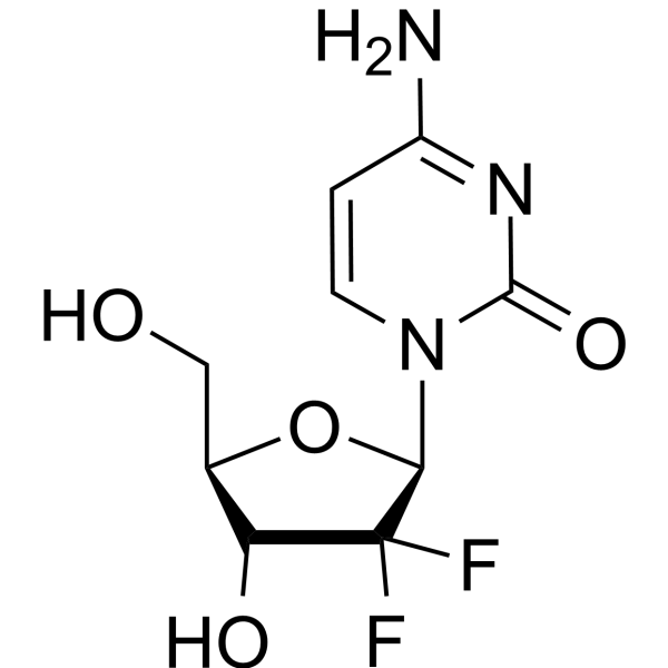
HY-17026
GemcitabineDNA Synthesis Inhibitor
Publications Citing Use of MedChemExpress (MCE) AZD-7648
More- Signal Transduct Target Ther. 2026 May 28;11(1):199. [Abstract]
- Cell. 2025 May 15;188(10):2637-2655.e31. [Abstract]
- Nat Methods. 2024 Mar;21(3):455-464. [Abstract]
- Nat Cell Biol. 2022 Nov;24(11):1666-1676. [Abstract]
- Nat Commun. 2025 Nov 3;16(1):9571. [Abstract]
- Nat Commun. 2025 Jul 15;16(1):6502. [Abstract]
- Nat Commun. 2025 May 8;16(1):4290. [Abstract]
- Nat Commun. 2022 Sep 8;13(1):5295. [Abstract]
- Nat Commun. 2022 Mar 24;13(1):1240. [Abstract]
- Trends Biotechnol. 2025 Aug 30:S0167-7799(25)00314-2. [Abstract]
- Nucleic Acids Res. 2025 Aug 11;53(15):gkaf767. [Abstract]
- Nucleic Acids Res. 2024 Dec 11;52(22):13561-13576. [Abstract]
- Theranostics. 2023 Aug 28;13(14):4745-4761. [Abstract]
- Adv Sci (Weinh). 2025 Jul;12(25):e2501934. [Abstract]
- Sci Adv. 2026 Jun 26;12(26):eadz3351. [Abstract]
- Mol Ther. 2026 Jul 20:S1525-0016(26)00602-7.
- EMBO J. 2023 Mar 15;42(6):e112094. [Abstract]
- Cell Rep. 2025 Jan 3;44(1):115141. [Abstract]
- Mol Cancer Ther. 2026 Jul 15:10.1158/1535-7163.MCT-26-0182.
- Mol Cancer Ther. 2024 Sep 4;23(9):1230-1240. [Abstract]
- Int J Mol Sci. 2023 Oct 18;24(20):15331. [Abstract]
- Sci Rep. 2023 Aug 1;13(1):12429. [Abstract]
- J Cell Mol Med. 2025 Oct;29(19):e70704. [Abstract]
- CRISPR J. 2025 Feb;8(1):60-70. [Abstract]
- DNA Repair. 2025 Sep:153:103889. [Abstract]
- DNA Repair. 2024 Jul:139:103689. [Abstract]
- PLoS One. 2026 Feb 12;21(2):e0341124. [Abstract]
- Technical University of Munich. 2026.
- bioRxiv. 2026 Jul 10:2026.07.10.737056.
- bioRxiv. 2026 Jun 22.
- SSRN. 2026 Jun 22.
- EANM Innovation. 2026 May 19;3:100274.
- University of Califomia San Francisco. 2026.
- bioRxiv. 2026 Mar 11.
- bioRxiv. 2026 Feb 17.
- bioRxiv. 2026 Jan 24.
- bioRxiv. 2025 Nov 29.
- Res Sq. 2025 Sep 11.
- bioRxiv. 2025 Sep 15.
- University of Wisconsin. 2025.
- bioRxiv. 2025 Aug 14.
- bioRxiv. 2025 Jun 17:2025.06.16.659978. [Abstract]
- bioRxiv. 2025 April 10.
- bioRxiv. 2025 Apr 18:2025.04.15.648906. [Abstract]
- bioRxiv. 2025 April 17.
- bioRxiv. 2025 March 15.
- bioRxiv. 2024 November 12.
- bioRxiv. 2024 Nov 6:2024.11.04.621884. [Abstract]
- bioRxiv. 2024 August 29.
- bioRxiv. 2024 Aug 12:2024.08.12.607571. [Abstract]
- Res Sq. 2024 Jun 25.
- University of British Columbia. 2024 Apr 30.
- bioRxiv. 2024 Feb 2.
- bioRxiv. 2024 Feb 1:2024.02.01.578476. [Abstract]
- bioRxiv. 2023 Aug 11:2023.08.10.552800. [Abstract]
- Res Sq. 2023 Apr 6:rs.3.rs-2688694. [Abstract]
- bioRxiv. 2023 Apr 14.
 Customer Validation & Images

-
Cell Proliferation/Viability Assay
-
Cell Proliferation/Viability Assay
-
Cell Proliferation/Viability Assay
-
Apoptosis Analysis
-
Cell Imaging/Staining


Biological Activity
Description
IC50 & Target
[1]|
PI3Kγ 1.37 μM (IC50) |
ATM 17.93 μM (IC50) |
DNA-PKcs 91.3 nM (IC50) |
Cellular Effect
|
Cell Line

|
Type | Value | Description | References |
|---|---|---|---|---|
| A549 | IC50 |
0.091 μM
Compound: Example 16; AZD7648
|
Inhibition of DNA-PK in human A549 cells assessed as reduction in irradiation-induced autophosphorylation at S2056 residue preincubated for 1 hr followed by 8 Gy irradiation and measured after 1 hr by ELISA
Inhibition of DNA-PK in human A549 cells assessed as reduction in irradiation-induced autophosphorylation at S2056 residue preincubated for 1 hr followed by 8 Gy irradiation and measured after 1 hr by ELISA
|
[PMID: 31851518] |
| B16-F10 | IC50 |
11.4 μM
Compound: AZD-7648
|
Synergistic antiproliferative activity against mouse B16-F10 cells assessed as reduction in cell viability incubated for 48 hrs in presence of doxorubicin by CCK-8 assay
Synergistic antiproliferative activity against mouse B16-F10 cells assessed as reduction in cell viability incubated for 48 hrs in presence of doxorubicin by CCK-8 assay
|
[PMID: 38587857] |
| B16-F10 | IC50 |
33.6 μM
Compound: AZD-7648
|
Antiproliferative activity against mouse B16-F10 cells assessed as reduction in cell viability incubated for 48 hrs by CCK-8 assay
Antiproliferative activity against mouse B16-F10 cells assessed as reduction in cell viability incubated for 48 hrs by CCK-8 assay
|
[PMID: 38587857] |
| CHO | IC50 |
>198 μM
Compound: Example 16; AZD7648
|
Inhibition of human ERG stably expressed in CHO cells at -90 mV holding potential by electrophysiological assay
Inhibition of human ERG stably expressed in CHO cells at -90 mV holding potential by electrophysiological assay
|
[PMID: 31851518] |
| CT26 | IC50 |
18.8 μM
Compound: AZD-7648
|
Antiproliferative activity against mouse CT26 cells assessed as reduction in cell viability incubated for 48 hrs by CCK-8 assay
Antiproliferative activity against mouse CT26 cells assessed as reduction in cell viability incubated for 48 hrs by CCK-8 assay
|
[PMID: 38587857] |
| CT26 | IC50 |
6.74 μM
Compound: AZD-7648
|
Synergistic antiproliferative activity against mouse CT26 cells assessed as reduction in cell viability incubated for 48 hrs in presence of doxorubicin by CCK-8 assay
Synergistic antiproliferative activity against mouse CT26 cells assessed as reduction in cell viability incubated for 48 hrs in presence of doxorubicin by CCK-8 assay
|
[PMID: 38587857] |
| HCT-116 | IC50 |
19.5 μM
Compound: AZD-7648
|
Antiproliferative activity against human HCT-116 cells assessed as inhibition of cell growth incubated for 48 hrs in presence of 100 nM doxorubicin by MTT assay
Antiproliferative activity against human HCT-116 cells assessed as inhibition of cell growth incubated for 48 hrs in presence of 100 nM doxorubicin by MTT assay
|
[PMID: 35468512] |
| HCT-116 | IC50 |
35.3 μM
Compound: AZD-7648
|
Antiproliferative activity against human HCT-116 cells assessed as inhibition of cell growth incubated for 48 hrs by MTT assay
Antiproliferative activity against human HCT-116 cells assessed as inhibition of cell growth incubated for 48 hrs by MTT assay
|
[PMID: 35468512] |
| HCT-116 | IC50 |
38.08 nM
Compound: 4; AZD7648
|
Inhibition of colony formation in human HCT-116 cells preincubated with compound for 1 hr followed by exposure to 2 Gy gamma irradiation and measured after 7 days by crystal violet staining based chemiluminescence assay
Inhibition of colony formation in human HCT-116 cells preincubated with compound for 1 hr followed by exposure to 2 Gy gamma irradiation and measured after 7 days by crystal violet staining based chemiluminescence assay
|
[PMID: 38117951] |
| HeLa | IC50 |
0.6 nM
Compound: Example 16; AZD7648
|
Inhibition of human HeLa cell-derived full length DNA-PK catalytic subunit using fluorescein-EPPLSQEAFADLWKK as substrate preincubated for 30 mins followed by substrate addition and measured after 40 mins TR-FRET assay
Inhibition of human HeLa cell-derived full length DNA-PK catalytic subunit using fluorescein-EPPLSQEAFADLWKK as substrate preincubated for 30 mins followed by substrate addition and measured after 40 mins TR-FRET assay
|
[PMID: 31851518] |
| HT-29 | IC50 |
>29 μM
Compound: Example 16; AZD7648
|
Inhibition of ATR in human HT-29 cells assessed as reduction in 4NQO-induced CHK1 phosphorylation at S345 residue preincubated for 1 hr followed by 4NQO addition and measured after 1 hr by Hoechst staining-based imaging analysis
Inhibition of ATR in human HT-29 cells assessed as reduction in 4NQO-induced CHK1 phosphorylation at S345 residue preincubated for 1 hr followed by 4NQO addition and measured after 1 hr by Hoechst staining-based imaging analysis
|
[PMID: 31851518] |
| HT-29 | IC50 |
17.9 μM
Compound: Example 16; AZD7648
|
Inhibition of ATM in human HT-29 cells assessed as reduction in irradiation-induced autophosphorylation at Ser1981 residue preincubated for 1 hr followed by 6 Gy irradiation and measured after 1 hr by Hoechst staining-based imaging analysis
Inhibition of ATM in human HT-29 cells assessed as reduction in irradiation-induced autophosphorylation at Ser1981 residue preincubated for 1 hr followed by 6 Gy irradiation and measured after 1 hr by Hoechst staining-based imaging analysis
|
[PMID: 31851518] |
| Jurkat | IC50 |
0.09 μM
Compound: AZD-7648
|
Synergistic antiproliferative activity against human Jurkat cells assessed as reduction in cell viability incubated for 48 hrs in presence of doxorubicin by CCK-8 assay
Synergistic antiproliferative activity against human Jurkat cells assessed as reduction in cell viability incubated for 48 hrs in presence of doxorubicin by CCK-8 assay
|
[PMID: 38587857] |
| Jurkat | IC50 |
12.3 μM
Compound: AZD-7648
|
Antiproliferative activity against human Jurkat cells assessed as reduction in cell viability incubated for 48 hrs by CCK-8 assay
Antiproliferative activity against human Jurkat cells assessed as reduction in cell viability incubated for 48 hrs by CCK-8 assay
|
[PMID: 38587857] |
| MDA-MB-231 | IC50 |
>30 μM
Compound: Example 16; AZD7648
|
Inhibition of mTOR/PI3Kalpha in human MDA-MB-231 cells assessed as reduction in AKT phosphorylation at S473 residue measured after 2 hrs by Hoechst staining-based imaging analysis
Inhibition of mTOR/PI3Kalpha in human MDA-MB-231 cells assessed as reduction in AKT phosphorylation at S473 residue measured after 2 hrs by Hoechst staining-based imaging analysis
|
[PMID: 31851518] |
| MDA-MB-468 | IC50 |
>30 μM
Compound: Example 16; AZD7648
|
Inhibition of PI3Kbeta in human MDA-MB-468 cells assessed as reduction in AKT phosphorylation at T208 residue measured after 2 hrs by QuantaBlu substrate based fluorescence assay
Inhibition of PI3Kbeta in human MDA-MB-468 cells assessed as reduction in AKT phosphorylation at T208 residue measured after 2 hrs by QuantaBlu substrate based fluorescence assay
|
[PMID: 31851518] |
In Vitro
AZD7648 (0-30 μM) is a potent radiosensitizer[1].
AZD7648 (3 μM) increases sensitivity to Doxorubicin (HY-15142A)[1].
AZD7648 (0.6 μM) enhances the activity of PARP inhibitor Olaparib (HY-10162)[1].
MedChemExpress (MCE) has not independently confirmed the accuracy of these methods. They are for reference only. Further protocols information, click here.
-
Cell Line:IR (ionizing radiation)-treated A549 cells or A549 cells
-
Concentration:0-30 μM
-
Incubation Time:48 h
-
Result:Arrested cell cycle at G2/M phase.
-
Cell Line:IR (ionizing radiation)-treated A549 cells or OAW42 cells treated with Doxorubicin
-
Concentration:0.03, 0.1, 0.3, 1, 3, 10 and 30 μM for A549; 3 μM for OAW42
-
Incubation Time:1 h for A549; 0.5, 2, 4, 8 and 16 h for OAW42
-
Result:Potently inhibited DNA-PKcs autophosphorylation at Ser2056. Downregulated pDNA-PKcs Ser2056, γH2AX Ser139 and pRPA32 Ser4/Ser8 phosphorylation at early time points (at 30 min, 2 h and 4 h). Resulted in increased levels of γH2AX and the apoptosis marker cleaved PARP1 compared with doxorubicin treatment alone at later time points (8 and 16 h).
In Vivo
MedChemExpress (MCE) has not independently confirmed the accuracy of these methods. They are for reference only.
-
Animal Model:Nude mice, A549 xenografts and NCI-H1299 xenografts[1]
-
Dosage:100 mg/kg
-
Administration:Oral administration, once daily for 5 days
-
Result:Induced tumour growth inhibition in combination with IR in A549 xenografts and induced tumour regression in combination with IR in NCI-H1299 xenografts.


Chemical Information
-
CAS No. 2230820-11-6
-
Appearance Solid
-
Molecular Weight 380.40
-
Formula C18H20N8O2
-
Color White to yellow
-
SMILES
CC1=CC2=NC=NN2C=C1NC3=NC=C(N(C)C(N4C5CCOCC5)=O)C4=N3
-
Shipping
Room temperature in continental US; may vary elsewhere.
-
Storage
Powder -20°C 3 years 4°C 2 years In solvent -80°C 6 months -20°C 1 month


Publications (57)
-
Journal Impact Factor
-
Most Recent
-
Signal Transduct Target Ther
Phosphoproteomics distinguishes disease-specific mechanisms for human phospholamban cardiomyopathy reversible by RNA therapy. [Abstract]2026 May 28;11(1):199. PMID: 42204136 -
Cell
2025 May 15;188(10):2637-2655.e31. PMID: 40209705 -
-
Nat Cell Biol
Intron-encoded cistronic transcripts for minimally invasive monitoring of coding and non-coding RNAs. [Abstract]2022 Nov;24(11):1666-1676. PMID: 36344775 -
Nat Commun
Unveiling the cut-and-repair cycle of designer nucleases in human stem and T cells via CLEAR-time dPCR. [Abstract]2025 Nov 3;16(1):9571. PMID: 41184307 -
Nat Commun
2025 Jul 15;16(1):6502. PMID: 40664653 -
Nat Commun
2025 May 8;16(1):4290. PMID: 40341582 -
Nat Commun
Multi-pathway DNA-repair reporters reveal competition between end-joining, single-strand annealing and homologous recombination at Cas9-induced DNA double-strand breaks. [Abstract]2022 Sep 8;13(1):5295. PMID: 36075911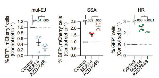
AZD-7648 purchased from MedChemExpress. Usage Cited in: Nat Commun. 2022 Sep 8;13(1):5295. [Abstract]
HEK 293T DSB-Spectrum_V3 cells were transfected with Cas9 and an sgRNA targeting either AAVS1 or BFP, followed by treatment with the DNA-PKcs inhibitors M3841 (2 µM) or AZD7648 (2 µM). At 72h after Cas9 transfection cells were analyzed by flow cytometry (n=4; mean ± SEM; One-way ANOVA, post-hoc Dunnett's).
-
Nat Commun
Harnessing DSB repair to promote efficient homology-dependent and -independent prime editing. [Abstract]2022 Mar 24;13(1):1240. PMID: 35332138 -
Trends Biotechnol
Highly efficient prime editors for mammalian genome editing based on porcine retrovirus reverse transcriptase. [Abstract]2025 Aug 30:S0167-7799(25)00314-2. PMID: 40885667 -
Nucleic Acids Res
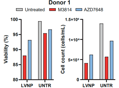
Gene editing in hematopoietic stem cells by co-delivery of Cas9/sgRNA ribonucleoprotein and templates for homology-directed repair in 'all-in-one' lentivirus-derived nanoparticles. [Abstract]2025 Aug 11;53(15):gkaf767. PMID: 40808298
AZD-7648 purchased from MedChemExpress. Usage Cited in: Nucleic Acids Res. 2025 Aug 11;53(15):gkaf767. [Abstract]
AZD7648 (1 µM; 48 h) showed less cytotoxicity in HSPCs relative to M3814 (2 µM; 48 h).
-
Nucleic Acids Res
Single-stranded DNA with internal base modifications mediates highly efficient knock-in in primary cells using CRISPR-Cas9. [Abstract]2024 Dec 11;52(22):13561-13576. PMID: 39569586 -
Theranostics
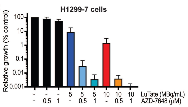
A genome-wide CRISPR/Cas9 screen identifies DNA-PK as a sensitiser to 177Lutetium-DOTA-octreotate radionuclide therapy. [Abstract]2023 Aug 28;13(14):4745-4761. PMID: 37771787
AZD-7648 purchased from MedChemExpress. Usage Cited in: Theranostics. 2023 Aug 28;13(14):4745-4761. [Abstract]
Cell growth response at Day 14, shown as % relative growth as compared to control of H1299-7 cells treated with 0, 5 or 10 MBq/mL LuTate alone, or in combination with 0.5 μM or 1 μM of AZD-7648.
-
Adv Sci (Weinh)
EccDNA-Driven VPS41 Amplification Alleviates Genotoxic Stress via Lysosomal KAI1 Degradation. [Abstract]2025 Jul;12(25):e2501934. PMID: 40271553 -
Sci Adv
Patient-derived organoids across cancers reveal conserved tumor heterogeneity and actionable therapeutic vulnerabilities. [Abstract]2026 Jun 26;12(26):eadz3351. PMID: 42361179 -
-
EMBO J
DNA-PKcs and ATM modulate mitochondrial ADP-ATP exchange as an oxidative stress checkpoint mechanism. [Abstract]2023 Mar 15;42(6):e112094. PMID: 36727301 -
Cell Rep
Dual α-globin-truncated erythropoietin receptor knockin restores hemoglobin production in α-thalassemia-derived erythroid cells. [Abstract]2025 Jan 3;44(1):115141. PMID: 39754719 -
-
Mol Cancer Ther
Radiation and chemo-sensitizing effects of DNA-PK inhibitors are proportional in tumors and normal tissues. [Abstract]2024 Sep 4;23(9):1230-1240. PMID: 38781104 -
Int J Mol Sci
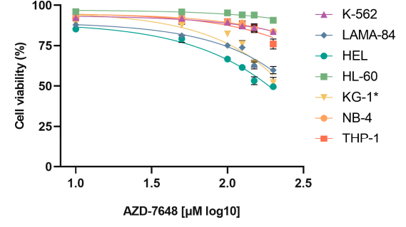
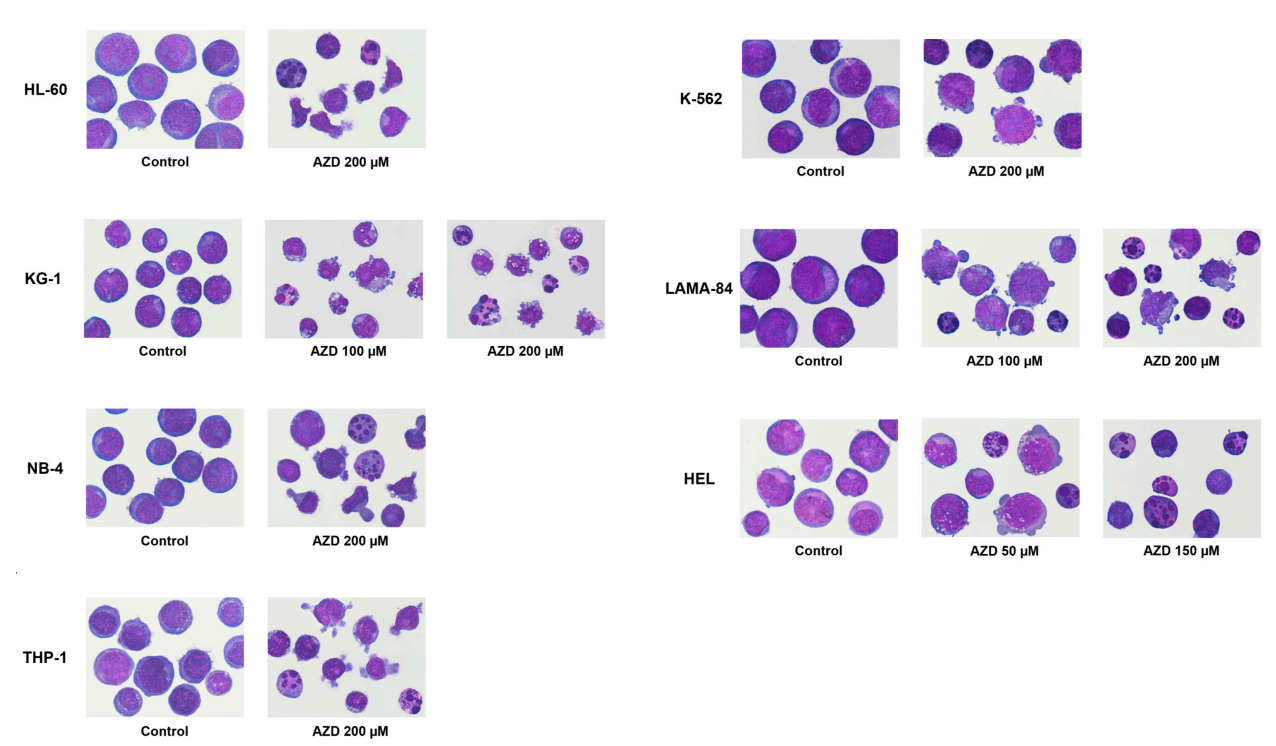
AZD-7648, a DNA-PK Inhibitor, Induces DNA Damage, Apoptosis, and Cell Cycle Arrest in Chronic and Acute Myeloid Leukemia Cells. [Abstract]2023 Oct 18;24(20):15331. PMID: 37895013
AZD-7648 purchased from MedChemExpress. Usage Cited in: Int J Mol Sci. 2023 Oct 18;24(20):15331. [Abstract]
Log dose–response viability curve of AZD-7648 after 24 or 48 (KG-1*) hours of treatment.
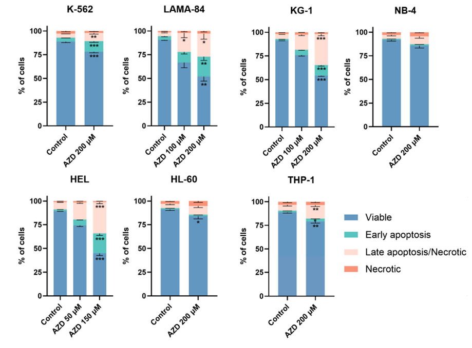
AZD-7648 purchased from MedChemExpress. Usage Cited in: Int J Mol Sci. 2023 Oct 18;24(20):15331. [Abstract]
Analysis of cell death induced by AZD-7648 (AZD,50-200 μM) in CML and AML cell lines after 24 or 48 (KG-1) hours of treatment.The type of cell death was identified using annexin V/propidium iodide staining and analyzed using flow cytometry. Data are expressed as a percentage (%) of viable, early apoptosis, late apoptosis/necrotic, and necrotic cells and represent mean ± SEM of 5 independent experiments. Statistical analyses were performed by comparison with control, using one-way ANOVA and Dunnett’s multiple comparisons test, Kruskal–Wallis and Dunn’s multiple comparisons test, unpaired t-test, or Mann–Whitney test.
AZD-7648 purchased from MedChemExpress. Usage Cited in: Int J Mol Sci. 2023 Oct 18;24(20):15331. [Abstract]
Analysis of cell death induced by AZD-7648 (AZD, 50-200 μM) in CML and AML cell lines after 24 or 48 (KG-1) hours of treatment. Cell morphology was observed by light microscopy (amplification: 1000×), and the most representative image was selected.
-
Sci Rep
DNA-PK inhibition extends the therapeutic effects of Top2 poisoning to non-proliferating cells, increasing activity at a cost. [Abstract]2023 Aug 1;13(1):12429. PMID: 37528151 -
J Cell Mol Med
BNIP3L/BNIP3-Mediated Mitophagy Contributes to the Maintenance of Ovarian Cancer Stem Cells. [Abstract]2025 Oct;29(19):e70704. PMID: 41078116 -
CRISPR J
CRISPR-Cas9-Mediated Correction of TSC2 Pathogenic Variants in iPSCs from Patients with Tuberous Sclerosis Complex Type 2. [Abstract]2025 Feb;8(1):60-70. PMID: 39654514 -
DNA Repair
2025 Sep:153:103889. PMID: 40913914 -
DNA Repair
DNA-PK inhibitor AZD7648 is a more portent radiosensitizer than PARP inhibitor Olaparib in BRCA1/2 deficient tumors. [Abstract]2024 Jul:139:103689. PMID: 38749239 -
PLoS One
Omics-aided design genome editing strategy for challenging human immortalized cell models. [Abstract]2026 Feb 12;21(2):e0341124. PMID: 41678468 -
-
-
-
-
-
-
-
-
-
-
-
-
-
-
bioRxiv
2025 Jun 17:2025.06.16.659978. PMID: 40667017 -
-
bioRxiv
2025 Apr 18:2025.04.15.648906. PMID: 40321211 -
-
-
-
bioRxiv
PAIRWISE: Deep Learning-based Prediction of Effective Personalized Drug Combinations in Cancer. [Abstract]2024 Nov 6:2024.11.04.621884. PMID: 39574568 -
-
bioRxiv
DNA-PKcs Inhibition Improves Sequential Gene Insertion of the Full-Length CFTR cDNA in Airway Stem Cells. [Abstract]2024 Aug 12:2024.08.12.607571. PMID: 39185207 -
-
-
-
bioRxiv
Single-Stranded DNA with Internal Base Modifications Mediates Highly Efficient Gene Insertion in Primary Cells. [Abstract]2024 Feb 1:2024.02.01.578476. PMID: 38352420 -
bioRxiv
Non-homologous end joining shapes the genomic rearrangement landscape of chromothripsis from mitotic errors. [Abstract]2023 Aug 11:2023.08.10.552800. PMID: 37609143 -
Res Sq
Haploinsufficiency of ZNF251 causes DNA-PKcs-dependent resistance to PARP inhibitors in BRCA1-mutated cancer cells. [Abstract]2023 Apr 6:rs.3.rs-2688694. PMID: 37066268 -


Solvent & Solubility
In Vitro:
DMSO : 5.83 mg/mL (15.33 mM; ultrasonic and warming and heat to 60°C; Hygroscopic DMSO has a significant impact on the solubility of product, please use newly opened DMSO)
Please refer to the solubility information to select the appropriate solvent. Once prepared, please aliquot and store the solution to prevent product inactivation from repeated freeze-thaw cycles.
Storage method and period of stock solution: -80°C, 6 months; -20°C, 1 month. When stored at -80°C, please use it within 6 months. When stored at -20°C, please use it within 1 month.
Please refer to the solubility information to select the appropriate solvent. Once prepared, please aliquot and store the solution to prevent product inactivation from repeated freeze-thaw cycles.
Storage method and period of stock solution: -80°C, 6 months; -20°C, 1 month. When stored at -80°C, please use it within 6 months. When stored at -20°C, please use it within 1 month.
Concentration (start) × Volume (start) = Concentration (final) × Volume (final)
In Vivo:
For the following dissolution methods, please prepare the working solution directly:
It is recommended to prepare fresh solutions and use them promptly within a short period of time.
The percentages shown for the solvents indicate their volumetric ratio in the final prepared solution. If precipitation or phase separation occurs during preparation, heat and/or sonication can be used to aid dissolution.
Add each solvent one by one: 0.5% HPMC/0.1% Tween-80 in Saline water
Solubility: 10 mg/mL (26.29 mM); Suspended solution; Need ultrasonic

Protocols
-
Apoptosis
Apoptosis, also called programmed cell death, is generally characterized by distinct morphological characteristics.
-
TUNEL staining for apoptotic DNA fragmentation
TUNEL staining detects DNA strand breaks by using terminal deoxynucleotidyl transferase to add labeled nucleotides to exposed 3′-OH DNA termini, generating either microscopic staining in fixed cells or tissue sections, or fluorescence/cytometric signal in cell suspensions. TUNEL positivity reflects DNA fragmentation but should not be interpreted alone as definitive apoptosis, because TUNEL can also label necrotic, autolytic, mechanically damaged, or DNA-repair-associated DNA breaks.
-
Cell Cytotoxicity Assay
Cytotoxicity assays are usually based on the assessment of cell membrane damage, which can also be indirectly detected by measuring cell viability. Detection methods include MTT assay, CKK-8 assay, LDH assay and ATP assay, etc.
-
Annexin V plus membrane-impermeant dye apoptosis staining
Annexin V-based apoptosis assays rely on the detection of phosphatidylserine (PS) externalization from the inner leaflet of the plasma membrane to the outer leaflet, an early biochemical hallmark of apoptosis. Fluorescently labeled Annexin V binds PS in a calcium-dependent manner, enabling identification of early apoptotic cells by flow cytometry or fluorescence microscopy. When combined with a membrane-impermeant DNA-binding dye (e. g. , propidium iodide), this approach allows discrimination between viable (Annexin V−/dye−), early apoptotic (Annexin V+/dye−), and late apoptotic or necrotic (Annexin V+/dye+) cell populations by assessing membrane integrity and PS exposure.
-
Apoptosis Solutions
Apoptosis is a regulated, generally non-lytic cell-death pathway that removes unwanted, damaged, infected, or abnormal cells through coordinated morphological changes, caspase activation, DNA fragmentation, and membrane remodeling. The intrinsic apoptosis pathway is controlled mainly by mitochondrial outer membrane permeabilization, BCL-2 family proteins, cytochrome c release, apoptosome formation, caspase-9 activation, and downstream executioner caspase-3/7 activation. The extrinsic apoptosis pathway is initiated by death receptors such as Fas, TNFR, and TRAIL receptors, which recruit adaptor proteins and activate caspase-8 before engaging executioner caspases or mitochondrial amplification through BID cleavage. Apoptosis is linked to many phenotypes, including cancer cell killing, tissue homeostasis, immune regulation, neurodegeneration, infection response, and treatment-induced cytotoxicity; unresolved questions include how apoptosis interacts with necroptosis, pyroptosis, ferroptos
-
Subcutaneous Cell-Line-Derived Xenograft
Subcutaneous cell-line-derived xenograft (CDX) models are established by implanting cultured human cancer cell lines into immunodeficient mice, where the injected cells form localized tumors that can be monitored in vivo as a measure of tumorigenic potential, growth kinetics, and treatment response. These models are widely used in oncology research because they allow reproducible tumor formation and enable comparative assessment of tumor growth between different cell lines or genetic manipulations in a controlled in vivo microenvironment. Subcutaneous implantation of cancer cells in immunodeficient mice is a standard approach for evaluating tumor growth behavior and therapeutic response across multiple cancer types, including prostate, esophageal, pancreatic, and colon cancer models.


Purity & Documentation
-
Data Sheet (279 KB)
-
SDS (396 KB)
- English - EN (396 KB)
- Français - FR (396 KB)
- Deutsch - DE (396 KB)
- Norwegian - NO (396 KB)
- Español - ES (396 KB)
- Swedish - SV (396 KB)
- Italian - IT (396 KB)
- Korean - KR (396 KB)
- Portuguese - PT (396 KB)
-
Handling Instructions (2659 KB)
References


Complete Stock Solution Preparation Table
Please refer to the solubility information to select the appropriate solvent. Once prepared, please aliquot and store the solution to prevent product inactivation from repeated freeze-thaw cycles.
Storage method and period of stock solution: -80°C, 6 months; -20°C, 1 month. When stored at -80°C, please use it within 6 months. When stored at -20°C, please use it within 1 month.
| Optional Solvent | Concentration Solvent Mass | 1 mg | 5 mg | 10 mg | 25 mg |
|---|---|---|---|---|---|
| DMSO | 1 mM | 2.6288 mL | 13.1441 mL | 26.2881 mL | 65.7203 mL |
| 5 mM | 0.5258 mL | 2.6288 mL | 5.2576 mL | 13.1441 mL | |
| 10 mM | 0.2629 mL | 1.3144 mL | 2.6288 mL | 6.5720 mL | |
| 15 mM | 0.1753 mL | 0.8763 mL | 1.7525 mL | 4.3814 mL |


AZD-7648 Related Classifications
HY-111783 Related Classifications
 Powered by Bioz