



AZD-7648 (GMP) is AZD-7648 (HY-111783) produced by using GMP guidelines. GMP small molecules works appropriately as an auxiliary reagent for cell therapy manufacture. AZD-7648 is a potent, orally active, selective DNA-PK inhibitor with an IC50 of 0.6 nM. AZD-7648 induces apoptosis and shows antitumor activity.
For research use only. We do not sell to patients.
- CAS No.: 2230820-11-6
- Formula: C18H20N8O2
- Molecular Weight:380.40
-
Storage:
Please store the product under the recommended conditions in the Certificate of Analysis.


To place orders, for customer services and technical support, please contact: MedChemExpress USA
Tel: 609-228-6898 E-mail: [email protected] [email protected]


-
 Biological Activity
-
Chemical Information
- Protocols
- Purity & Documentation
- References
-
 Help & FAQs


-
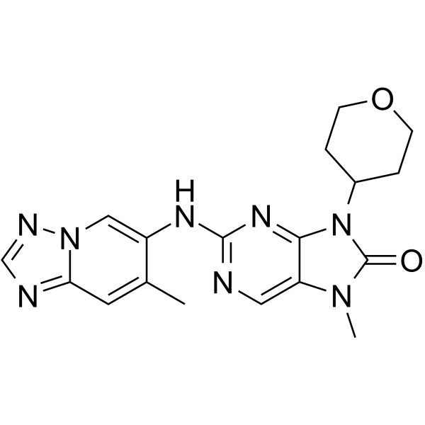
HY-111783
AZD-7648DNA-PK Inhibitor -
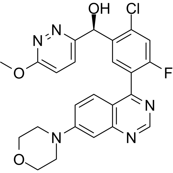
HY-101570
NedisertibDNA-PK Inhibitor -
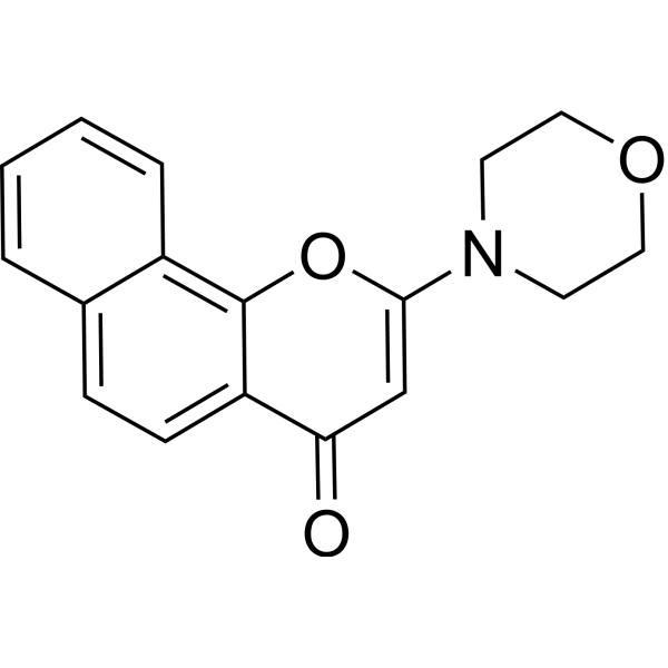
HY-15719
NU 7026DNA-PK Inhibitor -
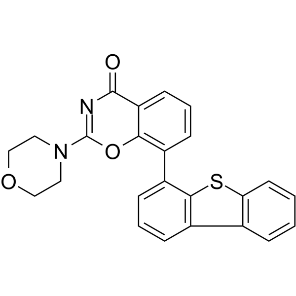
HY-101667
LTURM34DNA-PK Inhibitor -
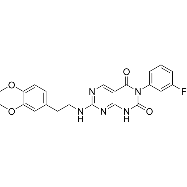
HY-122727
STL127705Ku 70/80 Heterodimer Protein Inhibitor -
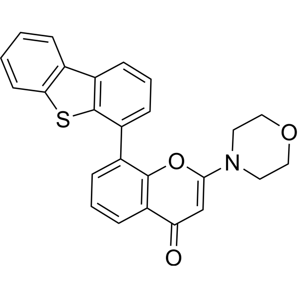
HY-11006
KU-57788DNA-PK Inhibitor -
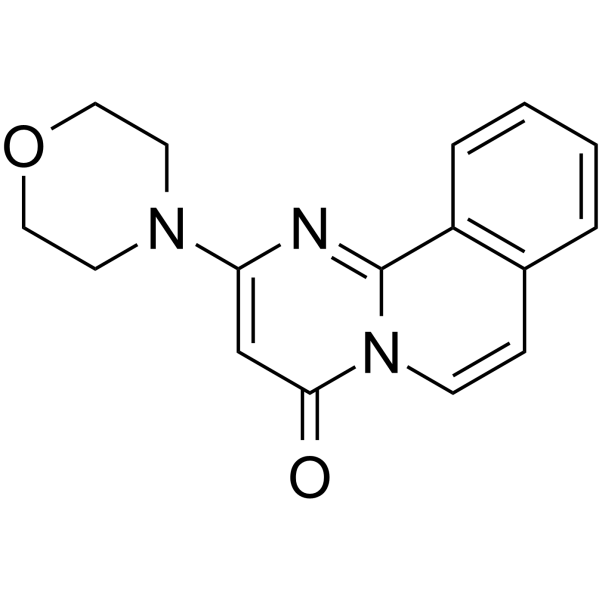
HY-19341
Compound 401DNA-PK Inhibitor -
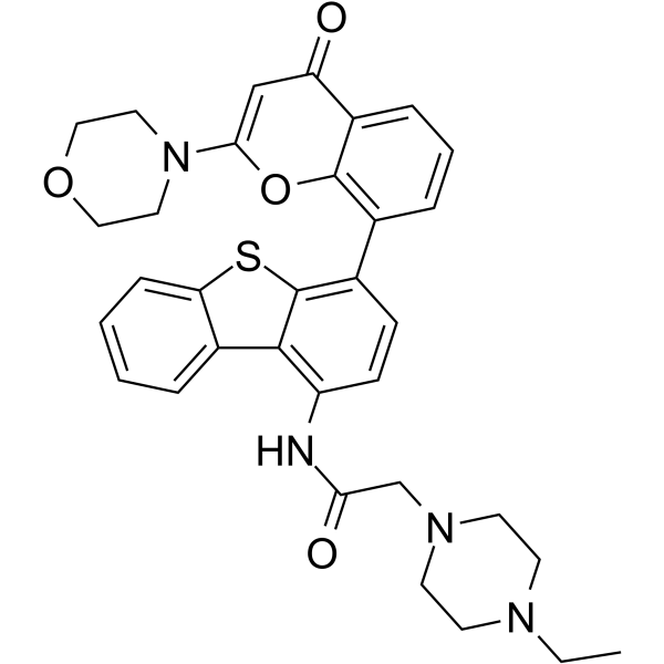
HY-13431
KU-0060648DNA-PK/PI3K Inhibitor -
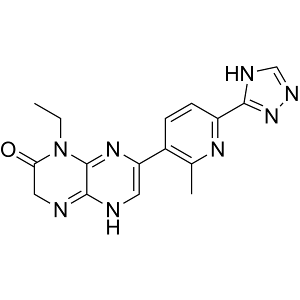
HY-16962
CC-115DNA-PK/mTOR Inhibitor -
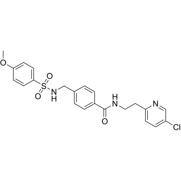
HY-19977
YU238259HDR Inhibtior -
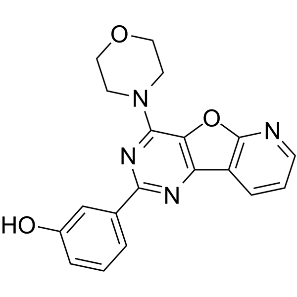
HY-10115
PI-103PI3K/mTOR Inhibitor -
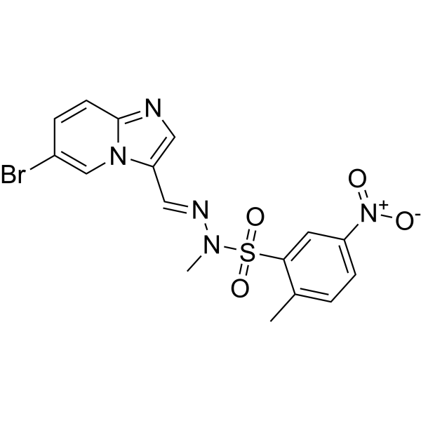
HY-107834
PIK-75DNA-PK/p110α Inhibitor


Biological Activity
Description
IC50 & Target
[1]|
PI3Kγ 1.37 (IC50) |
ATM 17.93 (IC50) |
DNA-PKcs 91.3 nM (IC50) |
Cellular Effect
|
Cell Line

|
Type | Value | Description | References |
|---|---|---|---|---|
| A549 | IC50 |
0.091 μM
Compound: Example 16; AZD7648
|
Inhibition of DNA-PK in human A549 cells assessed as reduction in irradiation-induced autophosphorylation at S2056 residue preincubated for 1 hr followed by 8 Gy irradiation and measured after 1 hr by ELISA
Inhibition of DNA-PK in human A549 cells assessed as reduction in irradiation-induced autophosphorylation at S2056 residue preincubated for 1 hr followed by 8 Gy irradiation and measured after 1 hr by ELISA
|
[PMID: 31851518] |
| B16-F10 | IC50 |
11.4 μM
Compound: AZD-7648
|
Synergistic antiproliferative activity against mouse B16-F10 cells assessed as reduction in cell viability incubated for 48 hrs in presence of doxorubicin by CCK-8 assay
Synergistic antiproliferative activity against mouse B16-F10 cells assessed as reduction in cell viability incubated for 48 hrs in presence of doxorubicin by CCK-8 assay
|
[PMID: 38587857] |
| B16-F10 | IC50 |
33.6 μM
Compound: AZD-7648
|
Antiproliferative activity against mouse B16-F10 cells assessed as reduction in cell viability incubated for 48 hrs by CCK-8 assay
Antiproliferative activity against mouse B16-F10 cells assessed as reduction in cell viability incubated for 48 hrs by CCK-8 assay
|
[PMID: 38587857] |
| CHO | IC50 |
>198 μM
Compound: Example 16; AZD7648
|
Inhibition of human ERG stably expressed in CHO cells at -90 mV holding potential by electrophysiological assay
Inhibition of human ERG stably expressed in CHO cells at -90 mV holding potential by electrophysiological assay
|
[PMID: 31851518] |
| CT26 | IC50 |
18.8 μM
Compound: AZD-7648
|
Antiproliferative activity against mouse CT26 cells assessed as reduction in cell viability incubated for 48 hrs by CCK-8 assay
Antiproliferative activity against mouse CT26 cells assessed as reduction in cell viability incubated for 48 hrs by CCK-8 assay
|
[PMID: 38587857] |
| CT26 | IC50 |
6.74 μM
Compound: AZD-7648
|
Synergistic antiproliferative activity against mouse CT26 cells assessed as reduction in cell viability incubated for 48 hrs in presence of doxorubicin by CCK-8 assay
Synergistic antiproliferative activity against mouse CT26 cells assessed as reduction in cell viability incubated for 48 hrs in presence of doxorubicin by CCK-8 assay
|
[PMID: 38587857] |
| HCT-116 | IC50 |
19.5 μM
Compound: AZD-7648
|
Antiproliferative activity against human HCT-116 cells assessed as inhibition of cell growth incubated for 48 hrs in presence of 100 nM doxorubicin by MTT assay
Antiproliferative activity against human HCT-116 cells assessed as inhibition of cell growth incubated for 48 hrs in presence of 100 nM doxorubicin by MTT assay
|
[PMID: 35468512] |
| HCT-116 | IC50 |
35.3 μM
Compound: AZD-7648
|
Antiproliferative activity against human HCT-116 cells assessed as inhibition of cell growth incubated for 48 hrs by MTT assay
Antiproliferative activity against human HCT-116 cells assessed as inhibition of cell growth incubated for 48 hrs by MTT assay
|
[PMID: 35468512] |
| HCT-116 | IC50 |
38.08 nM
Compound: 4; AZD7648
|
Inhibition of colony formation in human HCT-116 cells preincubated with compound for 1 hr followed by exposure to 2 Gy gamma irradiation and measured after 7 days by crystal violet staining based chemiluminescence assay
Inhibition of colony formation in human HCT-116 cells preincubated with compound for 1 hr followed by exposure to 2 Gy gamma irradiation and measured after 7 days by crystal violet staining based chemiluminescence assay
|
[PMID: 38117951] |
| HeLa | IC50 |
0.6 nM
Compound: Example 16; AZD7648
|
Inhibition of human HeLa cell-derived full length DNA-PK catalytic subunit using fluorescein-EPPLSQEAFADLWKK as substrate preincubated for 30 mins followed by substrate addition and measured after 40 mins TR-FRET assay
Inhibition of human HeLa cell-derived full length DNA-PK catalytic subunit using fluorescein-EPPLSQEAFADLWKK as substrate preincubated for 30 mins followed by substrate addition and measured after 40 mins TR-FRET assay
|
[PMID: 31851518] |
| HT-29 | IC50 |
>29 μM
Compound: Example 16; AZD7648
|
Inhibition of ATR in human HT-29 cells assessed as reduction in 4NQO-induced CHK1 phosphorylation at S345 residue preincubated for 1 hr followed by 4NQO addition and measured after 1 hr by Hoechst staining-based imaging analysis
Inhibition of ATR in human HT-29 cells assessed as reduction in 4NQO-induced CHK1 phosphorylation at S345 residue preincubated for 1 hr followed by 4NQO addition and measured after 1 hr by Hoechst staining-based imaging analysis
|
[PMID: 31851518] |
| HT-29 | IC50 |
17.9 μM
Compound: Example 16; AZD7648
|
Inhibition of ATM in human HT-29 cells assessed as reduction in irradiation-induced autophosphorylation at Ser1981 residue preincubated for 1 hr followed by 6 Gy irradiation and measured after 1 hr by Hoechst staining-based imaging analysis
Inhibition of ATM in human HT-29 cells assessed as reduction in irradiation-induced autophosphorylation at Ser1981 residue preincubated for 1 hr followed by 6 Gy irradiation and measured after 1 hr by Hoechst staining-based imaging analysis
|
[PMID: 31851518] |
| Jurkat | IC50 |
0.09 μM
Compound: AZD-7648
|
Synergistic antiproliferative activity against human Jurkat cells assessed as reduction in cell viability incubated for 48 hrs in presence of doxorubicin by CCK-8 assay
Synergistic antiproliferative activity against human Jurkat cells assessed as reduction in cell viability incubated for 48 hrs in presence of doxorubicin by CCK-8 assay
|
[PMID: 38587857] |
| Jurkat | IC50 |
12.3 μM
Compound: AZD-7648
|
Antiproliferative activity against human Jurkat cells assessed as reduction in cell viability incubated for 48 hrs by CCK-8 assay
Antiproliferative activity against human Jurkat cells assessed as reduction in cell viability incubated for 48 hrs by CCK-8 assay
|
[PMID: 38587857] |
| MDA-MB-231 | IC50 |
>30 μM
Compound: Example 16; AZD7648
|
Inhibition of mTOR/PI3Kalpha in human MDA-MB-231 cells assessed as reduction in AKT phosphorylation at S473 residue measured after 2 hrs by Hoechst staining-based imaging analysis
Inhibition of mTOR/PI3Kalpha in human MDA-MB-231 cells assessed as reduction in AKT phosphorylation at S473 residue measured after 2 hrs by Hoechst staining-based imaging analysis
|
[PMID: 31851518] |
| MDA-MB-468 | IC50 |
>30 μM
Compound: Example 16; AZD7648
|
Inhibition of PI3Kbeta in human MDA-MB-468 cells assessed as reduction in AKT phosphorylation at T208 residue measured after 2 hrs by QuantaBlu substrate based fluorescence assay
Inhibition of PI3Kbeta in human MDA-MB-468 cells assessed as reduction in AKT phosphorylation at T208 residue measured after 2 hrs by QuantaBlu substrate based fluorescence assay
|
[PMID: 31851518] |
In Vitro
AZD7648 (GMP) (0-30 μM) is a potent radiosensitizer[1].
AZD7648 (GMP) (3 μM) increases sensitivity to Doxorubicin (HY-15142A)[1].
AZD7648 (0.6 μM) enhances the activity of PARP inhibitor Olaparib (HY-10162)[1].
MedChemExpress (MCE) has not independently confirmed the accuracy of these methods. They are for reference only.
-
Cell Line:IR (ionizing radiation)-treated A549 cells or A549 cells
-
Concentration:0-30 μM
-
Incubation Time:48 h
-
Result:Arrested cell cycle at G2/M phase.
-
Cell Line:IR (ionizing radiation)-treated A549 cells or OAW42 cells treated with Doxorubicin
-
Concentration:0.03, 0.1, 0.3, 1, 3, 10 and 30 μM for A549; 3 μM for OAW42
-
Incubation Time:1 h for A549; 0.5, 2, 4, 8 and 16 h for OAW42
-
Result:Potently inhibited DNA-PKcs autophosphorylation at Ser2056. Downregulated pDNA-PKcs Ser2056, γH2AX Ser139 and pRPA32 Ser4/Ser8 phosphorylation at early time points (at 30 min, 2 h and 4 h). Resulted in increased levels of γH2AX and the apoptosis marker cleaved PARP1 compared with doxorubicin treatment alone at later time points (8 and 16 h).
In Vivo
MedChemExpress (MCE) has not independently confirmed the accuracy of these methods. They are for reference only.
-
Animal Model:Nude mice, A549 xenografts and NCI-H1299 xenografts[1]
-
Dosage:100 mg/kg
-
Administration:Oral administration, once daily for 5 days
-
Result:Induced tumour growth inhibition in combination with IR in A549 xenografts and induced tumour regression in combination with IR in NCI-H1299 xenografts.
Clinical Trial
| NCT Number | Sponsor | Condition | Start Date |
Phase
|
|---|---|---|---|---|
| NCT01329991 | Plexxikon| | 2011-05 | PHASE1 |


Chemical Information
-
CAS No. 2230820-11-6
-
Molecular Weight 380.40
-
Formula C18H20N8O2
-
SMILES
CC1=CC2=NC=NN2C=C1NC3=NC=C(N(C)C(N4C5CCOCC5)=O)C4=N3
-
Shipping
Room temperature in continental US; may vary elsewhere.
-
Storage
Please store the product under the recommended conditions in the Certificate of Analysis.


Purity & Documentation
References

Calculators
Concentration (start) × Volume (start) = Concentration (final) × Volume (final)


AZD-7648 (GMP) Related Classifications
HY-111783G Related Classifications
 Powered by Bioz