



Ambroxol
Based on 3 publication(s) in Google Scholar
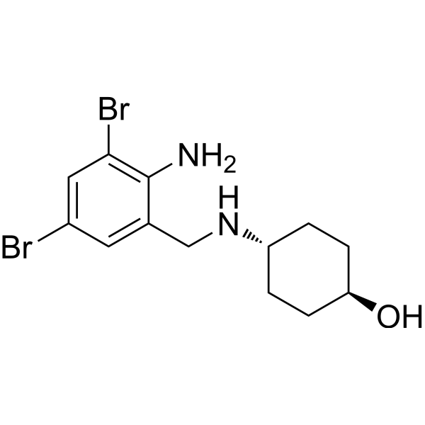
Ambroxol (NA-872), an active metabolite of the proagent Bromhexine, has potent expectorant effects. Ambroxol is a glucocerebrosidase (GCase) chaperone and increases glucocerebrosidase activity. Ambroxol induces lung autophagy and has the potential for Parkinson disease and neuronopathic Gaucher disease research.
For research use only. We do not sell to patients.
- Purity : 99.88%
- CAS No.: 18683-91-5
- Formula: C13H18Br2N2O
- Molecular Weight:378.10
-
Storage:Powder -20°C, 3 years , 4°C, 2 years ; In solvent -80°C, 2 years , -20°C, 1 year

To place orders, for customer services and technical support, please contact: MedChemExpress USA
Tel: 609-228-6898 E-mail: [email protected] [email protected]


-
 Biological Activity
-
Chemical Information
-
Solvent & Solubility
- Protocols
- Purity & Documentation
- References
-
 Help & FAQs


-

HY-100558
Bafilomycin A1V-ATPase Inhibitor -

HY-10219
RapamycinmTOR Inhibitor -
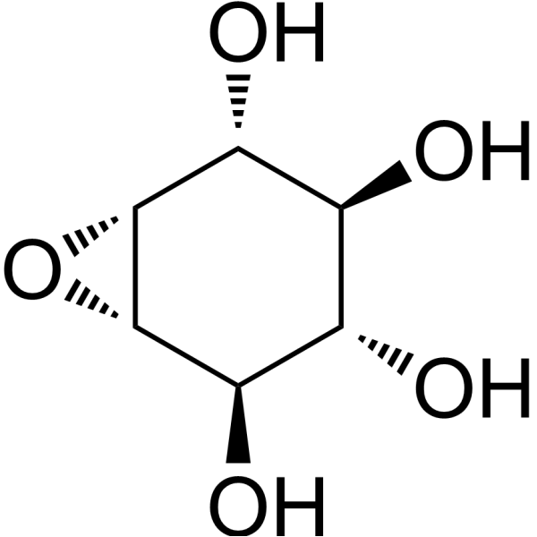
HY-100944
Conduritol B epoxideGcase Inhibitor -
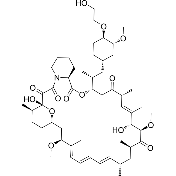
HY-10218
EverolimusmTORC Inhibitor -

HY-10071
Y-27632ROCK Inhibitor -
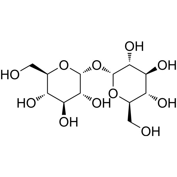
HY-N1132
D-(+)-TrehaloseDisaccharide -
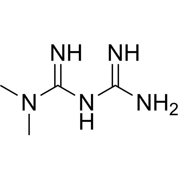
HY-B0627
MetforminAntihyperglycemic Agent -
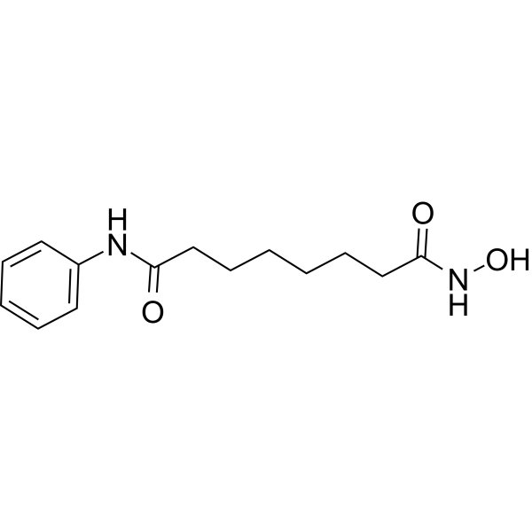
HY-10221
VorinostatHDAC Inhibitor -
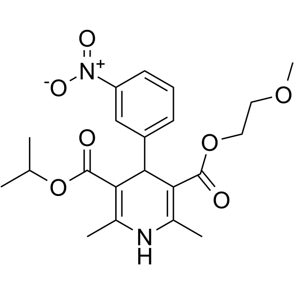
HY-B0265
NimodipineDihydropyridine Calcium Antagonist -
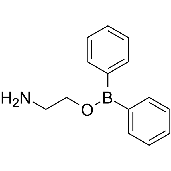
HY-W009724
2-Aminoethyl diphenylborinateIP3R Inhibitor -
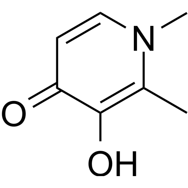
HY-B0568
DeferiproneFree iron chelating agent -
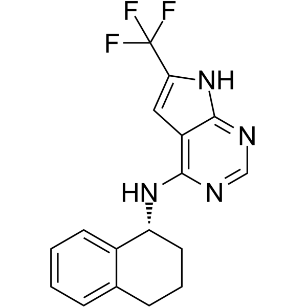
HY-152943
MTK458Mitophagy Activator
Publications Citing Use of MedChemExpress (MCE) Ambroxol
More

Biological Activity
Description
In Vivo
MedChemExpress (MCE) has not independently confirmed the accuracy of these methods. They are for reference only.
Clinical Trial
| NCT Number | Sponsor | Condition | Start Date |
Phase

|
|---|---|---|---|---|
| NCT01329991 | 2011-05 | PHASE1 |


Chemical Information
-
CAS No. 18683-91-5
-
Appearance Solid
-
Molecular Weight 378.10
-
Formula C13H18Br2N2O
-
Color White to off-white
-
SMILES
O[C@H]1CC[C@H](NCC2=CC(Br)=CC(Br)=C2N)CC1
-
Synonyms
NA-872
-
Shipping
Room temperature in continental US; may vary elsewhere.
-
Storage
Powder -20°C 3 years 4°C 2 years In solvent -80°C 2 years -20°C 1 year


Publications (3)
-
Journal Impact Factor
-
Most Recent
-
Cell Rep
TRESK channel contributes to depolarization-induced shunting inhibition and modulates epileptic seizures. [Abstract]2021 Jul 20;36(3):109404. PMID: 34289346 -
-
Evid Based Complement Alternat Med
Reineckia carnea Alleviates the Production of Inflammatory Cytokines and MUC5AC in Rats with Chronic Obstructive Pulmonary Disease. [Abstract]2022 Jun 16:2022:2135487. PMID: 35754687


Solvent & Solubility
In Vitro:
DMSO : 100 mg/mL (264.48 mM; Need ultrasonic; Hygroscopic DMSO has a significant impact on the solubility of product, please use newly opened DMSO)
Please refer to the solubility information to select the appropriate solvent. Once prepared, please aliquot and store the solution to prevent product inactivation from repeated freeze-thaw cycles.
Storage method and period of stock solution: -80°C, 2 years; -20°C, 1 year. When stored at -80°C, please use it within 2 years. When stored at -20°C, please use it within 1 year.
Please refer to the solubility information to select the appropriate solvent. Once prepared, please aliquot and store the solution to prevent product inactivation from repeated freeze-thaw cycles.
Storage method and period of stock solution: -80°C, 2 years; -20°C, 1 year. When stored at -80°C, please use it within 2 years. When stored at -20°C, please use it within 1 year.
Concentration (start) × Volume (start) = Concentration (final) × Volume (final)
In Vivo:
Select the appropriate dissolution method based on your experimental animal and administration route.
- For the following dissolution methods, please ensure to first prepare a clear stock solution using an In Vitro approach and then sequentially add co-solvents:
- To ensure reliable experimental results, the clarified stock solution can be appropriately stored based on storage conditions. As for the working solution for In Vivo experiments, it is recommended to prepare freshly and use it on the same day.
- The percentages shown for the solvents indicate their volumetric ratio in the final prepared solution. If precipitation or phase separation occurs during preparation, heat and/or sonication can be used to aid dissolution.
Add each solvent one by one: 5% DMSO 95% Saline
Solubility: 5 mg/mL (13.22 mM); Suspended solution; Need ultrasonic
Add each solvent one by one: 10% DMSO 40% PEG300 5% Tween-80 45% Saline
Solubility: ≥ 2.5 mg/mL (6.61 mM); Clear solution
This protocol yields a clear solution of ≥ 2.5 mg/mL (saturation unknown).
Taking 1 mL working solution as an example, add 100 μL DMSO stock solution (25.0 mg/mL) to 400 μL PEG300, and mix evenly; then add 50 μL Tween-80 and mix evenly; then add 450 μL Saline to adjust the volume to 1 mL.
Preparation of Saline: Dissolve 0.9 g sodium chloride in ddH₂O and dilute to 100 mL to obtain a clear Saline solution.
Add each solvent one by one: 10% DMSO 90% (20% SBE-β-CD in Saline)
Solubility: ≥ 2.5 mg/mL (6.61 mM); Clear solution
This protocol yields a clear solution of ≥ 2.5 mg/mL (saturation unknown).
Taking 1 mL working solution as an example, add 100 μL DMSO stock solution (25.0 mg/mL) to 900 μL 20% SBE-β-CD in Saline, and mix evenly.
Preparation of 20% SBE-β-CD in Saline (4°C, storage for one week): 2 g SBE-β-CD powder is dissolved in 10 mL Saline, completely dissolve until clear.
In Vivo Dissolution Calculator
Please enter the basic information of animal experiments:
-
-
-
-
Recommended: Prepare an additional quantity of animals to account for potential losses during experiments.
Please enter your animal formula composition:
-
%DMSO +
Recommended: Keep the proportion of DMSO in working solution below 2% if your animal is weak.
-
%+
-
+%Tween-80 + +
-
%Saline +
The co-solvents required include: DMSO, . All of co-solvents are available by MedChemExpress (MCE). , Tween 80. All of co-solvents are available by MedChemExpress (MCE).
Working solution concentration: 0.22 mg/mL
Method for preparing stock solution: mg drug dissolved in μL DMSO. Stock solution concentration: mg/mL.
1. Take μL DMSO stock solution;
2. Add μL .
μL , mix evenly;
3. Then add μL Tween 80, mix evenly;
4. Then add μL
Please ensure that the stock solution in the first step is dissolved to a clear state, and add co-solvents in sequence. You can use ultrasonic heating (ultrasonic cleaner, recommended frequency 20-40 kHz), vortexing, etc. to assist dissolution.


Purity & Documentation
-
Data Sheet (274 KB)
-
SDS (394 KB)
- English - EN (394 KB)
- Français - FR (394 KB)
- Deutsch - DE (394 KB)
- Norwegian - NO (394 KB)
- Español - ES (394 KB)
- Swedish - SV (394 KB)
- Italian - IT (394 KB)
- Korean - KR (394 KB)
- Portuguese - PT (394 KB)
-
Handling Instructions (2659 KB)
References
[1]. Vojo Deretic, et al. Enhancement of lung levels of antibiotics by ambroxol and bromhexine. Expert Opin Drug Metab Toxicol. 2019 Mar;15(3):213-218. [Content Brief]
[2]. Anna Migdalska-Richards, et al. Ambroxol effects in glucocerebrosidase and α-synuclein transgenic mice. Ann Neurol. 2016 Nov;80(5):766-775. [Content Brief]


Complete Stock Solution Preparation Table
Please refer to the solubility information to select the appropriate solvent. Once prepared, please aliquot and store the solution to prevent product inactivation from repeated freeze-thaw cycles.
Storage method and period of stock solution: -80°C, 2 years; -20°C, 1 year. When stored at -80°C, please use it within 2 years. When stored at -20°C, please use it within 1 year.
| Optional Solvent | Concentration Solvent Mass | 1 mg | 5 mg | 10 mg | 25 mg |
|---|---|---|---|---|---|
| DMSO | 1 mM | 2.6448 mL | 13.2240 mL | 26.4480 mL | 66.1201 mL |
| 5 mM | 0.5290 mL | 2.6448 mL | 5.2896 mL | 13.2240 mL | |
| 10 mM | 0.2645 mL | 1.3224 mL | 2.6448 mL | 6.6120 mL | |
| 15 mM | 0.1763 mL | 0.8816 mL | 1.7632 mL | 4.4080 mL | |
| 20 mM | 0.1322 mL | 0.6612 mL | 1.3224 mL | 3.3060 mL | |
| 25 mM | 0.1058 mL | 0.5290 mL | 1.0579 mL | 2.6448 mL | |
| 30 mM | 0.0882 mL | 0.4408 mL | 0.8816 mL | 2.2040 mL | |
| 40 mM | 0.0661 mL | 0.3306 mL | 0.6612 mL | 1.6530 mL | |
| 50 mM | 0.0529 mL | 0.2645 mL | 0.5290 mL | 1.3224 mL | |
| 60 mM | 0.0441 mL | 0.2204 mL | 0.4408 mL | 1.1020 mL | |
| 80 mM | 0.0331 mL | 0.1653 mL | 0.3306 mL | 0.8265 mL | |
| 100 mM | 0.0264 mL | 0.1322 mL | 0.2645 mL | 0.6612 mL |


Ambroxol Related Classifications
HY-B1039 Related Classifications
 Powered by Bioz