



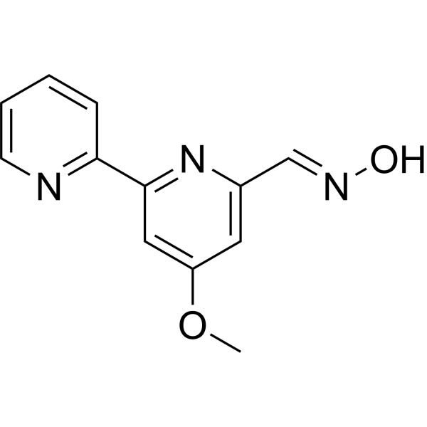
Caerulomycin A
Based on 3 publication(s) in Google Scholar
Caerulomycin A is an orally active immunomodulator and antimicrobial agent. Caerulomycin A targets Smad3, STAT1 and GATA-3. Caerulomycin A downregulates GATA-3 expression, inhibits Th2 cell differentiation and Th2 cytokine production, reduces IgE levels, and alleviates pulmonary inflammatory responses and eosinophil infiltration. Caerulomycin A ameliorates collagen-induced arthritis symptoms, reduces joint inflammation and synovitis, and decreases the levels of proinflammatory cytokines in joints. Caerulomycin A inhibits the growth of some filamentous fungi, yeasts and specific bacteria. Caerulomycin A can be used in research related to arthritis and asthma.
For research use only. We do not sell to patients.
- Purity : 98.0%
- CAS No.: 21802-37-9
- Formula: C12H11N3O2
- Molecular Weight:229.24
-
Storage:Powder -20°C, 3 years , 4°C, 2 years ; In solvent -80°C, 2 years , -20°C, 1 year

To place orders, for customer services and technical support, please contact: MedChemExpress USA
Tel: 609-228-6898 E-mail: [email protected] [email protected]


-
 Biological Activity
-
Chemical Information
-
Solvent & Solubility
- Protocols
- Purity & Documentation
- References
-
 Help & FAQs


-
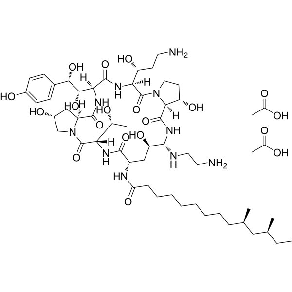
HY-17006
Caspofungin diacetateAntifungal Agent -
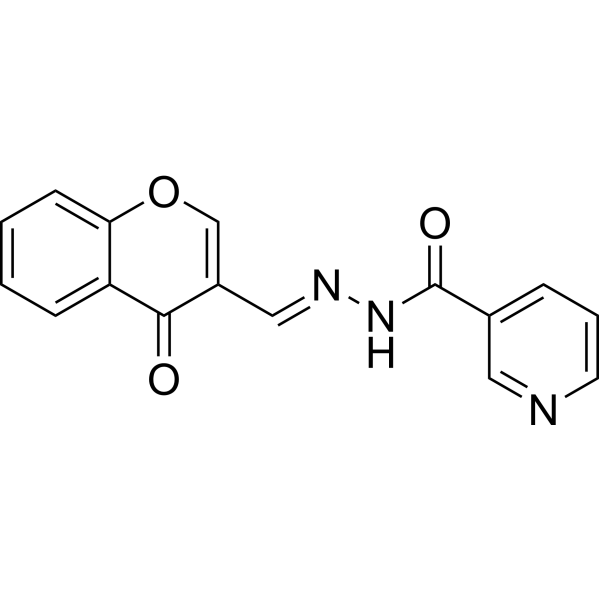
HY-101853
STAT5-IN-1STAT5 Inhibitor -
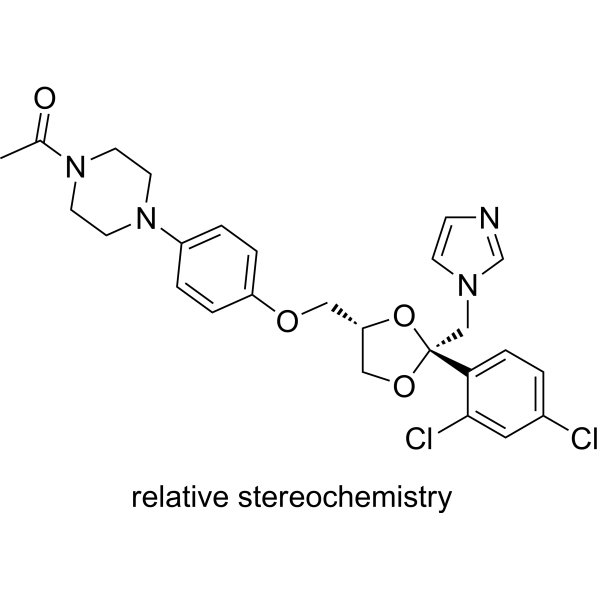
HY-B0105
KetoconazoleCYP3A4 Inhibitor -
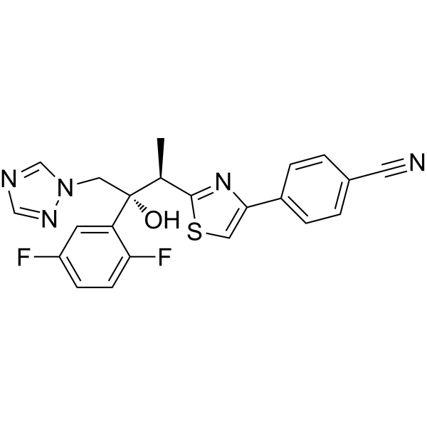
HY-14273
IsavuconazoleTriazole Antifungal -

HY-14273R
Isavuconazole (Standard)Triazole Antifungal -
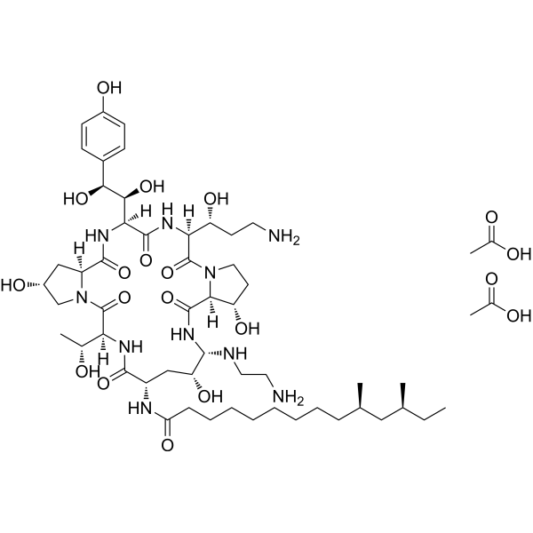
HY-17006R
Caspofungin diacetate (Standard)Antifungal Agent -
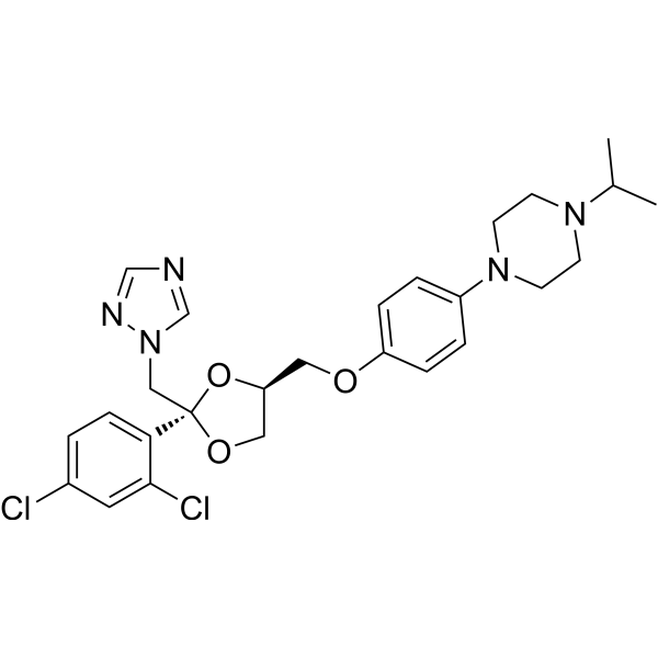
HY-B1790
TerconazoleFungal Inhibitor -
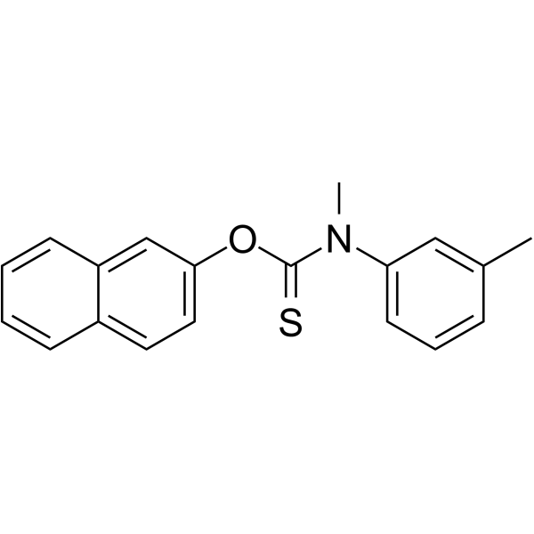
HY-B0370
TolnaftateFungal Inhibitor -
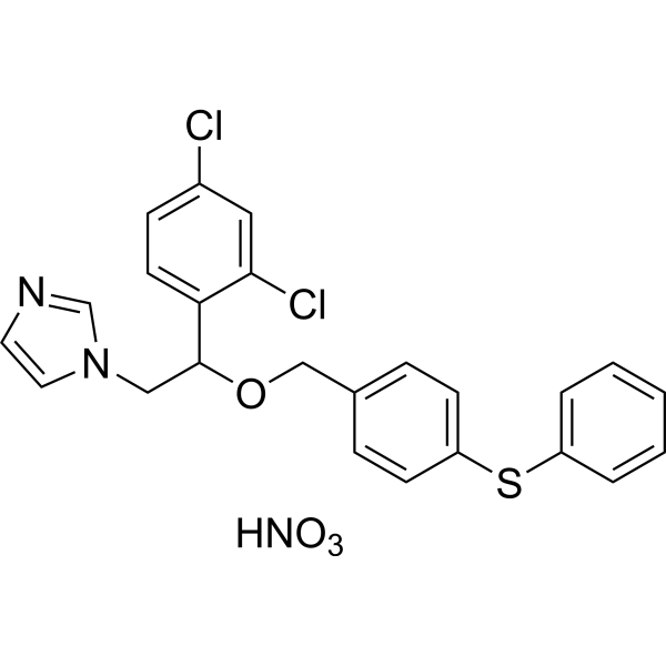
HY-B0359
Fenticonazole NitrateAntibiotic -

HY-B0105R
Ketoconazole (Standard)CYP3A4 Inhibitor -
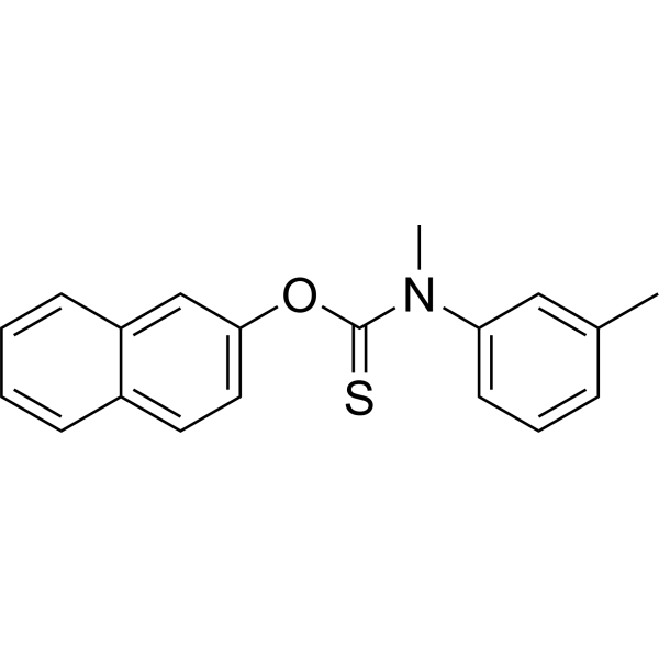
HY-B0370R
Tolnaftate (Standard)Fungal Inhibitor -
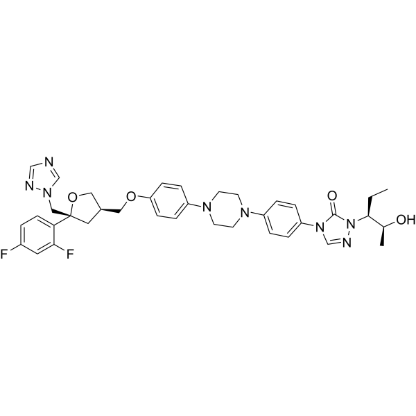
HY-17373
PosaconazoleAnti-fungal Agent
Publications Citing Use of MedChemExpress (MCE) Caerulomycin A
MoreAll Antibiotic Isoforms
More

Biological Activity
Description
IC50 & Target
Cellular Effect
|
Cell Line

|
Type | Value | Description | References |
|---|---|---|---|---|
| A549 | IC50 |
0.26 μM
Compound: 7
|
Cytotoxicity against human A549 cells after 24 hrs by SRB assay
Cytotoxicity against human A549 cells after 24 hrs by SRB assay
|
[PMID: 21770434] |
| HL-60 | IC50 |
0.71 μM
Compound: 7
|
Cytotoxicity against human HL60 cells after 72 hrs by MTT assay
Cytotoxicity against human HL60 cells after 72 hrs by MTT assay
|
[PMID: 21770434] |
| K562 | IC50 |
>50 μM
Compound: 7
|
Cytotoxicity against human K562 cells after 72 hrs by MTT assay
Cytotoxicity against human K562 cells after 72 hrs by MTT assay
|
[PMID: 21770434] |
| KB | IC50 |
>50 μM
Compound: 7
|
Cytotoxicity against human KB cells after 72 hrs by MTT assay
Cytotoxicity against human KB cells after 72 hrs by MTT assay
|
[PMID: 21770434] |
In Vitro
Caerulomycin A (0-0.31 μM; 5 days, 72 h) dose-dependently induces CD4+Foxp3+ Treg generation in non-polarizing conditions, synergistically enhances TGF-β-mediated Treg generation, suppresses Th1 and Th17 cell differentiation and cytokine secretion, reduces CD44 expression, and increases mitochondrial membrane potential in mouse naïve CD4+ T cells[1].
Caerulomycin A (0.04-0.15 μM; 48 h, 4 days) dose-dependently suppresses CD4+ effector T cell activation and alloreactive T cell proliferation in mouse cell cultures via generated Tregs (with or without TGF-β)[1].
Caerulomycin A (0.04-0.31 μM; 24 h pre-incubation) dose-dependently suppresses IFN-γ/IL-6-mediated STAT1 signaling by enhancing SOCS1 expression, reduces STAT1 target gene and protein expression, and enhances TGF-β-mediated Smad3 signaling in mouse CD4+ T cells[1].
Caerulomycin A (0.04-0.15 μM; 5 days) dose-dependently rescues mouse CD4+Foxp3+ Treg generation from IFN-γ-mediated suppression, and this Treg generation is strictly dependent on Smad3 activity[1].
Caerulomycin A (0.08-0.31 μM; 6 days) inhibits IL-4 expression in terminally differentiated mouse Th2 CD4+ T cells, with a concentration-dependent reduction in IL-4+ cell percentage over 6 days[2].
Caerulomycin A (0.08-0.31 μM; 6 days) concentration-dependently inhibits the differentiation of mouse naïve CD4+ T cells to Th2 cells, reducing both IL-4 expression (with significant inhibition at 0.31 μM) and GATA-3 expression in IL-4+ CD4+ T cells after 6 days of culture[2].
Caerulomycin A (5-100 μg/mL; up to 96 h) inhibits growth of diverse yeasts, filamentous fungi, uredospores, and bacteria in an agar dilution assay, with complete inhibition ranging from 5 μg/mL (for Candida albicans, Saccharomyces cerevisiae, Isaria cretacea, Rhizopus nigricans at 24 h) to 100 μg/mL (for Escherichia coli and Puccinia graminis var. tritici race 15B uredospore germination at all time points) when incubated at 28°C for up to 96 hours[3].
Caerulomycin A (50 μg/mL; 7-day fermentation) inhibits growth of Candida albicans in a disc diffusion assay, produced via a 7-day fermentation of Streptomyces caeruleus PRL 1687 in starch-Czapelt medium[3].
MedChemExpress (MCE) has not independently confirmed the accuracy of these methods. They are for reference only. Further protocols information, click here.
In Vivo
Caerulomycin A (1-10 mg/kg; p.o.; daily; 12 days) dose-dependently attenuates asthma symptoms in OVA-induced BALB/c mice, with the 10 mg/kg oral daily dose achieving significant suppression of lung inflammation, eosinophil infiltration, Th2 cytokine production, and IgE levels[2].
MedChemExpress (MCE) has not independently confirmed the accuracy of these methods. They are for reference only.
-
Animal Model:DBA/1 (male, 6-8 weeks of age, collagen-induced arthritis model)[1]
-
Dosage:1 mg/kg; 10 mg/kg
-
Administration:daily; 50 days
-
Result:Reduced arthritis clinical severity scores in a dose-dependent manner.
Showed significantly reduced inflammatory response and bone degeneration, with the 10 mg/kg dose showing the greatest reduction.
Significantly decreased knee joint proinflammatory cytokine levels (IFN-γ, TNF-α, IL-6).
Increased percentage of Tregs in draining lymph nodes, with the 10 mg/kg group reaching 13% CD4+Foxp3+ cells.
Significantly reduced IFN-γ and TNF-α secretion in in vitro restimulated lymph node cells with bovine collagen type II.
Significantly lowered serum levels of MMP-3.
-
Animal Model:BALB/c (female, 6-8 weeks old, OVA-induced asthma model)[2]
-
Dosage:1 mg/kg; 10 mg/kg
-
Administration:p.o.; daily; 12 days (days 13-24)
-
Result:Significantly reduced lung inflammation.
Reduced IL-5 and IL-13 levels in BAL fluid from.
Significantly reduced total IgE levels in BAL fluid.
Significantly reduced lung inflammation.
Significantly reduced total BAL fluid cell counts.


Chemical Information
-
CAS No. 21802-37-9
-
Appearance Solid
-
Molecular Weight 229.24
-
Formula C12H11N3O2
-
Color White to off-white
-
SMILES
COC1=CC(C2=NC=CC=C2)=NC(/C=N/O)=C1
-
Synonyms
Cerulomycin; Caerulomycin
-
Initial Source
Streptomyces caeruleus
-
Shipping
Room temperature in continental US; may vary elsewhere.
-
Storage
Powder -20°C 3 years 4°C 2 years In solvent -80°C 2 years -20°C 1 year


Publications (3)
-
Journal Impact Factor
-
Most Recent
-
Cell Res
Transient mechanical activation of the Piezo1 channel facilitates ex vivo expansion of hematopoietic stem cells. [Abstract]2026 Jan 9. PMID: 41507440 -
J Ovarian Res
IFN-γ in ovarian tumor microenvironment upregulates HLA-E expression and predicts a poor prognosis. [Abstract]2023 Nov 25;16(1):229. PMID: 38007483 -
Eur J Med Res
Efficacy of caerulomycin A in modulating macrophage polarization and cytokine response in a murine model of lipopolysaccharide-induced sepsis. [Abstract]2025 Jan 10;30(1):22. PMID: 39794821


Solvent & Solubility
In Vitro:
DMSO : 150 mg/mL (654.34 mM; Need ultrasonic; Hygroscopic DMSO has a significant impact on the solubility of product, please use newly opened DMSO)
Please refer to the solubility information to select the appropriate solvent. Once prepared, please aliquot and store the solution to prevent product inactivation from repeated freeze-thaw cycles.
Storage method and period of stock solution: -80°C, 2 years; -20°C, 1 year. When stored at -80°C, please use it within 2 years. When stored at -20°C, please use it within 1 year.
Please refer to the solubility information to select the appropriate solvent. Once prepared, please aliquot and store the solution to prevent product inactivation from repeated freeze-thaw cycles.
Storage method and period of stock solution: -80°C, 2 years; -20°C, 1 year. When stored at -80°C, please use it within 2 years. When stored at -20°C, please use it within 1 year.
Concentration (start) × Volume (start) = Concentration (final) × Volume (final)
In Vivo:
Select the appropriate dissolution method based on your experimental animal and administration route.
- For the following dissolution methods, please ensure to first prepare a clear stock solution using an In Vitro approach and then sequentially add co-solvents:
- To ensure reliable experimental results, the clarified stock solution can be appropriately stored based on storage conditions. As for the working solution for In Vivo experiments, it is recommended to prepare freshly and use it on the same day.
- The percentages shown for the solvents indicate their volumetric ratio in the final prepared solution. If precipitation or phase separation occurs during preparation, heat and/or sonication can be used to aid dissolution.
Add each solvent one by one: 10% DMSO 40% PEG300 5% Tween-80 45% Saline
Solubility: ≥ 2.5 mg/mL (10.91 mM); Clear solution
This protocol yields a clear solution of ≥ 2.5 mg/mL (saturation unknown).
Taking 1 mL working solution as an example, add 100 μL DMSO stock solution (25.0 mg/mL) to 400 μL PEG300, and mix evenly; then add 50 μL Tween-80 and mix evenly; then add 450 μL Saline to adjust the volume to 1 mL.
Preparation of Saline: Dissolve 0.9 g sodium chloride in ddH₂O and dilute to 100 mL to obtain a clear Saline solution.
Add each solvent one by one: 10% DMSO 90% (20% SBE-β-CD in Saline)
Solubility: ≥ 2.5 mg/mL (10.91 mM); Clear solution
This protocol yields a clear solution of ≥ 2.5 mg/mL (saturation unknown).
Taking 1 mL working solution as an example, add 100 μL DMSO stock solution (25.0 mg/mL) to 900 μL 20% SBE-β-CD in Saline, and mix evenly.
Preparation of 20% SBE-β-CD in Saline (4°C, storage for one week): 2 g SBE-β-CD powder is dissolved in 10 mL Saline, completely dissolve until clear.
In Vivo Dissolution Calculator
Please enter the basic information of animal experiments:
-
-
-
-
Recommended: Prepare an additional quantity of animals to account for potential losses during experiments.
Please enter your animal formula composition:
-
%DMSO +
Recommended: Keep the proportion of DMSO in working solution below 2% if your animal is weak.
-
%+
-
+%Tween-80 + +
-
%Saline +
The co-solvents required include: DMSO, . All of co-solvents are available by MedChemExpress (MCE). , Tween 80. All of co-solvents are available by MedChemExpress (MCE).
Working solution concentration: 0.22 mg/mL
Method for preparing stock solution: mg drug dissolved in μL DMSO. Stock solution concentration: mg/mL.
1. Take μL DMSO stock solution;
2. Add μL .
μL , mix evenly;
3. Then add μL Tween 80, mix evenly;
4. Then add μL
Please ensure that the stock solution in the first step is dissolved to a clear state, and add co-solvents in sequence. You can use ultrasonic heating (ultrasonic cleaner, recommended frequency 20-40 kHz), vortexing, etc. to assist dissolution.

Protocols
-
Research Protocol for Infectious Diseases
Infectious-disease experiments test how pathogens interact with host barriers, innate immune receptors, inflammatory signaling, pathogen replication, and tissue injury; pattern-recognition receptors such as TLRs, RIG-I-like receptors, NOD-like receptors, and inflammasomes detect microbial molecules and activate NF-κB, interferon, and cytokine responses. The central hypothesis is that infection severity reflects the balance between pathogen burden and host response: protective inflammation restricts pathogen growth, whereas excessive or mislocalized inflammation contributes to tissue damage and disease phenotype. Unresolved questions include which host pathways are protective versus pathogenic, why some infection models fail to translate to human disease, and which combined readouts best predict clinically relevant infection outcomes.
-
RNA extraction experimental
By lysing cells, releasing RNA, and removing impurities such as proteins and DNA, high-purity RNA products are finally obtained. The commonly used traditional method is the guanidine isothiocyanate/phenol/chloroform method (Trizol), which is suitable for a variety of animal materials including animal tissues, microorganisms, cultured cells, etc., and most plant materials.
-
Collagen-Induced Arthritis
Collagen-induced arthritis (CIA) is an autoimmune murine model of rheumatoid arthritis in which immunization with type II collagen (CII) emulsified in an adjuvant induces a T cell- and autoantibody-driven inflammatory arthritis characterized by synovial hyperplasia, immune cell infiltration, and joint destruction. The model typically relies on genetically susceptible mouse strains (e. g. , DBA/1) and reproduces key features of human rheumatoid arthritis, including anti-collagen immune responses and progressive joint inflammation. Disease onset generally occurs within ~3-4 weeks after immunization, depending on antigen/adjuvant combinations and protocol variation. The immunopathology is driven by adaptive immune activation against CII, leading to systemic and local joint inflammation mediated by pro-inflammatory cytokines and effector immune cells, making CIA a standard preclinical platform for evaluating immunomodulatory and anti-arthritic interventions.
-
Naïve CD4+ T-cell subset differentiation/polarization
Naïve CD4+ T-cell subset differentiation/polarization is an in vitro assay in which purified naïve CD4+ T cells are activated through TCR and CD28 costimulation and cultured with defined cytokines and neutralizing antibodies to generate Th0, Th1, Th2, Th17, or induced Treg-like populations. Differentiation is detected by subset-associated cytokines and transcription factors: IFN-γ/T-bet for Th1, IL-4/GATA3 for Th2, IL-17A/RORγt for Th17, and Foxp3 for induced Treg cells. The assay readout is usually generated by intracellular cytokine staining after restimulation, transcription-factor staining by flow cytometry, ELISA of secreted cytokines, or gene-expression analysis. The result reflects cytokine-directed lineage commitment or polarization rather than antigen-specific immune protection by itself.
-
Filamentous Fungal Mold Culture and Sporulation
Filamentous fungal mold culture and sporulation assays grow hyphae under defined nutritional and environmental conditions until asexual spores, commonly conidia, are produced; the main readouts are colony growth, sporulation onset, conidial yield, conidial morphology, viability, and, when relevant, downstream infectivity or stress phenotype.
-
Research Protocol for Inflammation-related Diseases
The NLRP3 inflammasome is a cytosolic innate immune signaling platform that integrates priming signals and danger-signal activation to promote caspase-1 activation, maturation of IL-1β and IL-18, and gasdermin D-mediated pyroptotic cell death. The core experimental logic is to determine whether inflammatory disease phenotypes are driven by increased NLRP3 expression, ASC-containing inflammasome assembly, caspase-1 cleavage, GSDMD cleavage, and extracellular release of IL-1β/IL-18 rather than by nonspecific cell injury alone. The pathway is strongly linked to inflammation-related disease phenotypes because monosodium urate crystals activate NALP3/NLRP3 inflammasome signaling in gout-like crystal inflammation, cholesterol crystals activate NLRP3 inflammasomes in atherogenesis models, and DSS-induced intestinal inflammation has been reported to involve NLRP3 inflammasome activity. However, experimental colitis studies also show context-dependent protective effects of NLRP3 inflammasome co
-
iPSC cell differentiation
Induced pluripotent stem cells (iPSCs) are a type of cell that has similar properties to embryonic stem cells through somatic cell reprogramming.


Purity & Documentation
-
Data Sheet (281 KB)
-
SDS (479 KB)
- English - EN (479 KB)
- Français - FR (479 KB)
- Deutsch - DE (479 KB)
- Norwegian - NO (479 KB)
- Español - ES (479 KB)
- Swedish - SV (479 KB)
- Italian - IT (479 KB)
- Korean - KR (479 KB)
- Portuguese - PT (479 KB)
-
Handling Instructions (2659 KB)
References
[1]. Gurram RK, et al. Caerulomycin A enhances transforming growth factor-β (TGF-β)-Smad3 protein signaling by suppressing interferon-γ (IFN-γ)-signal transducer and activator of transcription 1 (STAT1) protein signaling to expand regulatory T cells (Tregs). J Biol Chem. 2014;289(25):17515-17528. [Content Brief]
[2]. Kujur W, et al. Caerulomycin A inhibits Th2 cell activity: a possible role in the management of asthma. Sci Rep. 2015;5:15396. Published 2015 Oct 20. [Content Brief]
[3]. FUNK A, et al. Caerulomycin, a new antibiotic from Streptomyces caeruleus Baldacci. I. Production, isolation, assay, and biological properties. Can J Microbiol. 1959 Aug;5:317-21. [Content Brief]


Complete Stock Solution Preparation Table
Please refer to the solubility information to select the appropriate solvent. Once prepared, please aliquot and store the solution to prevent product inactivation from repeated freeze-thaw cycles.
Storage method and period of stock solution: -80°C, 2 years; -20°C, 1 year. When stored at -80°C, please use it within 2 years. When stored at -20°C, please use it within 1 year.
| Optional Solvent | Concentration Solvent Mass | 1 mg | 5 mg | 10 mg | 25 mg |
|---|---|---|---|---|---|
| DMSO | 1 mM | 4.3622 mL | 21.8112 mL | 43.6224 mL | 109.0560 mL |
| 5 mM | 0.8724 mL | 4.3622 mL | 8.7245 mL | 21.8112 mL | |
| 10 mM | 0.4362 mL | 2.1811 mL | 4.3622 mL | 10.9056 mL | |
| 15 mM | 0.2908 mL | 1.4541 mL | 2.9082 mL | 7.2704 mL | |
| 20 mM | 0.2181 mL | 1.0906 mL | 2.1811 mL | 5.4528 mL | |
| 25 mM | 0.1745 mL | 0.8724 mL | 1.7449 mL | 4.3622 mL | |
| 30 mM | 0.1454 mL | 0.7270 mL | 1.4541 mL | 3.6352 mL | |
| 40 mM | 0.1091 mL | 0.5453 mL | 1.0906 mL | 2.7264 mL | |
| 50 mM | 0.0872 mL | 0.4362 mL | 0.8724 mL | 2.1811 mL | |
| 60 mM | 0.0727 mL | 0.3635 mL | 0.7270 mL | 1.8176 mL | |
| 80 mM | 0.0545 mL | 0.2726 mL | 0.5453 mL | 1.3632 mL | |
| 100 mM | 0.0436 mL | 0.2181 mL | 0.4362 mL | 1.0906 mL |


Caerulomycin A Related Classifications
HY-114495 Related Classifications
 Powered by Bioz