



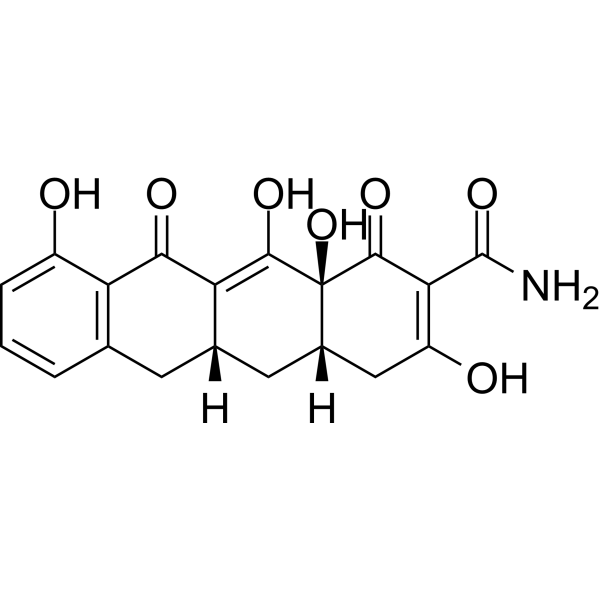
Incyclinide
Based on 12 publication(s) in Google Scholar
Incyclinide (CMT-3, COL-3) is a matrix metalloproteinase (MMP) inhibitor and inhibiting angiogenesis, tumor growth and invasion, and metastasis.
For research use only. We do not sell to patients.
- Purity : 98.51%
- CAS No.: 15866-90-7
- Formula: C19H17NO7
- Molecular Weight:371.34
-
Storage:Powder -20°C, 3 years , 4°C, 2 years ; In solvent -80°C, 2 years , -20°C, 1 year

To place orders, for customer services and technical support, please contact: MedChemExpress USA
Tel: 609-228-6898 E-mail: [email protected] [email protected]


-
 Biological Activity
-
Chemical Information
-
Solvent & Solubility
- Protocols
- Purity & Documentation
- References
-
 Help & FAQs


-
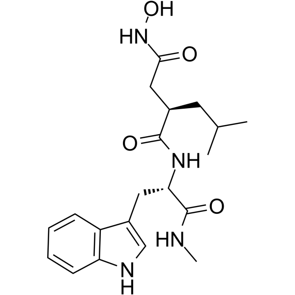
HY-15768
IlomastatMMP Inhibitor -

HY-12354
SB-3CTMMP2/9 Inhibitor -
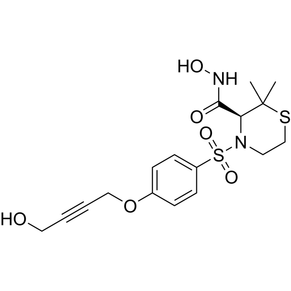
HY-119307
ApratastatTACE/MMPs Inhibitor -

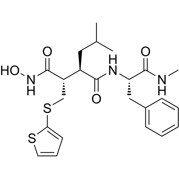
HY-12169
MarimastatMMP Inhibitor -

HY-Y0873
PEG300Neutral Polymer -
HY-13564
BatimastatMMP Inhibitor -

HY-P7109
EGF Protein, Human -

HY-15763
ErastinFerroptosis Inducer -

HY-15827
NSC 405020MMP14 Inhibitor -
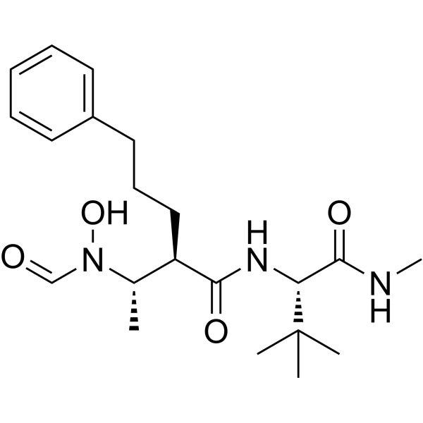
HY-19956
GI254023XADAM10/MMP9 Inhibitor -
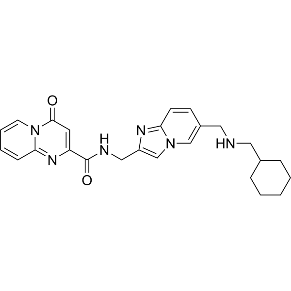
HY-134836
STM2457METTL3 Inhibitor -
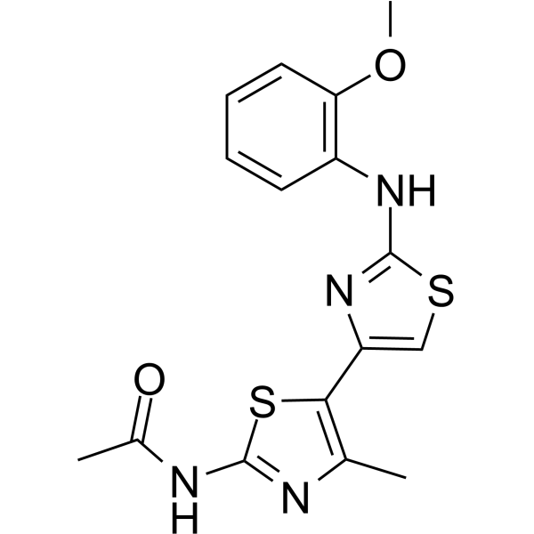
HY-103482
JNJ0966MMP-9 Inhibitor
Publications Citing Use of MedChemExpress (MCE) Incyclinide
More- Sci Adv. 2023 Jan 20;9(3):eadd3867. [Abstract]
- Proc Natl Acad Sci U S A. 2023 Apr 25;120(17):e2218522120. [Abstract]
- ACS Biomater Sci Eng. 2025 Feb 10;11(2):916-929. [Abstract]
- EMBO Rep. 2023 Dec 6;24(12):e57228. [Abstract]
- Cells. 2021 Aug 22;10(8):2163. [Abstract]
- Molecules. 2023 May 23;28(11):4262. [Abstract]
- Sci Rep. 2025 May 6;15(1):15762. [Abstract]
- In Vitro Model. 2025 Mar 7;4(1):59-69. [Abstract]
- bioRxiv. 2026 Jan 11:2026.01.09.698733. [Abstract]
- University of Minnesota. 2024.
- bioRxiv. 2023 Oct 12:2023.05.06.539698. [Abstract]
- Authorea. May 19, 2021.


Biological Activity
Description
In Vitro
Incyclinide has been shown to experimentally suppress prostate cancer, colon adenocarcinoma and melanoma invasiveness in cell culture. Adding incyclinide at final concentrations of 5 to 20 μM inhibits MT1-MMP gelatinolytic and caseinolytic activity, blocks MT1-MMPactivation of pro-MMP-2, and decreases invasiveness of HT-1080 fibrosarcoma cells[1]. Incyclinide is an especially effective inhibitor of the growth and viability of filamentous fungi. Most of the MICs of CMT-3 against filamentous fungi are found to be between 0.25 and 8 μg/mL, and the inhibition of viability of these fungi by incyclinide is routinely higher than 90%[2].
MedChemExpress (MCE) has not independently confirmed the accuracy of these methods. They are for reference only. Further protocols information, click here.
In Vivo
MedChemExpress (MCE) has not independently confirmed the accuracy of these methods. They are for reference only.
Clinical Trial
| NCT Number | Sponsor | Condition | Start Date |
Phase

|
|---|---|---|---|---|
| NCT01329991 | Plexxikon| | 2011-05 | PHASE1 |


Chemical Information
-
CAS No. 15866-90-7
-
Appearance Solid
-
Molecular Weight 371.34
-
Formula C19H17NO7
-
Color Light yellow to yellow
-
SMILES
O=C(C(C1=O)=C(O)C[C@]2([H])C[C@]3([H])CC4=C(C(C3=C(O)[C@@]21O)=O)C(O)=CC=C4)N
-
Synonyms
CMT-3; COL-3
-
Shipping
Room temperature in continental US; may vary elsewhere.
-
Storage
Powder -20°C 3 years 4°C 2 years In solvent -80°C 2 years -20°C 1 year


Publications (12)
-
Journal Impact Factor
-
Most Recent
-
Sci Adv
2023 Jan 20;9(3):eadd3867. PMID: 36662861 -
Proc Natl Acad Sci U S A
Computational drug discovery for castration-resistant prostate cancers through in vitro drug response modeling. [Abstract]2023 Apr 25;120(17):e2218522120. PMID: 37068243 -
ACS Biomater Sci Eng
2025 Feb 10;11(2):916-929. PMID: 39792458 -
EMBO Rep
Tetracyclines activate mitoribosome quality control and reduce ER stress to promote cell survival. [Abstract]2023 Dec 6;24(12):e57228. PMID: 37818824 -
Cells
The Chemically-Modified Tetracycline COL-3 and Its Parent Compound Doxycycline Prevent Microglial Inflammatory Responses by Reducing Glucose-Mediated Oxidative Stress. [Abstract]2021 Aug 22;10(8):2163. PMID: 34440932 -
Molecules
Scope and Limitations of Exploiting the Ability of the Chemosensitizer NV716 to Enhance the Activity of Tetracycline Derivatives against Pseudomonas aeruginosa. [Abstract]2023 May 23;28(11):4262. PMID: 37298737 -
Sci Rep
Pharmacological alternatives to oxytetracycline as potential treatment of flexural limb deformities in foals: a preliminary in vitro cell viability and proliferation study. [Abstract]2025 May 6;15(1):15762. PMID: 40328831 -
In Vitro Model
An in vitro model to measure the strength and stiffness of the extracellular matrix synthesized de novo by human fibroblasts. [Abstract]2025 Mar 7;4(1):59-69. PMID: 40160211 -
bioRxiv
Atypical tetracyclines promote longevity and ferroptotic neuroprotection via translation attenuation. [Abstract]2026 Jan 11:2026.01.09.698733. PMID: 41542485 -
-
bioRxiv
Omicron Spike confers enhanced infectivity and interferon resistance to SARS-CoV-2 in human nasal tissue. [Abstract]2023 Oct 12:2023.05.06.539698. PMID: 37425811 -


Solvent & Solubility
In Vitro:
DMSO : ≥ 100 mg/mL (269.29 mM; Hygroscopic DMSO has a significant impact on the solubility of product, please use newly opened DMSO)
* "≥" means soluble, but saturation unknown.
Please refer to the solubility information to select the appropriate solvent. Once prepared, please aliquot and store the solution to prevent product inactivation from repeated freeze-thaw cycles.
Storage method and period of stock solution: -80°C, 2 years; -20°C, 1 year. When stored at -80°C, please use it within 2 years. When stored at -20°C, please use it within 1 year.
Please refer to the solubility information to select the appropriate solvent. Once prepared, please aliquot and store the solution to prevent product inactivation from repeated freeze-thaw cycles.
Storage method and period of stock solution: -80°C, 2 years; -20°C, 1 year. When stored at -80°C, please use it within 2 years. When stored at -20°C, please use it within 1 year.
Concentration (start) × Volume (start) = Concentration (final) × Volume (final)
In Vivo:
Select the appropriate dissolution method based on your experimental animal and administration route.
- For the following dissolution methods, please ensure to first prepare a clear stock solution using an In Vitro approach and then sequentially add co-solvents:
- To ensure reliable experimental results, the clarified stock solution can be appropriately stored based on storage conditions. As for the working solution for In Vivo experiments, it is recommended to prepare freshly and use it on the same day.
- The percentages shown for the solvents indicate their volumetric ratio in the final prepared solution. If precipitation or phase separation occurs during preparation, heat and/or sonication can be used to aid dissolution.
Add each solvent one by one: 10% DMSO 40% PEG300 5% Tween-80 45% Saline
Solubility: 2.5 mg/mL (6.73 mM); Suspended solution; Need ultrasonic
This protocol yields a suspended solution of 2.5 mg/mL. Suspended solution can be used for oral and intraperitoneal injection.
Taking 1 mL working solution as an example, add 100 μL DMSO stock solution (25.0 mg/mL) to 400 μL PEG300, and mix evenly; then add 50 μL Tween-80 and mix evenly; then add 450 μL Saline to adjust the volume to 1 mL.
Preparation of Saline: Dissolve 0.9 g sodium chloride in ddH₂O and dilute to 100 mL to obtain a clear Saline solution.
In Vivo Dissolution Calculator
Please enter the basic information of animal experiments:
-
-
-
-
Recommended: Prepare an additional quantity of animals to account for potential losses during experiments.
Please enter your animal formula composition:
-
%DMSO +
Recommended: Keep the proportion of DMSO in working solution below 2% if your animal is weak.
-
%+
-
+%Tween-80 + +
-
%Saline +
The co-solvents required include: DMSO, . All of co-solvents are available by MedChemExpress (MCE). , Tween 80. All of co-solvents are available by MedChemExpress (MCE).
Working solution concentration: 0.22 mg/mL
Method for preparing stock solution: mg drug dissolved in μL DMSO. Stock solution concentration: mg/mL.
1. Take μL DMSO stock solution;
2. Add μL .
μL , mix evenly;
3. Then add μL Tween 80, mix evenly;
4. Then add μL
Please ensure that the stock solution in the first step is dissolved to a clear state, and add co-solvents in sequence. You can use ultrasonic heating (ultrasonic cleaner, recommended frequency 20-40 kHz), vortexing, etc. to assist dissolution.

Protocols
-
Mitochondrial membrane-potential fluorescent assay
Mitochondrial membrane potential fluorescent assays estimate ΔΨm in living cells using lipophilic cationic dyes such as TMRM, TMRE, rhodamine 123, and JC-1, which accumulate in mitochondria according to membrane polarization; loss of signal after FCCP or CCCP treatment is interpreted as mitochondrial depolarization. TMRM/TMRE and rhodamine 123 are commonly used for semi-quantitative live-cell microscopy or flow cytometry, while JC-1 can report a shift from red aggregate fluorescence to green monomer fluorescence during depolarization; interpretation requires controls because dye concentration, quenching mode, cell type, dye efflux, and mitochondrial mass can affect fluorescence independently of ΔΨm.
-
Human pluripotent stem cell endothelial-cell differentiation
Human pluripotent stem cell endothelial differentiation is based on stepwise developmental patterning: early activation of WNT/GSK3β inhibition promotes mesodermal or vascular progenitor entry, followed by endothelial specification using VEGF-related signaling, BMP4, FGF2, Notch modulation, or cAMP depending on the published protocol. Endothelial differentiation is read out by acquisition of CD31, CD34, VE-cadherin/CD144, KDR/VEGFR2, vWF, Tie2, NOS3, acetylated LDL uptake, tube/network formation, barrier function, and in vivo vessel-forming capacity where tested.
-
Vascular/Branching Fractal Analysis
Vascular/branching fractal analysis quantifies the geometric complexity of vessel trees or vascular networks from segmented 2D images, commonly by converting vessels into binary and/or skeletonized maps and estimating fractal dimension using box-counting or related approaches. Fractal dimension is interpreted as an image-derived readout of vascular branching complexity, space filling, or density, and has been applied to retinal photographs, fluorescein angiography, OCT angiography, capillary perfusion maps, and in vitro Matrigel angiogenesis networks. The assay readout is generated from vessel-positive pixels after image preprocessing, vessel segmentation, binarization, and optional skeletonization; reported outputs include fractal dimension, vessel density, branchpoint density, endpoint density, vessel length density, tortuosity, and generation-based branching metrics when VESGEN-style analysis is used. The biological interpretation is limited to quantitative vascular patterning and s
-
Mitochondrial membrane-potential and mitochondrial mass staining
Mitochondrial membrane potential staining measures the electrochemical polarization across the mitochondrial inner membrane in live cells using lipophilic cationic fluorescent probes; early rhodamine-based work showed that selective mitochondrial dye accumulation is lost when the mitochondrial transmembrane potential is dissipated. JC-1 reports mitochondrial polarization by shifting from green monomer fluorescence to red J-aggregate fluorescence as dye concentration increases within energized mitochondria; therefore, the red/green fluorescence ratio is used as a relative readout of mitochondrial membrane potential. TMRE or TMRM staining provides a single-channel relative readout because these cationic rhodamine esters accumulate in polarized mitochondria, and lower fluorescence indicates reduced mitochondrial polarization when acquisition and dye-loading conditions are controlled. Mitochondrial mass staining is commonly performed with MitoTracker Green FM or related MitoTracker dyes as
-
Endothelial Tube Formation Assay
Endothelial tube formation assay evaluates the ability of endothelial cells to attach, migrate, align, and organize into capillary-like networks when cultured on gelled basement membrane extract or Matrigel; the readout is the morphology and quantity of tube-like networks, which reflects an in vitro endothelial morphogenesis step related to angiogenesis. Basement membrane extract/Matrigel provides laminin-rich extracellular matrix cues that support endothelial differentiation into capillary-like structures, but it can contain biologically active growth factors, so growth-factor-reduced matrix is preferred when testing defined angiogenic stimulators or inhibitors.
-
Patient-Derived Xenograft (PDX)
Patient-derived xenograft (PDX) models are generated by engrafting primary human tumor tissue directly into immunodeficient mice, allowing in vivo propagation of patient tumor biology without initial in vitro adaptation. These models are used to preserve key histopathological and molecular characteristics of the original tumor and enable assessment of tumor growth dynamics and therapeutic response in a living organism. The biological readout is tumor engraftment and subsequent growth in the murine host, which reflects the ability of human tumor cells to survive, vascularize, and expand in an immunocompromised microenvironment.
-
Patient-Derived Orthotopic Xenograft (PDOX)
Patient-derived orthotopic xenograft (PDOX) modeling implants fresh patient tumor tissue or patient-derived tumor cells into the anatomically corresponding organ or tissue site of immunodeficient mice, usually by surgical orthotopic implantation, to preserve patient tumor histology, local microenvironmental context, invasion, metastatic behavior, and treatment-response features better than subcutaneous implantation. PDOX readouts include tumor engraftment, orthotopic tumor growth, local invasion, metastasis, recurrence after resection, histologic similarity to the donor tumor, biomarker retention, molecular concordance, survival, and response or resistance to therapy. PDOX models are used for preclinical drug testing and individualized therapy evaluation, but engraftment success varies by tumor type and specimen quality.
-
Cell invasion
Cell invasion is the ability of cells to migrate from one area to another via the extracellular matrix. Cell invasion is the response of normal and cancer cells to chemical and mechanical stimuli. Before migrating to a new region, the extracellular matrix is degraded by proteases within the cell. Cell invasion often occurs during wound repair, vascularization and inflammation, abnormal tissue invasion, and tumor cell metastasis.
-
Subcutaneous Cell-Line-Derived Xenograft
Subcutaneous cell-line-derived xenograft (CDX) models are established by implanting cultured human cancer cell lines into immunodeficient mice, where the injected cells form localized tumors that can be monitored in vivo as a measure of tumorigenic potential, growth kinetics, and treatment response. These models are widely used in oncology research because they allow reproducible tumor formation and enable comparative assessment of tumor growth between different cell lines or genetic manipulations in a controlled in vivo microenvironment. Subcutaneous implantation of cancer cells in immunodeficient mice is a standard approach for evaluating tumor growth behavior and therapeutic response across multiple cancer types, including prostate, esophageal, pancreatic, and colon cancer models.
-
Endothelial Cell Migration/Angiogenic Sprouting Assay
Endothelial cell migration and angiogenic sprouting assays are in vitro (and partially ex vivo-adapted) functional models that quantify the ability of endothelial cells to undergo coordinated migration, extracellular matrix invasion, and multicellular organization into capillary-like sprouts in response to pro-angiogenic stimuli such as VEGF, bFGF, or conditioned microenvironments. These assays are used to model early angiogenic events including tip-cell formation, directional migration, and lumen-like sprout extension, which collectively reflect angiogenic activation and vascular morphogenesis processes observed in vivo.
-
Orthotopic Cell-Line Xenograft
Orthotopic cell-line xenograft models involve implantation of human cancer cell lines into the anatomically corresponding organ of immunodeficient mice to reproduce tumor growth within a native microenvironment, enabling more clinically relevant tumor behavior compared with subcutaneous models. These models are widely used because orthotopic placement better recapitulates tumor progression, including invasion and metastatic spread, which are often underrepresented in heterotopic implantation systems. Compared with conventional xenografts, orthotopic implantation is described as more technically complex but provides improved simulation of tumor-microenvironment interactions and metastatic behavior, making it particularly valuable for translational oncology research. Surgical orthotopic implantation approaches have been emphasized as enabling faithful reproduction of clinical cancer features, including metastasis and disease progression patterns that align with the tumor’s organ of origi


Purity & Documentation
-
Data Sheet (278 KB)
-
SDS (252 KB)
- English - EN (252 KB)
- Français - FR (252 KB)
- Deutsch - DE (252 KB)
- Norwegian - NO (252 KB)
- Español - ES (252 KB)
- Swedish - SV (252 KB)
- Italian - IT (252 KB)
- Korean - KR (252 KB)
- Portuguese - PT (252 KB)
-
Handling Instructions (2659 KB)
References
[1]. Lee HM, et al. CMT-3, a non-antimicrobial tetracycline (TC), inhibits MT1-MMPactivity: relevance to cancer. Curr Med Chem. 2001 Feb;8(3):257-60. [Content Brief]
[2]. Liu Y, A chemically modified tetracycline (CMT-3) is a new antifungal agent. Antimicrob Agents Chemother. 2002 May;46(5):1447-54. [Content Brief]
[3]. Bildt MM, et al. CMT-3 inhibits orthodontic tooth displacement in the rat. Arch Oral Biol. 2007 Jun;52(6):571-8. [Content Brief]


Complete Stock Solution Preparation Table
Please refer to the solubility information to select the appropriate solvent. Once prepared, please aliquot and store the solution to prevent product inactivation from repeated freeze-thaw cycles.
Storage method and period of stock solution: -80°C, 2 years; -20°C, 1 year. When stored at -80°C, please use it within 2 years. When stored at -20°C, please use it within 1 year.
| Optional Solvent | Concentration Solvent Mass | 1 mg | 5 mg | 10 mg | 25 mg |
|---|---|---|---|---|---|
| DMSO | 1 mM | 2.6929 mL | 13.4647 mL | 26.9295 mL | 67.3237 mL |
| 5 mM | 0.5386 mL | 2.6929 mL | 5.3859 mL | 13.4647 mL | |
| 10 mM | 0.2693 mL | 1.3465 mL | 2.6929 mL | 6.7324 mL | |
| 15 mM | 0.1795 mL | 0.8976 mL | 1.7953 mL | 4.4882 mL | |
| 20 mM | 0.1346 mL | 0.6732 mL | 1.3465 mL | 3.3662 mL | |
| 25 mM | 0.1077 mL | 0.5386 mL | 1.0772 mL | 2.6929 mL | |
| 30 mM | 0.0898 mL | 0.4488 mL | 0.8976 mL | 2.2441 mL | |
| 40 mM | 0.0673 mL | 0.3366 mL | 0.6732 mL | 1.6831 mL | |
| 50 mM | 0.0539 mL | 0.2693 mL | 0.5386 mL | 1.3465 mL | |
| 60 mM | 0.0449 mL | 0.2244 mL | 0.4488 mL | 1.1221 mL | |
| 80 mM | 0.0337 mL | 0.1683 mL | 0.3366 mL | 0.8415 mL | |
| 100 mM | 0.0269 mL | 0.1346 mL | 0.2693 mL | 0.6732 mL |


Incyclinide Related Classifications
HY-13648 Related Classifications
 Powered by Bioz