



Adenosine-d13

Based on 1 Customer Validation
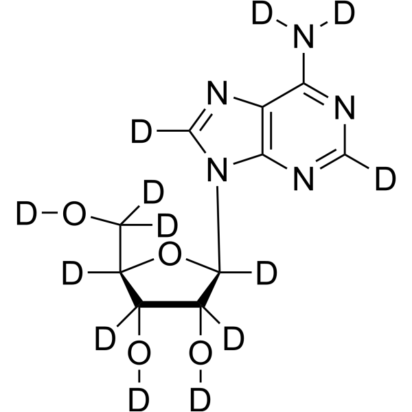
Adenosine-d13 (Adenine riboside-d13; D-Adenosine-d13) is deuterium labeled Adenosine (HY-B0228). Adenosine (Adenine riboside), a ubiquitous endogenous autacoid, acts through the enrollment of four G protein-coupled receptors: A1, A2A, A2B, and A3. Adenosine affects almost all aspects of cellular physiology, including neuronal activity, vascular function, platelet aggregation, and blood cell regulation.
商品は「研究用試薬」です。人や動物の医療用・臨床診断用・食品用の製品ではありません。
研究用途以外に使用した場合、当社は一切の責任を負いかねます。
- 純度 : 96.6%
- 分子式: C10D13N5O4
- 分子量:280.32
-
保管条件:Powder -20°C, 3 years , 4°C, 2 years ; In solvent -80°C, 6 months , -20°C, 1 month

To place orders, for customer services and technical support, please contact: MedChemExpress USA
Tel: 609-228-6898 E-mail: [email protected] [email protected]


-
 生物活性
生物活性
-
化学情報
- プロトコル
- 純度とドキュメンテーション
- 参考文献
-
 Help & FAQs


Endogenous Metabolite アイソフォーム固有の製品をすべて表示
More

生物活性
製品説明
体外実験
Stable heavy isotopes of hydrogen, carbon, and other elements have been incorporated into drug molecules, largely as tracers for quantitation during the drug development process. Deuteration has gained attention because of its potential to affect the pharmacokinetic and metabolic profiles of drugs[1].
MedChemExpress (MCE) has not independently confirmed the accuracy of these methods. They are for reference only. Further protocols information, click here.
アプリケーション
1. This compound can be used as a tracer
2. This compound can be used as an internal standard for quantitative analysis by NMR, GC-MS, or LC-MS.


化学情報
-
非標識Cas 58-61-7
-
性状 Solid
-
分子量 280.32
-
分子式 C10D13N5O4
-
Color White to off-white
-
SMILES
[2H]N([2H])C1=NC([2H])=NC2=C1N=C([2H])N2[C@]3([2H])O[C@@](C([2H])([2H])O[2H])([2H])[C@](O[2H])([2H])[C@]3([2H])O[2H]
-
別名
Adenine riboside-d13; D-Adenosine-d13
-
輸送条件
Room temperature in continental US; may vary elsewhere.
-
保管条件
Powder -20°C 3 years 4°C 2 years In solvent -80°C 6 months -20°C 1 month

プロトコル
-
Neuronal voltage-sensitive dye imaging
Neuronal voltage-sensitive dye imaging detects membrane-potential-dependent optical changes from dyes associated with neuronal membranes, enabling optical recording of electrical activity from single neurons, dendrites, axons, spines, or neuronal populations in brain slices and cultured neurons. VSD signals are typically reported as fractional fluorescence or absorbance changes over baseline, such as ΔF/F or ΔI/I, and published protocols use high-speed cameras or photodiode arrays because neuronal voltage signals occur on millisecond time scales. Fast VSD imaging can be applied at two common scales: bulk staining of brain slices to measure circuit-level spatiotemporal activity, and single-cell loading or biolistic delivery to record membrane-potential transients from individual neuronal compartments. Optical signals should be interpreted as membrane-potential-related readouts, and validation by simultaneous electrophysiology or pharmacological controls is recommended when the experimen


純度とドキュメンテーション
-
データシート (275 KB)
-
SDS (251 KB)
- English - EN (251 KB)
- Français - FR (251 KB)
- Deutsch - DE (251 KB)
- Norwegian - NO (251 KB)
- Español - ES (251 KB)
- Swedish - SV (251 KB)
- Italian - IT (251 KB)
- Korean - KR (251 KB)
- Portuguese - PT (251 KB)
-
取扱説明書 (2659 KB)
参考文献
[1]. Russak EM, et al. Impact of Deuterium Substitution on the Pharmacokinetics of Pharmaceuticals. Ann Pharmacother. 2019 Feb;53(2):211-216. [Content Brief]
[2]. Zhou XT, et al. Inhibition of autophagy enhances adenosine induced apoptosis in human hepatoblastoma HepG2 cells. Oncol Rep. 2019;41(2):829-838. [Content Brief]
[3]. Eltzschig HK. Adenosine: an old drug newly discovered. Anesthesiology. 2009;111(4):904-915. [Content Brief]
[4]. Borea PA, Gessi S, Merighi S, Vincenzi F, Varani K. Pharmacology of Adenosine Receptors: The State of the Art. Physiol Rev. 2018;98(3):1591-1625. [Content Brief]
[5]. Fredholm BB. Adenosine, an endogenous distress signal, modulates tissue damage and repair. Cell Death Differ. 2007;14(7):1315-1323. [Content Brief]

Calculators
濃度 (開始) × 体積 (開始) = 濃度 (終了) × 体積 (終了)


Adenosine-d13 関連分類
HY-B0228S12 関連分類
 Powered by Bioz