



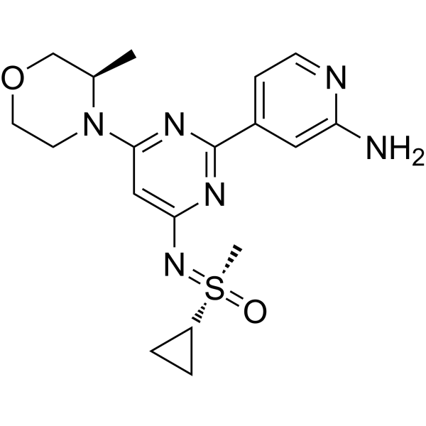
Alnodesertib

Based on 1 Customer Validation
ART0380 is a potent, selective and orally active ATR kinase inhibitor. ART0380 potently inhibits human ATR-ATRIP complex with an IC50 of 51.7 nM. ART0380 binds the ATP pocket of the ATR-ATRIP complex, blocks ATR-dependent Chk1 serine 345 phosphorylation, and induces cell cycle disorder and DNA damage. ART0380 demonstrates potent and selective antitumor activity in preclinical models with varying types of ataxia-telangiectasia mutated (ATM) gene aberrancy. ART0380 can be used for the research of cancer, such as colorectal cancer and prostate cancer.
For research use only. We do not sell to patients.
- Purity : 99.90%
- CAS No.: 2267316-76-5
- Formula: C18H24N6O2S
- Molecular Weight:388.49
-
Storage:Powder -20°C, 3 years , 4°C, 2 years ; In solvent -80°C, 6 months , -20°C, 1 month

To place orders, for customer services and technical support, please contact: MedChemExpress USA
Tel: 609-228-6898 E-mail: [email protected] [email protected]


-
 Biological Activity
-
Chemical Information
-
Solvent & Solubility
- Protocols
- Purity & Documentation
- References
-
 Help & FAQs


-
HY-13902
BerzosertibATR Inhibitor -
HY-19323
CeralasertibATR Inhibitor -
HY-136270
GartisertibATR Inhibitor -
HY-139609
CamonsertibATR Inhibitor -
HY-111451
TuvusertibATR Inhibitor -
HY-101566
ElimusertibATR Inhibitor -
HY-109566
ElrelesertibATM Kinase Inhibitor -
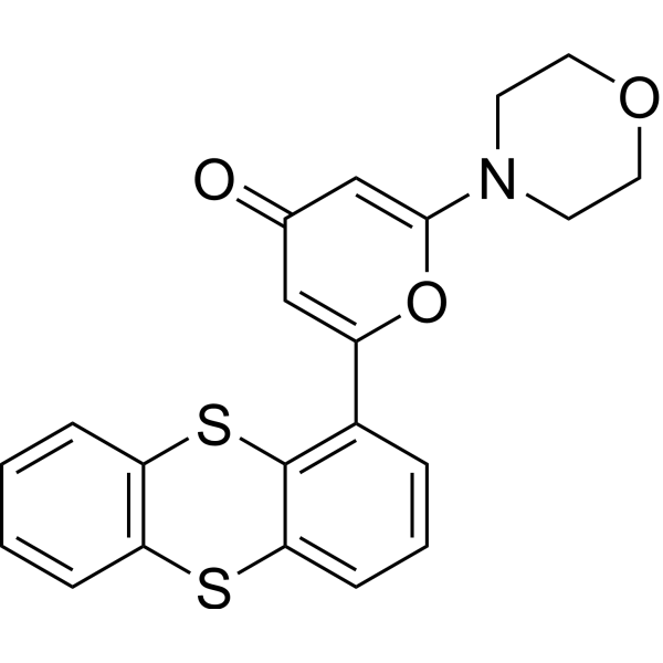
HY-12016
KU-55933ATM Inhibitor -
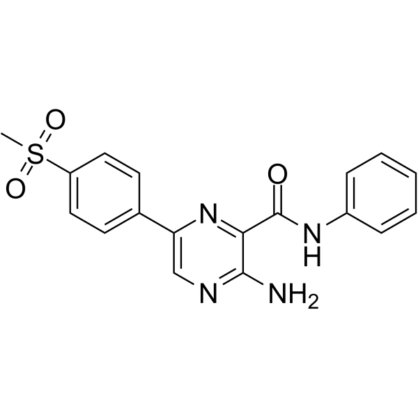
HY-14731
VE-821ATR Inhibitor -
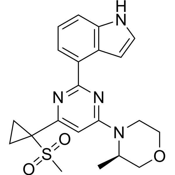
HY-15557
AZ20ATR Inhibitor -
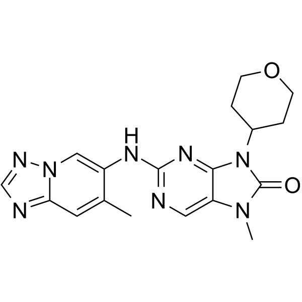
HY-111783
AZD-7648DNA-PK Inhibitor -

HY-10583
Y-27632 dihydrochlorideROCK Inhibitor
All DNA/RNA Synthesis Isoforms
More

Biological Activity
Description
IC50 & Target
[1]|
Chk1 |
Cellular Effect
|
Cell Line

|
Type | Value | Description | References |
|---|---|---|---|---|
| HEK293 | IC50 |
7.9 μM
Compound: ART0380
|
Antiproliferative activity against HEK293 cells assessed as inhibition of cell growth incubated for 48 hrs by CCK8 assay
Antiproliferative activity against HEK293 cells assessed as inhibition of cell growth incubated for 48 hrs by CCK8 assay
|
[PMID: 38955347] |
In Vitro
ART0380 potently inhibits the catalytic activity of the purified human ATR-ATRIP complex with an IC50 of 51.7 nM[1].
ART0380 potently inhibits ATR-dependent pChk1 (Ser345) phosphorylation in HT-29 colorectal adenocarcinoma cells with an EC50 of 0.022 μM[1].
ART0380 exhibits minimal inhibition of mTOR pathway-mediated pRPS6 (Ser235/236) phosphorylation in HT-29 colorectal adenocarcinoma cells, with an EC50 range of 6.0 μM, indicating high selectivity for ATR over mTOR[1].
ART0380 (7 days) potently inhibits proliferation in ATM loss-of-function cancer cell lines (NCI-H23, Granta-519) and replication stress-prone LoVo cells, with weaker activity in normal CCD-18Co colon fibroblasts, showing EC50 values of 0.13, 0.44, 1.0, and 6.4 μM, respectively[1].
ART0380 (0.001 to 1 μM; 7-10 days) exhibits preferential antiproliferative activity in ATM knockout isogenic cell lines (NCI-H460, Calu-6, PC-3) compared to their ATM-proficient parental counterparts, with EC50 values ranging from 0.045-0.213 μmol/L in ATM KO cells and 0.170-1.215 μM in parental cells[1].
ART0380 (0.3-1 μM; 24-48 h) induces cell cycle disorder and increases DNA damage (measured via γH2AX foci) in ATM knockout NCI-H460 cells[1].
MedChemExpress (MCE) has not independently confirmed the accuracy of these methods. They are for reference only. Further protocols information, click here.
-
Cell Line:NCI-H460 ATM KO and parental wild-type cells
-
Concentration:0.3, 1 μM
-
Incubation Time:48 h (continuous); 24 h on/24 h off (intermittent)
-
Result:Induced G1-phase accumulation in ATM KO cells with continuous 48-hour treatment.
Caused arrested S-phase accumulation and increased gH2AX foci in geminin-positive cells in ATM KO cells with intermittent 24-hour on/24-hour off schedule, indicating elevated DNA damage.
In Vivo
ART0380 (100 mg/kg; p.o.; once daily or b.i.d., 3 days on/4 days off.) induces tumor regression in an ATM loss-of-function lung adenocarcinoma PDX model in NSG mice[1].
ART0380 (10-100 mg/kg; p.o.; daily) produces dose-dependent tumor growth inhibition in LoVo colorectal adenocarcinoma xenografts in CD-1 nude mice[1].
ART0380 (100 mg/kg; p.o.; twice daily for 3 days followed by 4 days off) produces significant tumor growth inhibition in ATM-aberrant colorectal cancer PDX models with varying degrees of ATM protein loss, but not in wild-type ATM PDX models, in NSG mice[1].
MedChemExpress (MCE) has not independently confirmed the accuracy of these methods. They are for reference only.
-
Animal Model:oVo colorectal adenocarcinoma xenografts CD-1 nude (female, 6-12 weeks old)[1]
-
Dosage:10 mg/kg; 30 mg/kg; 50 mg/kg; 100 mg/kg
-
Administration:P.o.; daily
-
Result:Induced dose-dependent tumor growth inhibition, with 50 mg/kg and 100 mg/kg producing 84% and 96% tumor growth inhibition, respectively.
Reduced pChk1 Ser345 levels in tumor xenografts after treatment combined with gemcitabine-induced DNA damage.
Clinical Trial
| NCT Number | Sponsor | Condition | Start Date |
Phase
|
|---|---|---|---|---|
| NCT01329991 | Plexxikon| | 2011-05 | PHASE1 |


Chemical Information
-
CAS No. 2267316-76-5
-
Appearance Solid
-
Molecular Weight 388.49
-
Formula C18H24N6O2S
-
Color White to off-white
-
SMILES
C[C@@H]1COCCN1C2=NC(C3=CC=NC(N)=C3)=NC(N=[S@](C)(C4CC4)=O)=C2
-
Synonyms
ART0380
-
Shipping
Room temperature in continental US; may vary elsewhere.
-
Storage
Powder -20°C 3 years 4°C 2 years In solvent -80°C 6 months -20°C 1 month


Solvent & Solubility
In Vitro:
DMSO : 100 mg/mL (257.41 mM; Need ultrasonic; Hygroscopic DMSO has a significant impact on the solubility of product, please use newly opened DMSO)
Please refer to the solubility information to select the appropriate solvent. Once prepared, please aliquot and store the solution to prevent product inactivation from repeated freeze-thaw cycles.
Storage method and period of stock solution: -80°C, 6 months; -20°C, 1 month. When stored at -80°C, please use it within 6 months. When stored at -20°C, please use it within 1 month.
Please refer to the solubility information to select the appropriate solvent. Once prepared, please aliquot and store the solution to prevent product inactivation from repeated freeze-thaw cycles.
Storage method and period of stock solution: -80°C, 6 months; -20°C, 1 month. When stored at -80°C, please use it within 6 months. When stored at -20°C, please use it within 1 month.
Concentration (start) × Volume (start) = Concentration (final) × Volume (final)
In Vivo:
Select the appropriate dissolution method based on your experimental animal and administration route.
- For the following dissolution methods, please ensure to first prepare a clear stock solution using an In Vitro approach and then sequentially add co-solvents:
- To ensure reliable experimental results, the clarified stock solution can be appropriately stored based on storage conditions. As for the working solution for In Vivo experiments, it is recommended to prepare freshly and use it on the same day.
- The percentages shown for the solvents indicate their volumetric ratio in the final prepared solution. If precipitation or phase separation occurs during preparation, heat and/or sonication can be used to aid dissolution.
Add each solvent one by one: 10% DMSO 40% PEG300 5% Tween-80 45% Saline
Solubility: ≥ 5 mg/mL (12.87 mM); Clear solution
This protocol yields a clear solution of ≥ 5 mg/mL (saturation unknown).
Taking 1 mL working solution as an example, add 100 μL DMSO stock solution (50.0 mg/mL) to 400 μL PEG300, and mix evenly; then add 50 μL Tween-80 and mix evenly; then add 450 μL Saline to adjust the volume to 1 mL.
Preparation of Saline: Dissolve 0.9 g sodium chloride in ddH₂O and dilute to 100 mL to obtain a clear Saline solution.
Add each solvent one by one: 10% DMSO 90% (20% SBE-β-CD in Saline)
Solubility: ≥ 5 mg/mL (12.87 mM); Clear solution
This protocol yields a clear solution of ≥ 5 mg/mL (saturation unknown).
Taking 1 mL working solution as an example, add 100 μL DMSO stock solution (50.0 mg/mL) to 900 μL 20% SBE-β-CD in Saline, and mix evenly.
Preparation of 20% SBE-β-CD in Saline (4°C, storage for one week): 2 g SBE-β-CD powder is dissolved in 10 mL Saline, completely dissolve until clear.
In Vivo Dissolution Calculator
Please enter the basic information of animal experiments:
-
-
-
-
Recommended: Prepare an additional quantity of animals to account for potential losses during experiments.
Please enter your animal formula composition:
-
%DMSO +
Recommended: Keep the proportion of DMSO in working solution below 2% if your animal is weak.
-
%+
-
+%Tween-80 + +
-
%Saline +
The co-solvents required include: DMSO, . All of co-solvents are available by MedChemExpress (MCE). , Tween 80. All of co-solvents are available by MedChemExpress (MCE).
Working solution concentration: 0.22 mg/mL
Method for preparing stock solution: mg drug dissolved in μL DMSO. Stock solution concentration: mg/mL.
1. Take μL DMSO stock solution;
2. Add μL .
μL , mix evenly;
3. Then add μL Tween 80, mix evenly;
4. Then add μL
Please ensure that the stock solution in the first step is dissolved to a clear state, and add co-solvents in sequence. You can use ultrasonic heating (ultrasonic cleaner, recommended frequency 20-40 kHz), vortexing, etc. to assist dissolution.

Protocols
-
Kinase activity and phosphorylation assays
Kinase activity assays measure the ability of kinases to transfer phosphate groups from ATP to specific substrates, while phosphorylation assays detect the presence and levels of phosphorylated proteins. Common methods include radiolabeled ATP incorporation (e. g. ,), ADP release detection via bioluminescence (e. g. ,[3]), enzyme-linked immunosorbent assays (ELISA) for phospho-specific epitopes (e. g. ,[6]), and microtiter-based formats for high-throughput screening (e. g. ,[8]). The ADP-Glo assay quantifies kinase activity by measuring ADP produced during phosphorylation using a luciferase-based system. Radiometric assays involve autoradiography or scintillation counting after incorporation of 32P-labeled ATP into substrate proteins. ELISA-based approaches rely on phospho-specific antibodies to detect activated kinases in cell lysates or purified samples.
-
Western Blot
Western blotting (WB) is a commonly used experimental method in molecular biology, biochemistry, and immunogenetics for identifying and quantifying target proteins. It combines gel electrophoresis with immunoassay, enabling researchers to analyze protein expression, post-translational modifications, and molecular weight.
-
Cell Cytotoxicity Assay
Cytotoxicity assays are usually based on the assessment of cell membrane damage, which can also be indirectly detected by measuring cell viability. Detection methods include MTT assay, CKK-8 assay, LDH assay and ATP assay, etc.
-
Flow cytometric DNA-content cell-cycle staining
Flow cytometric DNA-content cell-cycle staining measures the fluorescence intensity of DNA-bound fluorochromes in single cells or nuclei to estimate DNA content distributions, allowing assignment of populations to G0/G1, S, and G2/M phases by DNA histogram deconvolution. Propidium iodide (PI) intercalates into DNA, and PI fluorescence is proportional to cellular DNA content when staining is performed under conditions that make DNA accessible and minimize non-DNA signal. Cells with G2/M DNA content are expected to show approximately twice the fluorescence intensity of G0/G1 cells, while S-phase cells occupy intermediate fluorescence values. PI-based DNA-content analysis can also detect cells with fractional DNA content, often reported as sub-G1, when DNA fragmentation and extraction during staining reduce retained DNA signal in apoptotic cells. DAPI is an alternative DNA fluorochrome for univariate DNA-content analysis, while bivariate approaches combining DNA content with proliferation
-
BrdU Incorporation Assay
Bromodeoxyuridine (BrdU) incorporation assay is based on the principle that BrdU, a thymidine analog, is incorporated into newly synthesized DNA during the S phase of the cell cycle, thereby serving as a marker of DNA replication and cellular proliferation. Incorporated BrdU can be detected using anti-BrdU antibodies following DNA denaturation, enabling visualization or quantification of proliferating cells through immunochemical detection methods such as immunofluorescence or immunohistochemistry.
-
Protocol for Cell Cycle
Cell-cycle analysis by flow cytometry measures DNA content in single cells to estimate the fraction of cells in G0/G1, S, and G2/M phases. Propidium iodide intercalates into DNA, and after RNA removal with RNase, fluorescence intensity reflects cellular DNA content: 2N cells are assigned to G0/G1, cells between 2N and 4N to S phase, and 4N cells to G2/M. DNA-content analysis alone cannot reliably separate G0 from G1 or G2 from M. Ki-67 can distinguish quiescent G0 cells from cycling cells, EdU or BrdU incorporation marks active DNA synthesis in S phase, and phospho-histone H3 staining identifies mitotic cells within the 4N population.
-
Protocol for Kinase activity and phosphorylation assays
Kinase activity assays measure transfer of phosphate from ATP to a protein or peptide substrate, generating phosphorylated substrate, ADP, or incorporated radiolabeled phosphate as the readout; phosphorylation assays measure site-specific phosphorylation in cells or tissues as a proxy for kinase-pathway activation, inhibition, or substrate regulation. Phosphorylation can be detected by phospho-specific Western blot, immunoprecipitation kinase assay, phospho-immunofluorescence, phospho-flow cytometry, luminescent ADP detection, radiolabeled ATP incorporation, or reporter-based pathway assays, and these readouts can be applied to cancer cells, primary neurons, mouse tumors, organoids, inflammatory macrophages, ferroptosis studies, and mitophagy studies when the kinase target is biologically relevant.
-
Subcutaneous Cell-Line-Derived Xenograft
Subcutaneous cell-line-derived xenograft (CDX) models are established by implanting cultured human cancer cell lines into immunodeficient mice, where the injected cells form localized tumors that can be monitored in vivo as a measure of tumorigenic potential, growth kinetics, and treatment response. These models are widely used in oncology research because they allow reproducible tumor formation and enable comparative assessment of tumor growth between different cell lines or genetic manipulations in a controlled in vivo microenvironment. Subcutaneous implantation of cancer cells in immunodeficient mice is a standard approach for evaluating tumor growth behavior and therapeutic response across multiple cancer types, including prostate, esophageal, pancreatic, and colon cancer models.
-
Genotoxicity/Mutagenicity Study
The bacterial reverse mutation assay detects point mutations that restore amino-acid prototrophy in auxotrophic Salmonella typhimurium or Escherichia coli tester strains; after exposure to a test article, mutagenic activity is read out as an increased number of revertant colonies on minimal agar compared with the vehicle control. The assay uses tester strains with different mutation targets so that base-substitution and frameshift mutagens can be detected, and testing is performed with and without exogenous mammalian metabolic activation because some chemicals require biotransformation to become mutagenic.


Purity & Documentation
-
Data Sheet (279 KB)
-
SDS (254 KB)
- English - EN (254 KB)
- Français - FR (254 KB)
- Deutsch - DE (254 KB)
- Norwegian - NO (254 KB)
- Español - ES (254 KB)
- Swedish - SV (254 KB)
- Italian - IT (254 KB)
- Korean - KR (254 KB)
- Portuguese - PT (254 KB)
-
Handling Instructions (2659 KB)
References


Complete Stock Solution Preparation Table
Please refer to the solubility information to select the appropriate solvent. Once prepared, please aliquot and store the solution to prevent product inactivation from repeated freeze-thaw cycles.
Storage method and period of stock solution: -80°C, 6 months; -20°C, 1 month. When stored at -80°C, please use it within 6 months. When stored at -20°C, please use it within 1 month.
| Optional Solvent | Concentration Solvent Mass | 1 mg | 5 mg | 10 mg | 25 mg |
|---|---|---|---|---|---|
| DMSO | 1 mM | 2.5741 mL | 12.8703 mL | 25.7407 mL | 64.3517 mL |
| 5 mM | 0.5148 mL | 2.5741 mL | 5.1481 mL | 12.8703 mL | |
| 10 mM | 0.2574 mL | 1.2870 mL | 2.5741 mL | 6.4352 mL | |
| 15 mM | 0.1716 mL | 0.8580 mL | 1.7160 mL | 4.2901 mL | |
| 20 mM | 0.1287 mL | 0.6435 mL | 1.2870 mL | 3.2176 mL | |
| 25 mM | 0.1030 mL | 0.5148 mL | 1.0296 mL | 2.5741 mL | |
| 30 mM | 0.0858 mL | 0.4290 mL | 0.8580 mL | 2.1451 mL | |
| 40 mM | 0.0644 mL | 0.3218 mL | 0.6435 mL | 1.6088 mL | |
| 50 mM | 0.0515 mL | 0.2574 mL | 0.5148 mL | 1.2870 mL | |
| 60 mM | 0.0429 mL | 0.2145 mL | 0.4290 mL | 1.0725 mL | |
| 80 mM | 0.0322 mL | 0.1609 mL | 0.3218 mL | 0.8044 mL | |
| 100 mM | 0.0257 mL | 0.1287 mL | 0.2574 mL | 0.6435 mL |


Alnodesertib Related Classifications
HY-157941 Related Classifications
 Powered by Bioz