



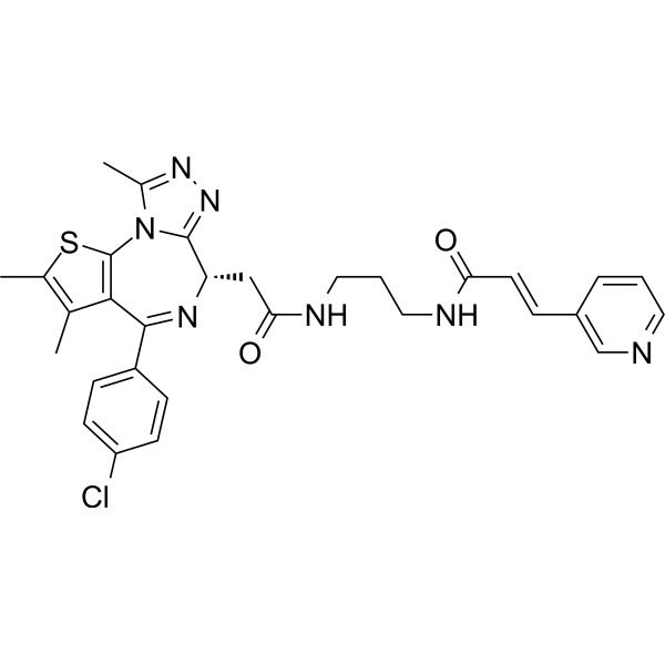
BRD4/NAMPT-IN-1

Based on 1 Customer Validation
BRD4/NAMPT-IN-1 (Compound A2) shows strong inhibitory effects on NAMPT and BRD4 (IC50=35 nM (NAMPT) and 58 nM (BRD4)). BRD4/NAMPT-IN-1 inhibits the growth and migration of hepatocellular carcinoma cells and promotes apoptosis. BRD4/NAMPT-IN-1 also shows potent anticancer effects in HCCLM3 xenograft mouse model, with no obvious toxic effects.
For research use only. We do not sell to patients.
- Purity : 99.45%
- CAS No.: 3049218-28-9
- Formula: C30H30ClN7O2S
- Molecular Weight:588.12
-
Storage:Powder -20°C, 3 years , 4°C, 2 years ; In solvent -80°C, 6 months , -20°C, 1 month

To place orders, for customer services and technical support, please contact: MedChemExpress USA
Tel: 609-228-6898 E-mail: [email protected] [email protected]


-
 Biological Activity
-
Chemical Information
-
Solvent & Solubility
- Protocols
- Purity & Documentation
- References
-
 Help & FAQs




Biological Activity
Description
IC50 & Target
[1]|
BRD4(BD1BD2) 58 nM (IC50) |
BRD4 (BD1) 12 nM (IC50) |
BRD4 (BD2) 41 nM (IC50) |
Cellular Effect
|
Cell Line

|
Type | Value | Description | References |
|---|---|---|---|---|
| HCCLM3 | IC50 |
5.44 μM
Compound: A2
|
Antiproliferative activity against human HCCLM3 cells incubated for 120 hrs by MTT assay
Antiproliferative activity against human HCCLM3 cells incubated for 120 hrs by MTT assay
|
[PMID: 38691889] |
| Hep 3B2 | IC50 |
2.37 μM
Compound: A2
|
Antiproliferative activity against human Hep3B cells incubated for 120 hrs by MTT assay
Antiproliferative activity against human Hep3B cells incubated for 120 hrs by MTT assay
|
[PMID: 38691889] |
| Huh-7 | IC50 |
6.49 μM
Compound: A2
|
Antiproliferative activity against human Huh-7 cells incubated for 120 hrs by MTT assay
Antiproliferative activity against human Huh-7 cells incubated for 120 hrs by MTT assay
|
[PMID: 38691889] |
| LX-2 | IC50 |
9.51 μM
Compound: A2
|
Antiproliferative activity against human LX2 cells incubated for 120 hrs by MTT assay
Antiproliferative activity against human LX2 cells incubated for 120 hrs by MTT assay
|
[PMID: 38691889] |
In Vitro
BRD4/NAMPT-IN-1 exhibits IC50 values of 12 nM for BRD4(BD1) and 41 nM for BRD4(BD2) against other members of the BET family[1].
BRD4/NAMPT-IN-1 inhibits the proliferation of cancer cells with IC50 of 2.37 μM (Hep3B), 6.49 μM (Huh7), 5.44 μM (HCCLM3) and 9.51 μM (LX-2), respectively[1].
BRD4/NAMPT-IN-1 (1-10 μM; 72 h) on Hep3B cells shows that: 1: it can inhibit the expression of oncogenes up-regulated by BRD4, and at the same time reduces the levels of NAPRT and NAMPT. 2: It significantly increases cell arrest at G0/G1 phase. 3: It dose-dependently induces apoptosis. 4: It dose-dependently inhibits the migratory ability of the cells[1].
BRD4/NAMPT-IN-1 (1-10 μM; 72 h) dose-dependently reduces NAD + concentration in Hep3B cells and HCCLM3 cells[1].
MedChemExpress (MCE) has not independently confirmed the accuracy of these methods. They are for reference only. Further protocols information, click here.
-
Cell Line:Hep3B cells
-
Concentration:1; 5; 10 μM
-
Incubation Time:72 h
-
Result:The apoptosis rate induced was significantly higher than that of the control FK866 (HY-50876) and JQ1 (HY-13030) at the same dose.
-
Cell Line:Hep3B cells
-
Concentration:1; 5; 10 μM
-
Incubation Time:72 h
-
Result:Significantly increased the accumulation of Hep3B cells at the G0/G1 stage over the commonly used hepatocellular carcinoma therapeutic agents FK866 (HY-50876) and JQ1 (HY-13030).
In Vivo
MedChemExpress (MCE) has not independently confirmed the accuracy of these methods. They are for reference only.
-
Animal Model:HCCLM3 xenograft nude mice [1]
-
Dosage:40 mg/kg/day and 80 mg/kg/day
-
Administration:i.p.; 27day
-
Result:Inhibited the growth of HCCLM3 tumors significantly in both groups at two different doses, with significant decreases in tumor volume and weight.
In the 40 mg/kg dose group, the tumor growth inhibition rate reached 37.20%, and in the 80 mg/kg dose group, the tumor growth inhibition rate reached 58.17%.
Showed no significant weight loss or other significant toxic side effects.


Chemical Information
-
CAS No. 3049218-28-9
-
Appearance Solid
-
Molecular Weight 588.12
-
Formula C30H30ClN7O2S
-
Color White to off-white
-
SMILES
ClC(C=C1)=CC=C1C2=N[C@@H](CC(NCCCNC(/C=C/C3=CN=CC=C3)=O)=O)C4=NN=C(C)N4C5=C2C(C)=C(C)S5
-
Shipping
Room temperature in continental US; may vary elsewhere.
-
Storage
Powder -20°C 3 years 4°C 2 years In solvent -80°C 6 months -20°C 1 month


Solvent & Solubility
In Vitro:
DMSO : 100 mg/mL (170.03 mM; Need ultrasonic; Hygroscopic DMSO has a significant impact on the solubility of product, please use newly opened DMSO)
Please refer to the solubility information to select the appropriate solvent. Once prepared, please aliquot and store the solution to prevent product inactivation from repeated freeze-thaw cycles.
Storage method and period of stock solution: -80°C, 6 months; -20°C, 1 month. When stored at -80°C, please use it within 6 months. When stored at -20°C, please use it within 1 month.
Please refer to the solubility information to select the appropriate solvent. Once prepared, please aliquot and store the solution to prevent product inactivation from repeated freeze-thaw cycles.
Storage method and period of stock solution: -80°C, 6 months; -20°C, 1 month. When stored at -80°C, please use it within 6 months. When stored at -20°C, please use it within 1 month.
Concentration (start) × Volume (start) = Concentration (final) × Volume (final)
In Vivo:
Select the appropriate dissolution method based on your experimental animal and administration route.
- For the following dissolution methods, please ensure to first prepare a clear stock solution using an In Vitro approach and then sequentially add co-solvents:
- To ensure reliable experimental results, the clarified stock solution can be appropriately stored based on storage conditions. As for the working solution for In Vivo experiments, it is recommended to prepare freshly and use it on the same day.
- The percentages shown for the solvents indicate their volumetric ratio in the final prepared solution. If precipitation or phase separation occurs during preparation, heat and/or sonication can be used to aid dissolution.
Add each solvent one by one: 10% DMSO 40% PEG300 5% Tween-80 45% Saline
Solubility: ≥ 2.5 mg/mL (4.25 mM); Clear solution
This protocol yields a clear solution of ≥ 2.5 mg/mL (saturation unknown).
Taking 1 mL working solution as an example, add 100 μL DMSO stock solution (25.0 mg/mL) to 400 μL PEG300, and mix evenly; then add 50 μL Tween-80 and mix evenly; then add 450 μL Saline to adjust the volume to 1 mL.
Preparation of Saline: Dissolve 0.9 g sodium chloride in ddH₂O and dilute to 100 mL to obtain a clear Saline solution.
Add each solvent one by one: 10% DMSO 90% (20% SBE-β-CD in Saline)
Solubility: ≥ 2.5 mg/mL (4.25 mM); Clear solution
This protocol yields a clear solution of ≥ 2.5 mg/mL (saturation unknown).
Taking 1 mL working solution as an example, add 100 μL DMSO stock solution (25.0 mg/mL) to 900 μL 20% SBE-β-CD in Saline, and mix evenly.
Preparation of 20% SBE-β-CD in Saline (4°C, storage for one week): 2 g SBE-β-CD powder is dissolved in 10 mL Saline, completely dissolve until clear.
In Vivo Dissolution Calculator
Please enter the basic information of animal experiments:
-
-
-
-
Recommended: Prepare an additional quantity of animals to account for potential losses during experiments.
Please enter your animal formula composition:
-
%DMSO +
Recommended: Keep the proportion of DMSO in working solution below 2% if your animal is weak.
-
%+
-
+%Tween-80 + +
-
%Saline +
The co-solvents required include: DMSO, . All of co-solvents are available by MedChemExpress (MCE). , Tween 80. All of co-solvents are available by MedChemExpress (MCE).
Working solution concentration: 0.22 mg/mL
Method for preparing stock solution: mg drug dissolved in μL DMSO. Stock solution concentration: mg/mL.
1. Take μL DMSO stock solution;
2. Add μL .
μL , mix evenly;
3. Then add μL Tween 80, mix evenly;
4. Then add μL
Please ensure that the stock solution in the first step is dissolved to a clear state, and add co-solvents in sequence. You can use ultrasonic heating (ultrasonic cleaner, recommended frequency 20-40 kHz), vortexing, etc. to assist dissolution.

Protocols
-
Annexin V plus membrane-impermeant dye apoptosis staining
Annexin V-based apoptosis assays rely on the detection of phosphatidylserine (PS) externalization from the inner leaflet of the plasma membrane to the outer leaflet, an early biochemical hallmark of apoptosis. Fluorescently labeled Annexin V binds PS in a calcium-dependent manner, enabling identification of early apoptotic cells by flow cytometry or fluorescence microscopy. When combined with a membrane-impermeant DNA-binding dye (e. g. , propidium iodide), this approach allows discrimination between viable (Annexin V−/dye−), early apoptotic (Annexin V+/dye−), and late apoptotic or necrotic (Annexin V+/dye+) cell populations by assessing membrane integrity and PS exposure.
-
Cell migration
Cell migration is a method that plays an important role in wound healing, cell differentiation, embryonic development, etc.
-
Liver Cancer Modeling
Liver cancer can be classified into primary liver cancer and secondary liver cancer. Secondary liver cancer is the metastatic liver cancer. Primary liver cancer includes hepatocellular carcinoma (HCC), intrahepatic cholangiocarcinoma (ICC) and fibrolamellar HCC, of which HCC is the most common form, accounting for approximately 90% of primary liver cancers[1]. HCC mouse models include chemical agent-induced models, transplanted tumor models, and genetic engineered models.
-
How to Choose the Right Model Animal
Choosing the right model animal is a validity-driven decision in which the species, strain, sex, age, genetic background, disease-induction method, outcome measures, and welfare burden must match the scientific question rather than laboratory tradition or convenience. A model should be selected by judging face validity, construct validity, and predictive validity: whether it resembles the human phenotype, whether it reproduces relevant mechanisms, and whether results are likely to predict human biology or treatment response. Animal studies often fail to translate because of species differences, weak disease resemblance, poor experimental design, inadequate reporting, publication bias, and underuse of randomization, blinding, and sample-size justification. Unresolved questions include how to rank competing models objectively, how much human-disease complexity must be reproduced for a given objective, and when non-animal systems such as organoids, ex vivo tissue, or computational models
-
Apoptosis
Apoptosis, also called programmed cell death, is generally characterized by distinct morphological characteristics.
-
TUNEL staining for apoptotic DNA fragmentation
TUNEL staining detects DNA strand breaks by using terminal deoxynucleotidyl transferase to add labeled nucleotides to exposed 3′-OH DNA termini, generating either microscopic staining in fixed cells or tissue sections, or fluorescence/cytometric signal in cell suspensions. TUNEL positivity reflects DNA fragmentation but should not be interpreted alone as definitive apoptosis, because TUNEL can also label necrotic, autolytic, mechanically damaged, or DNA-repair-associated DNA breaks.
-
Cell Cytotoxicity Assay
Cytotoxicity assays are usually based on the assessment of cell membrane damage, which can also be indirectly detected by measuring cell viability. Detection methods include MTT assay, CKK-8 assay, LDH assay and ATP assay, etc.
-
Patient-Derived Xenograft (PDX)
Patient-derived xenograft (PDX) models are generated by engrafting primary human tumor tissue directly into immunodeficient mice, allowing in vivo propagation of patient tumor biology without initial in vitro adaptation. These models are used to preserve key histopathological and molecular characteristics of the original tumor and enable assessment of tumor growth dynamics and therapeutic response in a living organism. The biological readout is tumor engraftment and subsequent growth in the murine host, which reflects the ability of human tumor cells to survive, vascularize, and expand in an immunocompromised microenvironment.
-
Apoptosis Solutions
Apoptosis is a regulated, generally non-lytic cell-death pathway that removes unwanted, damaged, infected, or abnormal cells through coordinated morphological changes, caspase activation, DNA fragmentation, and membrane remodeling. The intrinsic apoptosis pathway is controlled mainly by mitochondrial outer membrane permeabilization, BCL-2 family proteins, cytochrome c release, apoptosome formation, caspase-9 activation, and downstream executioner caspase-3/7 activation. The extrinsic apoptosis pathway is initiated by death receptors such as Fas, TNFR, and TRAIL receptors, which recruit adaptor proteins and activate caspase-8 before engaging executioner caspases or mitochondrial amplification through BID cleavage. Apoptosis is linked to many phenotypes, including cancer cell killing, tissue homeostasis, immune regulation, neurodegeneration, infection response, and treatment-induced cytotoxicity; unresolved questions include how apoptosis interacts with necroptosis, pyroptosis, ferroptos
-
Splenic/Portal-Vein Liver Metastasis Xenograft
Splenic and portal-vein liver metastasis xenograft models deliver tumor cells into the portal circulation so that cells reach the liver first and form hepatic metastatic lesions; splenic injection uses the spleen as an access route to the portal system, while direct portal-vein injection introduces cells into the portal vein without requiring splenectomy. The assay detects liver colonization, intrahepatic tumor growth, tumor distribution, treatment response, survival, and liver-metastasis microenvironment changes; readouts include bioluminescence or fluorescence imaging, gross liver nodule counts, liver weight or tumor burden, histology, and survival.
-
Subcutaneous Cell-Line-Derived Xenograft
Subcutaneous cell-line-derived xenograft (CDX) models are established by implanting cultured human cancer cell lines into immunodeficient mice, where the injected cells form localized tumors that can be monitored in vivo as a measure of tumorigenic potential, growth kinetics, and treatment response. These models are widely used in oncology research because they allow reproducible tumor formation and enable comparative assessment of tumor growth between different cell lines or genetic manipulations in a controlled in vivo microenvironment. Subcutaneous implantation of cancer cells in immunodeficient mice is a standard approach for evaluating tumor growth behavior and therapeutic response across multiple cancer types, including prostate, esophageal, pancreatic, and colon cancer models.
-
Orthotopic Cell-Line Xenograft
Orthotopic cell-line xenograft models involve implantation of human cancer cell lines into the anatomically corresponding organ of immunodeficient mice to reproduce tumor growth within a native microenvironment, enabling more clinically relevant tumor behavior compared with subcutaneous models. These models are widely used because orthotopic placement better recapitulates tumor progression, including invasion and metastatic spread, which are often underrepresented in heterotopic implantation systems. Compared with conventional xenografts, orthotopic implantation is described as more technically complex but provides improved simulation of tumor-microenvironment interactions and metastatic behavior, making it particularly valuable for translational oncology research. Surgical orthotopic implantation approaches have been emphasized as enabling faithful reproduction of clinical cancer features, including metastasis and disease progression patterns that align with the tumor’s organ of origi
-
Intraperitoneal/Peritoneal Dissemination Xenograft
Intraperitoneal (IP) or peritoneal dissemination xenograft models are based on the introduction of human cancer cells into the peritoneal cavity of immunodeficient mice, where they attach to peritoneal surfaces, form multicellular aggregates or spheroids, and progressively generate disseminated tumor nodules that mimic advanced peritoneal metastatic disease. These models are widely used to study ovarian cancer progression, tumor-microenvironment interactions, and intraperitoneal therapeutic responses, often incorporating bioluminescence or fluorescence imaging to longitudinally monitor tumor burden in vivo. The biological principle relies on the capacity of tumor cells such as SKOV3 or related ovarian carcinoma lines to survive in suspension, aggregate within ascites-like fluid, adhere to mesothelial surfaces, and invade peritoneal organs, thereby recapitulating human peritoneal carcinomatosis patterns observed in advanced disease.


Purity & Documentation
-
Data Sheet (277 KB)
-
SDS (393 KB)
- English - EN (393 KB)
- Français - FR (393 KB)
- Deutsch - DE (393 KB)
- Norwegian - NO (393 KB)
- Español - ES (393 KB)
- Swedish - SV (393 KB)
- Italian - IT (393 KB)
- Korean - KR (393 KB)
- Portuguese - PT (393 KB)
-
Handling Instructions (2659 KB)
References


Complete Stock Solution Preparation Table
Please refer to the solubility information to select the appropriate solvent. Once prepared, please aliquot and store the solution to prevent product inactivation from repeated freeze-thaw cycles.
Storage method and period of stock solution: -80°C, 6 months; -20°C, 1 month. When stored at -80°C, please use it within 6 months. When stored at -20°C, please use it within 1 month.
| Optional Solvent | Concentration Solvent Mass | 1 mg | 5 mg | 10 mg | 25 mg |
|---|---|---|---|---|---|
| DMSO | 1 mM | 1.7003 mL | 8.5017 mL | 17.0033 mL | 42.5083 mL |
| 5 mM | 0.3401 mL | 1.7003 mL | 3.4007 mL | 8.5017 mL | |
| 10 mM | 0.1700 mL | 0.8502 mL | 1.7003 mL | 4.2508 mL | |
| 15 mM | 0.1134 mL | 0.5668 mL | 1.1336 mL | 2.8339 mL | |
| 20 mM | 0.0850 mL | 0.4251 mL | 0.8502 mL | 2.1254 mL | |
| 25 mM | 0.0680 mL | 0.3401 mL | 0.6801 mL | 1.7003 mL | |
| 30 mM | 0.0567 mL | 0.2834 mL | 0.5668 mL | 1.4169 mL | |
| 40 mM | 0.0425 mL | 0.2125 mL | 0.4251 mL | 1.0627 mL | |
| 50 mM | 0.0340 mL | 0.1700 mL | 0.3401 mL | 0.8502 mL | |
| 60 mM | 0.0283 mL | 0.1417 mL | 0.2834 mL | 0.7085 mL | |
| 80 mM | 0.0213 mL | 0.1063 mL | 0.2125 mL | 0.5314 mL | |
| 100 mM | 0.0170 mL | 0.0850 mL | 0.1700 mL | 0.4251 mL |


BRD4/NAMPT-IN-1 Related Classifications
HY-161515 Related Classifications
 Powered by Bioz