



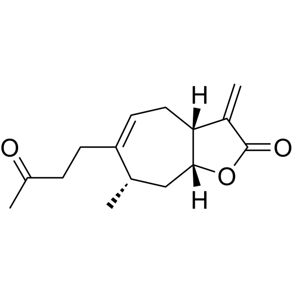
Tomentosin
Based on 1 publication(s) in Google Scholar
Tomentosin is an orally active natural sesquiterpenoid lactone. Tomentosin exhibits multiple activities such as anti-tumor, anti-fungal, anti-inflammatory, anti-oxidant and neuroprotective effects. Tomentosin can inhibit tumor cell proliferation, migration and invasion, and induce apoptosis. Tomentosin can be used in the research of tumors, inflammation and nervous system diseases.
For research use only. We do not sell to patients.
- Purity : 98.95%
- CAS No.: 33649-15-9
- Formula: C15H20O3
- Molecular Weight:248.32
-
Storage:
-20°C, protect from light
* In solvent : -80°C, 6 months; -20°C, 1 month (protect from light)

To place orders, for customer services and technical support, please contact: MedChemExpress USA
Tel: 609-228-6898 E-mail: [email protected] [email protected]


-
 Biological Activity
-
Chemical Information
-
Solvent & Solubility
- Protocols
- Purity & Documentation
- References
-
 Help & FAQs


Publications Citing Use of MedChemExpress (MCE) Tomentosin
More

Biological Activity
Description
IC50 & Target
[2]|
IL-6 |
IL-1β |
TLR2 |
TLR4 |
Cellular Effect
|
Cell Line

|
Type | Value | Description | References |
|---|---|---|---|---|
| HT-29 | IC50 |
56.5 μM
Compound: 9
|
Cytostatic activity against human HT-29 cells incubated for 72 hrs by alamar blue reagent based resazurin metabolism assay
Cytostatic activity against human HT-29 cells incubated for 72 hrs by alamar blue reagent based resazurin metabolism assay
|
[PMID: 26218649] |
| MCF7 | IC50 |
>50 μg/mL
Compound: 17
|
Antiproliferative activity against human MCF7 cells after 48 hrs by MTT assay
Antiproliferative activity against human MCF7 cells after 48 hrs by MTT assay
|
[PMID: 21109433] |
| MCF7 | IC50 |
26.6 μM
Compound: 9
|
Cytostatic activity against human MCF7 cells incubated for 6 days by alamar blue reagent based resazurin metabolism assay
Cytostatic activity against human MCF7 cells incubated for 6 days by alamar blue reagent based resazurin metabolism assay
|
[PMID: 26218649] |
| RAW264.7 | IC50 |
15.7 μM
Compound: 9
|
Anti-inflammatory activity in mouse RAW264.7 cells assessed as inhibition of LPS-induced thromboxane B2 production incubated for 48 hrs by ELISA method
Anti-inflammatory activity in mouse RAW264.7 cells assessed as inhibition of LPS-induced thromboxane B2 production incubated for 48 hrs by ELISA method
|
[PMID: 26218649] |
| RAW264.7 | IC50 |
2 μM
Compound: 11
|
Inhibition of LPS-induced NO production in mouse RAW264.7 cells incubated for 24 hrs by Griess reagent based assay
Inhibition of LPS-induced NO production in mouse RAW264.7 cells incubated for 24 hrs by Griess reagent based assay
|
[PMID: 27276091] |
| RAW264.7 | IC50 |
7.14 μM
Compound: 32
|
Antiinflammatory activity in mouse RAW264.7 cells assessed as inhibition of LPS-induced NO production pretreated for 30 mins before LPS challenge measured 24 hrs after LPS challenge by Griess reaction method
Antiinflammatory activity in mouse RAW264.7 cells assessed as inhibition of LPS-induced NO production pretreated for 30 mins before LPS challenge measured 24 hrs after LPS challenge by Griess reaction method
|
[PMID: 21924800] |
| RAW264.7 | IC50 |
7.3 μM
Compound: 9
|
Anti-inflammatory activity in mouse RAW264.7 cells assessed as inhibition of LPS-induced prostaglandin E2 production incubated for 48 hrs by ELISA method
Anti-inflammatory activity in mouse RAW264.7 cells assessed as inhibition of LPS-induced prostaglandin E2 production incubated for 48 hrs by ELISA method
|
[PMID: 26218649] |
| RAW264.7 | IC50 |
9.15 μM
Compound: 12
|
Inhibition of LPS-induced NO production in mouse RAW264.7 cells measured after 24 hrs
Inhibition of LPS-induced NO production in mouse RAW264.7 cells measured after 24 hrs
|
[PMID: 20515062] |
In Vitro
Tomentosin (0-100 µM; 24-72 h) inhibits the proliferation, migration and invasion, induces apoptosis, and increases ROS levels and decreases mitochondrial membrane potential in pancreatic cancer cells PANC-1 and MIA PaCa-2[1].
MedChemExpress (MCE) has not independently confirmed the accuracy of these methods. They are for reference only.
-
Cell Line:PANC-1 and MIA PaCa-2 cells
-
Concentration:5, 10, 15, 20, 25, 30, 40, 50, 75 and 100 μM
-
Incubation Time:24, 48 and 72 h
-
Result:Inhibited proliferation of pancreatic cancer cells in a dose- and time-dependent manner.
Had IC50 values of 147.62 μM, 31.11 μM and 22.87 μM for 24, 48 and 72 h in PANC-1 cells.
Had IC50 values of 98.08 μM, 33.93 μM and 22.14 μM for 24, 48 and 72 h in MIA PaCa-2 cells.
-
Cell Line:PANC-1 cells
-
Concentration:31.11 μM
-
Incubation Time:48 h
-
Result:Increased the levels of cleaved Caspase-3, and -9.
In Vivo
Tomentosin (12.5-50 mg/kg; oral gavage; 10 days) improves cardiac dysfunction in a mouse model[3].
MedChemExpress (MCE) has not independently confirmed the accuracy of these methods. They are for reference only.
-
Animal Model:Male C57/BL6 mice (23-35 g, 9-12 weeks old) treated MPTP (HY-W114750)[2]
-
Dosage:25 and 50 mg/kg
-
Administration:15 days
-
Result:Significantly prevented weight loss.
Improved motor function (evidenced by better performance in rota-rod test, grasping test, and pole-climbing test).
Reduced levels of proinflammatory cytokines (TNF-α, IL-1β, IL-6), ROS, and MPO, enhanced SOD activity.
Inhibited the expression of TLR-4, TLR-2, and NF-κB in brain tissues, and prevented glial cell damage and neuronal loss as shown by histopathological analysis.
-
Animal Model:Male Balb/c mice (28-33 g, 8-10 weeks old) with LPS (HY-D1056)[3]
-
Dosage:12.5, 25 and 50 mg/kg
-
Administration:Oral gavage (i.g.); 10 days
-
Result:Mitigated cardiac injury and histopathological damage.
Significantly reduced Troponin and CK-MB levels.
Alleviated oxidative stress, and suppressed inflammatory responses.
Inhibited NF-κB activation while enhancing Nrf-2 expression.
- Annexin V plus membrane-impermeant dye apoptosis staining
- BrdU Incorporation Assay
- Cell migration
- Somatic Cell Culture
- CFSE Dye Dilution Proliferation Assay
- Dye-dilution cell tracking and proliferation staining
- CCK-8/WST-8 Cell Proliferation Assay
- Protocol for Cell Cycle
- Research Protocol for Inflammation-related Diseases
- Cell Counting-Based Growth Curve Assay
- Colony Formation (Clonogenic) Assay
- Cell Viability Determination by MTT Colorimetric Assay
- EdU Incorporation Assay (Click Chemistry-Based DNA Synthesis Measurement)
- Ki-67 Immunostaining Proliferation Assay
- Detection of Apoptosis
- TUNEL staining for apoptotic DNA fragmentation
- PCNA Immunodetection Proliferation Assay
- Protocol for Cell Counting and Cell Density Analysis
- Apoptosis Solutions
- Cell invasion
- MTT Cell Proliferation Assay


Chemical Information
-
CAS No. 33649-15-9
-
Appearance Solid
-
Molecular Weight 248.32
-
Formula C15H20O3
-
Color White to off-white
-
SMILES
C=C1[C@](CC=C2CCC(C)=O)([H])[C@@](OC1=O)([H])C[C@@H]2C
-
Structure Classification
-
Shipping
Room temperature in continental US; may vary elsewhere.
-
Storage
-20°C, protect from light
* In solvent : -80°C, 6 months; -20°C, 1 month (protect from light)


Publications (1)
-
Journal Impact Factor
-
Most Recent
-
Naunyn Schmiedebergs Arch Pharmacol
A sesquiterpene lactone, tomentosin, as a novel anticancer agent: orchestrating apoptosis, autophagy, and ER stress in colorectal cancer. [Abstract]2025 Apr 11. PMID: 40214725


Solvent & Solubility
In Vitro:
DMSO : 50 mg/mL (201.35 mM; Need ultrasonic; Hygroscopic DMSO has a significant impact on the solubility of product, please use newly opened DMSO)
Please refer to the solubility information to select the appropriate solvent. Once prepared, please aliquot and store the solution to prevent product inactivation from repeated freeze-thaw cycles.
Storage method and period of stock solution: -80°C, 6 months; -20°C, 1 month (protect from light). When stored at -80°C, please use it within 6 months. When stored at -20°C, please use it within 1 month.
Please refer to the solubility information to select the appropriate solvent. Once prepared, please aliquot and store the solution to prevent product inactivation from repeated freeze-thaw cycles.
Storage method and period of stock solution: -80°C, 6 months; -20°C, 1 month (protect from light). When stored at -80°C, please use it within 6 months. When stored at -20°C, please use it within 1 month.
Concentration (start) × Volume (start) = Concentration (final) × Volume (final)
In Vivo:
Select the appropriate dissolution method based on your experimental animal and administration route.
- For the following dissolution methods, please ensure to first prepare a clear stock solution using an In Vitro approach and then sequentially add co-solvents:
- To ensure reliable experimental results, the clarified stock solution can be appropriately stored based on storage conditions. As for the working solution for In Vivo experiments, it is recommended to prepare freshly and use it on the same day.
- The percentages shown for the solvents indicate their volumetric ratio in the final prepared solution. If precipitation or phase separation occurs during preparation, heat and/or sonication can be used to aid dissolution.
Add each solvent one by one: 10% DMSO 40% PEG300 5% Tween-80 45% Saline
Solubility: ≥ 1.25 mg/mL (5.03 mM); Clear solution
This protocol yields a clear solution of ≥ 1.25 mg/mL (saturation unknown).
Taking 1 mL working solution as an example, add 100 μL DMSO stock solution (12.5 mg/mL) to 400 μL PEG300, and mix evenly; then add 50 μL Tween-80 and mix evenly; then add 450 μL Saline to adjust the volume to 1 mL.
Preparation of Saline: Dissolve 0.9 g sodium chloride in ddH₂O and dilute to 100 mL to obtain a clear Saline solution.
Add each solvent one by one: 10% DMSO 90% (20% SBE-β-CD in Saline)
Solubility: ≥ 1.25 mg/mL (5.03 mM); Clear solution
This protocol yields a clear solution of ≥ 1.25 mg/mL (saturation unknown).
Taking 1 mL working solution as an example, add 100 μL DMSO stock solution (12.5 mg/mL) to 900 μL 20% SBE-β-CD in Saline, and mix evenly.
Preparation of 20% SBE-β-CD in Saline (4°C, storage for one week): 2 g SBE-β-CD powder is dissolved in 10 mL Saline, completely dissolve until clear.
In Vivo Dissolution Calculator
Please enter the basic information of animal experiments:
-
-
-
-
Recommended: Prepare an additional quantity of animals to account for potential losses during experiments.
Please enter your animal formula composition:
-
%DMSO +
Recommended: Keep the proportion of DMSO in working solution below 2% if your animal is weak.
-
%+
-
+%Tween-80 + +
-
%Saline +
The co-solvents required include: DMSO, . All of co-solvents are available by MedChemExpress (MCE). , Tween 80. All of co-solvents are available by MedChemExpress (MCE).
Working solution concentration: 0.22 mg/mL
Method for preparing stock solution: mg drug dissolved in μL DMSO. Stock solution concentration: mg/mL. * In solvent : -80°C, 6 months; -20°C, 1 month (protect from light)
1. Take μL DMSO stock solution;
2. Add μL .
μL , mix evenly;
3. Then add μL Tween 80, mix evenly;
4. Then add μL
Please ensure that the stock solution in the first step is dissolved to a clear state, and add co-solvents in sequence. You can use ultrasonic heating (ultrasonic cleaner, recommended frequency 20-40 kHz), vortexing, etc. to assist dissolution.


Purity & Documentation
-
Data Sheet (281 KB)
-
SDS (252 KB)
- English - EN (252 KB)
- Français - FR (252 KB)
- Deutsch - DE (252 KB)
- Norwegian - NO (252 KB)
- Español - ES (252 KB)
- Swedish - SV (252 KB)
- Italian - IT (252 KB)
- Korean - KR (252 KB)
- Portuguese - PT (252 KB)
-
Handling Instructions (2659 KB)
References
[1]. Güçlü E, et al. Tomentosin induces apoptosis in pancreatic cancer cells through increasing reactive oxygen species and decreasing mitochondrial membrane potential. Toxicol In Vitro. 2022 Oct;84:105458. [Content Brief]
[2]. Fan Y, et al. Tomentosin Reduces Behavior Deficits and Neuroinflammatory Response in MPTP-Induced Parkinson's Disease in Mice. J Environ Pathol Toxicol Oncol. 2021;40(1):75-84. [Content Brief]
[3]. Güvenç M, et al. Tomentosin mitigates the LPS induced cardiac injury by regulating Nrf-2/Nf-κβ pathway in mice. Eur J Pharmacol. 2025 Jun 5;996:177589. [Content Brief]
[4]. Cafarchia C, et al. Attività fungistatica di un sesquiterpene lattone (tomentosina) isolato dai fiori freschi di Inula viscosa (Asteraceae) della regione Puglia [Fungistatic activity of a sesquiterpene lactone (tomentosin) isolated from fresh Inula viscosa (Asteraceae) flowers from the Puglia region]. Parassitologia. 2001 Sep;43(3):117-21. Italian. [Content Brief]


Complete Stock Solution Preparation Table
Please refer to the solubility information to select the appropriate solvent. Once prepared, please aliquot and store the solution to prevent product inactivation from repeated freeze-thaw cycles.
Storage method and period of stock solution: -80°C, 6 months; -20°C, 1 month (protect from light). When stored at -80°C, please use it within 6 months. When stored at -20°C, please use it within 1 month.
| Optional Solvent | Concentration Solvent Mass | 1 mg | 5 mg | 10 mg | 25 mg |
|---|---|---|---|---|---|
| DMSO | 1 mM | 4.0271 mL | 20.1353 mL | 40.2706 mL | 100.6765 mL |
| 5 mM | 0.8054 mL | 4.0271 mL | 8.0541 mL | 20.1353 mL | |
| 10 mM | 0.4027 mL | 2.0135 mL | 4.0271 mL | 10.0677 mL | |
| 15 mM | 0.2685 mL | 1.3424 mL | 2.6847 mL | 6.7118 mL | |
| 20 mM | 0.2014 mL | 1.0068 mL | 2.0135 mL | 5.0338 mL | |
| 25 mM | 0.1611 mL | 0.8054 mL | 1.6108 mL | 4.0271 mL | |
| 30 mM | 0.1342 mL | 0.6712 mL | 1.3424 mL | 3.3559 mL | |
| 40 mM | 0.1007 mL | 0.5034 mL | 1.0068 mL | 2.5169 mL | |
| 50 mM | 0.0805 mL | 0.4027 mL | 0.8054 mL | 2.0135 mL | |
| 60 mM | 0.0671 mL | 0.3356 mL | 0.6712 mL | 1.6779 mL | |
| 80 mM | 0.0503 mL | 0.2517 mL | 0.5034 mL | 1.2585 mL | |
| 100 mM | 0.0403 mL | 0.2014 mL | 0.4027 mL | 1.0068 mL |


Tomentosin Related Classifications
HY-N8284 Related Classifications
 Powered by Bioz