



Isomangiferin
Based on 4 publication(s) in Google Scholar
Isomangiferin is an orally active xanthone C-glucoside, and its chemical structure is similar to Mangiferin (HY-N0290). Isomangiferin is an effective VEGFR-2 kinase inhibitor, which can induces cell apoptosis, inhibit the growth, metastasis and angiogenesis of breast cancer. Isomangiferin exerts anti-inflammatory effects by inhibiting the HMGB1/NLRP3/NF-κB signaling pathway, thereby improving the renal function indicators of diabetic mice. Isomangiferin exhibits inhibitory effects on various bacteria and herpes simplex virus type 1 (HSV-1). Isomangiferin promotes the migration and osteogenic differentiation of bone marrow mesenchymal stem cells (BMSCs) and reduces cell apoptosis and the production of ROS by activating the AMPK/ACC pathway, thereby facilitating fracture healing.
Nos produits utilisent uniquement pour la recherche. Nous ne vendons pas aux patients.
- Pureté : 99.60%
- CAS No.: 24699-16-9
- Formule: C19H18O11
- Masse moléculaire:422.34
-
Stockage:
4°C, protect from light
* In solvent : -80°C, 6 months; -20°C, 1 month (protect from light)

To place orders, for customer services and technical support, please contact: MedChemExpress USA
Tel: 609-228-6898 E-mail: [email protected] [email protected]


-
 Activité biologique
-
Chemical Information
-
Solvant et solubilité
- Protocole
- Pureté et documentation
- Références
-
 Help & FAQs


-

HY-N0772R
Isomangiferin (Standard)Standard of Isomangiferin -
HY-10964
VadimezanVascular Disrupting Agent -
HY-109025
Baloxavir marboxilInfluenza A/B Inhibitor -
HY-107201
β-CyclodextrinInfluenza Virus Inhibitor -
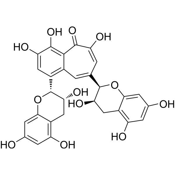
HY-N0243
TheaflavinEndogenous Metabolite -
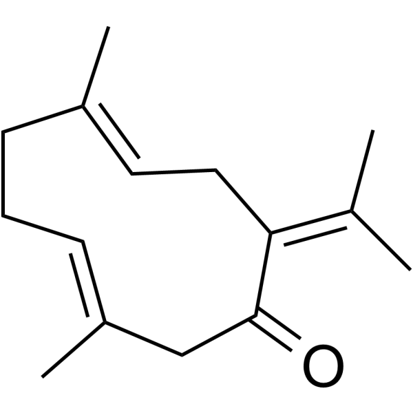
HY-N0440
GermacroneAnti-viral/Cancer/Inflammatory Agent -
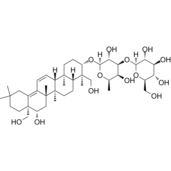
HY-N0247
Saikosaponin B1Hedgehog Inhibitor -
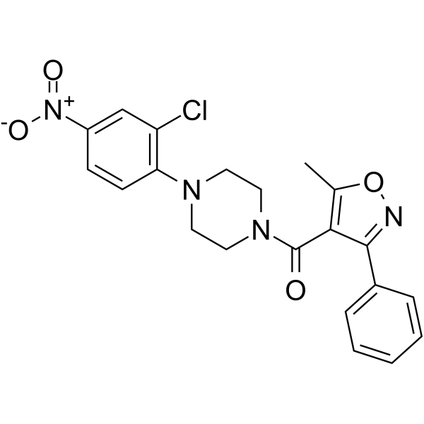
HY-50001
NucleozinInfluenza Virus Inhibitor -

HY-109025R
Baloxavir marboxil (Standard)Influenza A/B Inhibitor -
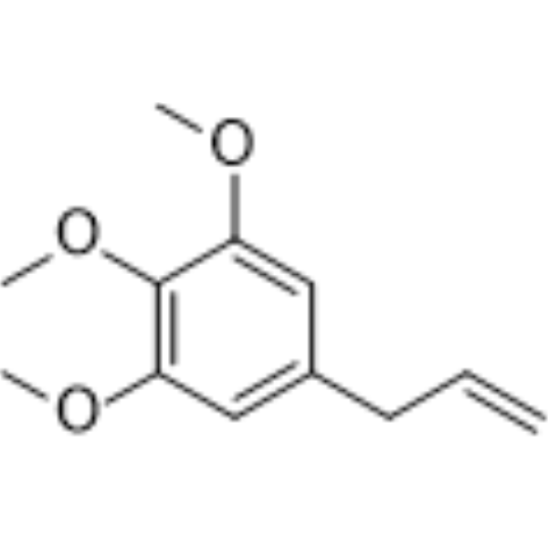
HY-N6807
ElemicinSCD Inhibitor -

HY-N0440R
Germacrone (Standard)Standard of Germacrone -
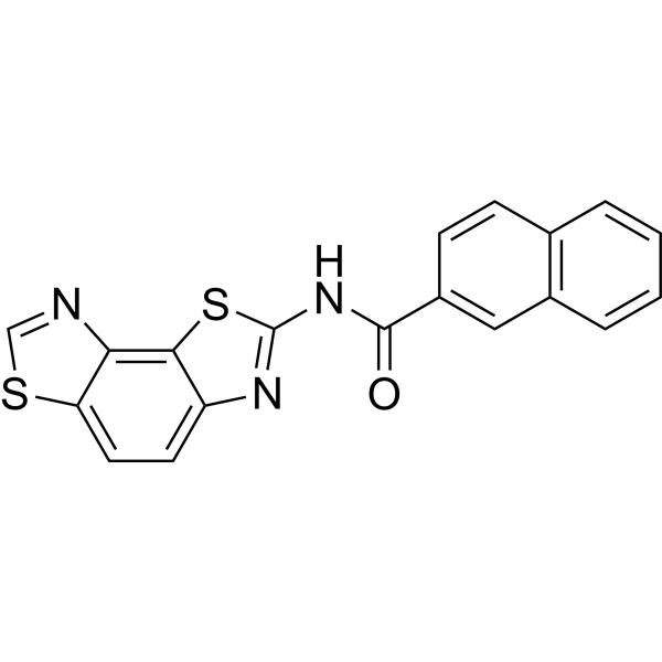
HY-101950
KIN1148IRF3 Agonist
Publications Citing Use of MedChemExpress (MCE) Isomangiferin
MoreVoir tous les produits spécifiques à Isoform VEGFR
MoreVoir tous les produits spécifiques à Isoform AMPK
More

Activité biologique
Description
In Vitro
Isomangiferin inhibits HSV-1 that exceeds Acyclovir (HY-17422), Idoxuridine (HY-B0307), and Cyclocytidine (HY-N0093) in log by 0.27-0.50 in log determination and exhibits the average plaque reduction rate of 69.5%[1].
Isomangiferin shows better antibacterial potential than Mangiferin against all the tested strain including B. subtilis, S. aureus, K. pneumoniae, S. Setubal, E. coli with MICs ≤ 250 μg/mL[2].
Isomangiferin shows poor cytotoxicity towards Brine shrimp nauplii with a LD50 of 768.92 mg/mL[2].
Isomangiferin (2.5-10 μM, 24 h) promotes osteogenic differentiation and migration of H2O2-treated bone marrow mesenchymal stem cells (BMSCs), reduces apoptosis and reactive oxygen species production, and activates the AMP-activated protein kinase/acetyl-CoA carboxylase (AMPK/ACC) pathway[3].
Isomangiferin (1 μM, 48 h) decreases the viability of breast cancer cells (MDA-MB-231, T47D, MCF7, SKBR3, 4T1) with IC50s of approximately 1 μM, , while the IC50 for the MCF-10A cells exceeded 100 μM[5].
Isomangiferin (1 μM, 5 days) inhibits microvessel sprouting and vascular tubulogenesis of endothelial cells[5].
Isomangiferin (0-5 μM, 1-36 h) inhibits migration and invasion and induces breast cancer cell apoptosis and impairs cell adhesion in MDA-MB-231 cells through VEGFR-2-mediated signaling pathway[5].
MedChemExpress (MCE) has not independently confirmed the accuracy of these methods. They are for reference only. Further protocols information, click here.
-
Cell Line:BMSCs stimulated with H2O2
-
Concentration:2.5, 5 and 10 μM
-
Incubation Time:24 h
-
Result:Dose-dependently restored cell viability.
-
Cell Line:BMSCs stimulated with H2O2
-
Concentration:2.5, 5 and 10 μM
-
Incubation Time:24 h
-
Result:Restored the migration ability by 2.1 times at 10 μM.
-
Cell Line:BMSCs stimulated with H2O2
-
Concentration:2.5, 5 and 10 μM
-
Incubation Time:24 h
-
Result:Reduced the apoptosis rate to 11.3% at 10 μM.
-
Cell Line:BMSCs stimulated with H2O2
-
Concentration:2.5, 5 and 10 μM
-
Incubation Time:24 h
-
Result:Restored the ALP activity to 90% of its normal level at 10 μM.
Reduced the level of ROS by 60% at 10 μM.
-
Cell Line:BMSCs stimulated with H2O2
-
Concentration:2.5, 5 and 10 μM
-
Incubation Time:24 h
-
Result:Induced the upregulation of Runx2 and BMP2 expression.
Downregulated the pro-apoptotic protein Bax, upregulated the anti-apoptotic protein Bcl-2 and inhibited the activation of caspase-3.
Significantly increase the levels of p-AMPK and p-ACC (Ser79).
-
Cell Line:MDA-MB-231 cells
-
Concentration:0, 0.5, 1, 2.5 and 5 μM
-
Incubation Time:6 h
-
Result:Showed dose-dependent inhibition, and almost completely inhibited migration at 5 μM.
-
Cell Line:MDA-MB-231 cells
-
Concentration:0, 0.5, 1, 2.5 and 5 μM
-
Incubation Time:8 to 10 h
-
Result:Showed dose-dependent inhibition, and almost completely inhibited invasion at 5 μM.
-
Cell Line:MDA-MB-231 cells
-
Concentration:0, 0.5, 1, 2.5 and 5 μM
-
Incubation Time:36 h
-
Result:Increased the apoptosis rate from 7.15% to 13.06% at a concentration of 5 μM.
-
Cell Line:MDA-MB-231 cells
-
Concentration:0, 0.5, 1, 2.5 μM
-
Incubation Time:24 h
-
Result:Activated caspase-3 and PARP cleavage.
In Vivo
Isomangiferin (10 mg/kg, i.p., once daily for a month) inhibits breast cancer growth and blocks the VEGFR-2 pathway in the xenograft model[5].
MedChemExpress (MCE) has not independently confirmed the accuracy of these methods. They are for reference only.
-
Animal Model:Type 2 diabetes mellitus (T2DM) model established in mice (C57BL/KsJ db/db mice, male) and male db/m mice[4]
-
Dosage:10 and 20 mg/kg
-
Administration:Oral administration (p.o.), once daily for 12 weeks
-
Result:Reduced UA and Cr levels in Db/Db mice.
Reduced serum insulin levels and improved lipid profiles in Db/Db mice.
Attenuated kidney damage.
Reduced inflammatory cytokine levels in serum and the kidney.
Inhibited HMGB1/NACHT leucine-rich repeat- and PYD-containing 3 (NLRP3)/NF-κB pathway in Db/Db mice.
-
Animal Model:MDA-MB-231 induced xenograft model established in five-week-old male BALB/c nude mice[5]
-
Dosage:10 mg/kg
-
Administration:Intraperitoneal injection (i.p.), once daily for a month
-
Result:Significantly reduced the volume and weight of the tumor.
Significantly reduced the expression of p-VEGFR2 and CD31. Reduced the microvessel density (MVD) within the tumor tissue and the number of p-VEGFR2 positive cells.


Chemical Information
-
CAS No. 24699-16-9
-
Appearance Solid
-
Masse moléculaire 422.34
-
Formule C19H18O11
-
Color Light yellow to green yellow
-
SMILES
OC1=CC(O)=C2C(OC3=CC(O)=C(O)C=C3C2=O)=C1[C@@H]([C@@H]([C@@H](O)[C@@H]4O)O)O[C@@H]4CO
-
Structure Classification
-
Initial Source
-
Livraison
Room temperature in continental US; may vary elsewhere.
-
Stockage
4°C, protect from light
* In solvent : -80°C, 6 months; -20°C, 1 month (protect from light)


Publications (4)
-
Journal Impact Factor
-
Most Recent
-
Food Chem
Effects of sun drying combined with baking processes on the flavor quality of Chongqing Tuocha raw tea. [Abstract]2025 Dec 30:497:146992. PMID: 41285060 -
Food Chem
Flavonoid-mediated metabolic underpinning quality variation in red bud-sport pear mutants. [Abstract]2025 Oct 15:489:144992. PMID: 40466530 -
Vet Microbiol
The Chinese medicine monomer Schisandrin C inhibits PRRSV infection by regulating the OGT-PI3K/AKT/mTOR signaling pathway. [Abstract]2026 May:316:110992. PMID: 41865607 -


Solvant et solubilité
In Vitro:
DMSO : 50 mg/mL (118.39 mM; Need ultrasonic; Hygroscopic DMSO has a significant impact on the solubility of product, please use newly opened DMSO)
Please refer to the solubility information to select the appropriate solvent. Once prepared, please aliquot and store the solution to prevent product inactivation from repeated freeze-thaw cycles.
Storage method and period of stock solution: -80°C, 6 months; -20°C, 1 month (protect from light). When stored at -80°C, please use it within 6 months. When stored at -20°C, please use it within 1 month.
Please refer to the solubility information to select the appropriate solvent. Once prepared, please aliquot and store the solution to prevent product inactivation from repeated freeze-thaw cycles.
Storage method and period of stock solution: -80°C, 6 months; -20°C, 1 month (protect from light). When stored at -80°C, please use it within 6 months. When stored at -20°C, please use it within 1 month.
Concentration (start) × Volume (start) = Concentration (final) × Volume (final)
In Vivo:
Select the appropriate dissolution method based on your experimental animal and administration route.
- For the following dissolution methods, please ensure to first prepare a clear stock solution using an In Vitro approach and then sequentially add co-solvents:
- To ensure reliable experimental results, the clarified stock solution can be appropriately stored based on storage conditions. As for the working solution for In Vivo experiments, it is recommended to prepare freshly and use it on the same day.
- The percentages shown for the solvents indicate their volumetric ratio in the final prepared solution. If precipitation or phase separation occurs during preparation, heat and/or sonication can be used to aid dissolution.
Add each solvent one by one: 10% DMSO 40% PEG300 5% Tween-80 45% Saline
Solubility: ≥ 2.5 mg/mL (5.92 mM); Clear solution
This protocol yields a clear solution of ≥ 2.5 mg/mL (saturation unknown).
Taking 1 mL working solution as an example, add 100 μL DMSO stock solution (25.0 mg/mL) to 400 μL PEG300, and mix evenly; then add 50 μL Tween-80 and mix evenly; then add 450 μL Saline to adjust the volume to 1 mL.
Preparation of Saline: Dissolve 0.9 g sodium chloride in ddH₂O and dilute to 100 mL to obtain a clear Saline solution.
Add each solvent one by one: 10% DMSO 90% (20% SBE-β-CD in Saline)
Solubility: ≥ 2.5 mg/mL (5.92 mM); Clear solution
This protocol yields a clear solution of ≥ 2.5 mg/mL (saturation unknown).
Taking 1 mL working solution as an example, add 100 μL DMSO stock solution (25.0 mg/mL) to 900 μL 20% SBE-β-CD in Saline, and mix evenly.
Preparation of 20% SBE-β-CD in Saline (4°C, storage for one week): 2 g SBE-β-CD powder is dissolved in 10 mL Saline, completely dissolve until clear.
In Vivo Dissolution Calculator
Please enter the basic information of animal experiments:
-
-
-
-
Recommended: Prepare an additional quantity of animals to account for potential losses during experiments.
Please enter your animal formula composition:
-
%DMSO +
Recommended: Keep the proportion of DMSO in working solution below 2% if your animal is weak.
-
%+
-
+%Tween-80 + +
-
%Saline +
The co-solvents required include: DMSO, . All of co-solvents are available by MedChemExpress (MCE). , Tween 80. All of co-solvents are available by MedChemExpress (MCE).
Working solution concentration: 0.22 mg/mL
Method for preparing stock solution: mg drug dissolved in μL DMSO. Stock solution concentration: mg/mL. * In solvent : -80°C, 6 months; -20°C, 1 month (protect from light)
1. Take μL DMSO stock solution;
2. Add μL .
μL , mix evenly;
3. Then add μL Tween 80, mix evenly;
4. Then add μL
Please ensure that the stock solution in the first step is dissolved to a clear state, and add co-solvents in sequence. You can use ultrasonic heating (ultrasonic cleaner, recommended frequency 20-40 kHz), vortexing, etc. to assist dissolution.

Protocole
-
Human pluripotent stem cell endothelial-cell differentiation
Human pluripotent stem cell endothelial differentiation is based on stepwise developmental patterning: early activation of WNT/GSK3β inhibition promotes mesodermal or vascular progenitor entry, followed by endothelial specification using VEGF-related signaling, BMP4, FGF2, Notch modulation, or cAMP depending on the published protocol. Endothelial differentiation is read out by acquisition of CD31, CD34, VE-cadherin/CD144, KDR/VEGFR2, vWF, Tie2, NOS3, acetylated LDL uptake, tube/network formation, barrier function, and in vivo vessel-forming capacity where tested.
-
Vascular/Branching Fractal Analysis
Vascular/branching fractal analysis quantifies the geometric complexity of vessel trees or vascular networks from segmented 2D images, commonly by converting vessels into binary and/or skeletonized maps and estimating fractal dimension using box-counting or related approaches. Fractal dimension is interpreted as an image-derived readout of vascular branching complexity, space filling, or density, and has been applied to retinal photographs, fluorescein angiography, OCT angiography, capillary perfusion maps, and in vitro Matrigel angiogenesis networks. The assay readout is generated from vessel-positive pixels after image preprocessing, vessel segmentation, binarization, and optional skeletonization; reported outputs include fractal dimension, vessel density, branchpoint density, endpoint density, vessel length density, tortuosity, and generation-based branching metrics when VESGEN-style analysis is used. The biological interpretation is limited to quantitative vascular patterning and s
-
Annexin V plus membrane-impermeant dye apoptosis staining
Annexin V-based apoptosis assays rely on the detection of phosphatidylserine (PS) externalization from the inner leaflet of the plasma membrane to the outer leaflet, an early biochemical hallmark of apoptosis. Fluorescently labeled Annexin V binds PS in a calcium-dependent manner, enabling identification of early apoptotic cells by flow cytometry or fluorescence microscopy. When combined with a membrane-impermeant DNA-binding dye (e. g. , propidium iodide), this approach allows discrimination between viable (Annexin V−/dye−), early apoptotic (Annexin V+/dye−), and late apoptotic or necrotic (Annexin V+/dye+) cell populations by assessing membrane integrity and PS exposure.
-
Cell migration
Cell migration is a method that plays an important role in wound healing, cell differentiation, embryonic development, etc.
-
ROS/oxidative-stress fluorescent staining
ROS/oxidative-stress fluorescent staining uses cell-permeant fluorogenic probes that become fluorescent after oxidation inside cells or tissues; commonly used examples include DCFH-DA/DCFDA for broad cellular oxidant detection, DHE for superoxide-related signal detection, MitoSOX for mitochondrial superoxide-related signal detection, and CellROX probes for oxidative-stress-associated fluorescence readouts. The assay detects probe oxidation rather than a single ROS species unless the probe and analysis method have been chemically validated for that species. DCFH-DA enters cells, is deacetylated by intracellular esterases to DCFH, and produces fluorescent DCF after oxidation, so the readout is used as an operational measure of total cellular oxidative stress rather than a species-specific ROS measurement. DHE and MitoSOX can report superoxide-related oxidation, but red fluorescence alone can include non-specific ethidium-like oxidation products; HPLC or optimized spectral approaches are
-
Research Protocol for Inflammation-related Diseases
The NLRP3 inflammasome is a cytosolic innate immune signaling platform that integrates priming signals and danger-signal activation to promote caspase-1 activation, maturation of IL-1β and IL-18, and gasdermin D-mediated pyroptotic cell death. The core experimental logic is to determine whether inflammatory disease phenotypes are driven by increased NLRP3 expression, ASC-containing inflammasome assembly, caspase-1 cleavage, GSDMD cleavage, and extracellular release of IL-1β/IL-18 rather than by nonspecific cell injury alone. The pathway is strongly linked to inflammation-related disease phenotypes because monosodium urate crystals activate NALP3/NLRP3 inflammasome signaling in gout-like crystal inflammation, cholesterol crystals activate NLRP3 inflammasomes in atherogenesis models, and DSS-induced intestinal inflammation has been reported to involve NLRP3 inflammasome activity. However, experimental colitis studies also show context-dependent protective effects of NLRP3 inflammasome co
-
Breast Cancer Modeling
Breast cancer is a heterogeneous cancer, and it has been distinguished into four subtypes: luminal A, luminal B, HER2-positive and basal-like. Molecular mutations, epigenetic alterations, hormone exposure and immune microenvironment are related to the progression of breast cancer.
-
Apoptosis
Apoptosis, also called programmed cell death, is generally characterized by distinct morphological characteristics.
-
TUNEL staining for apoptotic DNA fragmentation
TUNEL staining detects DNA strand breaks by using terminal deoxynucleotidyl transferase to add labeled nucleotides to exposed 3′-OH DNA termini, generating either microscopic staining in fixed cells or tissue sections, or fluorescence/cytometric signal in cell suspensions. TUNEL positivity reflects DNA fragmentation but should not be interpreted alone as definitive apoptosis, because TUNEL can also label necrotic, autolytic, mechanically damaged, or DNA-repair-associated DNA breaks.
-
Mesenchymal stromal/stem cell osteogenic differentiation
Mesenchymal stromal/stem cells can be induced toward an osteoblast-like lineage in vitro by culture in osteogenic medium containing dexamethasone, ascorbic acid or ascorbate-2-phosphate, and β-glycerophosphate; the differentiation process is commonly evaluated by alkaline phosphatase activity, osteogenic marker expression, collagenous matrix formation, and calcium-rich matrix mineralization. The main readouts are alkaline phosphatase activity as an early osteogenic marker and Alizarin Red S staining as a calcium-deposit readout for mineralized extracellular matrix; Alizarin Red S can be inspected microscopically or extracted and measured colorimetrically at 405 nm.
-
Endothelial Tube Formation Assay
Endothelial tube formation assay evaluates the ability of endothelial cells to attach, migrate, align, and organize into capillary-like networks when cultured on gelled basement membrane extract or Matrigel; the readout is the morphology and quantity of tube-like networks, which reflects an in vitro endothelial morphogenesis step related to angiogenesis. Basement membrane extract/Matrigel provides laminin-rich extracellular matrix cues that support endothelial differentiation into capillary-like structures, but it can contain biologically active growth factors, so growth-factor-reduced matrix is preferred when testing defined angiogenic stimulators or inhibitors.
-
Apoptosis Solutions
Apoptosis is a regulated, generally non-lytic cell-death pathway that removes unwanted, damaged, infected, or abnormal cells through coordinated morphological changes, caspase activation, DNA fragmentation, and membrane remodeling. The intrinsic apoptosis pathway is controlled mainly by mitochondrial outer membrane permeabilization, BCL-2 family proteins, cytochrome c release, apoptosome formation, caspase-9 activation, and downstream executioner caspase-3/7 activation. The extrinsic apoptosis pathway is initiated by death receptors such as Fas, TNFR, and TRAIL receptors, which recruit adaptor proteins and activate caspase-8 before engaging executioner caspases or mitochondrial amplification through BID cleavage. Apoptosis is linked to many phenotypes, including cancer cell killing, tissue homeostasis, immune regulation, neurodegeneration, infection response, and treatment-induced cytotoxicity; unresolved questions include how apoptosis interacts with necroptosis, pyroptosis, ferroptos
-
Orthotopic Cell-Line Xenograft
Orthotopic cell-line xenograft models involve implantation of human cancer cell lines into the anatomically corresponding organ of immunodeficient mice to reproduce tumor growth within a native microenvironment, enabling more clinically relevant tumor behavior compared with subcutaneous models. These models are widely used because orthotopic placement better recapitulates tumor progression, including invasion and metastatic spread, which are often underrepresented in heterotopic implantation systems. Compared with conventional xenografts, orthotopic implantation is described as more technically complex but provides improved simulation of tumor-microenvironment interactions and metastatic behavior, making it particularly valuable for translational oncology research. Surgical orthotopic implantation approaches have been emphasized as enabling faithful reproduction of clinical cancer features, including metastasis and disease progression patterns that align with the tumor’s organ of origi
-
Endothelial Cell Migration/Angiogenic Sprouting Assay
Endothelial cell migration and angiogenic sprouting assays are in vitro (and partially ex vivo-adapted) functional models that quantify the ability of endothelial cells to undergo coordinated migration, extracellular matrix invasion, and multicellular organization into capillary-like sprouts in response to pro-angiogenic stimuli such as VEGF, bFGF, or conditioned microenvironments. These assays are used to model early angiogenic events including tip-cell formation, directional migration, and lumen-like sprout extension, which collectively reflect angiogenic activation and vascular morphogenesis processes observed in vivo.


Pureté et documentation
-
Fiche technique (292 KB)
-
SDS (393 KB)
- English - EN (393 KB)
- Français - FR (393 KB)
- Deutsch - DE (393 KB)
- Norwegian - NO (393 KB)
- Español - ES (393 KB)
- Swedish - SV (393 KB)
- Italian - IT (393 KB)
- Korean - KR (393 KB)
- Portuguese - PT (393 KB)
-
Instruction de manipulation (2659 KB)
Références
[1]. Zheng MS, et al. Antiviral effect of mangiferin and isomangiferin on herpes simplex virus. Chin Med J (Engl). 1990 Feb;103(2):160-5. [Content Brief]
[2]. Ishaque M, et al. Xanthone C-glycosides isomers purified from Dryopteris ramosa (Hope) C. Chr. with bactericidal and cytotoxic prospects. Saudi J Biol Sci. 2022 Feb;29(2):1191-1196. [Content Brief]
[3]. Gao B, et al. Isomangiferin promotes the migration and osteogenic differentiation of rat bone marrow mesenchymal stem cells. Open Life Sci. 2024 Jul 20;19(1):20220884. doi: 10.1515/biol-2022-0884. PMID: 39035458; PMCID: PMC11259999. [Content Brief]
[4]. Yue S, et al. Isomangiferin Attenuates Renal Injury in Diabetic Mice via Inhibiting Inflammation. Diabetes Metab Syndr Obes. 2020 Nov 10;13:4273-4280. [Content Brief]
[5]. Wang B, et al. Isomangiferin, a Novel Potent Vascular Endothelial Growth Factor Receptor 2 Kinase Inhibitor, Suppresses Breast Cancer Growth, Metastasis and Angiogenesis. J Breast Cancer. 2018 Mar;21(1):11-20. [Content Brief]


Complete Stock Solution Preparation Table
Please refer to the solubility information to select the appropriate solvent. Once prepared, please aliquot and store the solution to prevent product inactivation from repeated freeze-thaw cycles.
Storage method and period of stock solution: -80°C, 6 months; -20°C, 1 month (protect from light). When stored at -80°C, please use it within 6 months. When stored at -20°C, please use it within 1 month.
| Optional Solvent | Concentration Solvent Mass | 1 mg | 5 mg | 10 mg | 25 mg |
|---|---|---|---|---|---|
| DMSO | 1 mM | 2.3678 mL | 11.8388 mL | 23.6776 mL | 59.1940 mL |
| 5 mM | 0.4736 mL | 2.3678 mL | 4.7355 mL | 11.8388 mL | |
| 10 mM | 0.2368 mL | 1.1839 mL | 2.3678 mL | 5.9194 mL | |
| 15 mM | 0.1579 mL | 0.7893 mL | 1.5785 mL | 3.9463 mL | |
| 20 mM | 0.1184 mL | 0.5919 mL | 1.1839 mL | 2.9597 mL | |
| 25 mM | 0.0947 mL | 0.4736 mL | 0.9471 mL | 2.3678 mL | |
| 30 mM | 0.0789 mL | 0.3946 mL | 0.7893 mL | 1.9731 mL | |
| 40 mM | 0.0592 mL | 0.2960 mL | 0.5919 mL | 1.4799 mL | |
| 50 mM | 0.0474 mL | 0.2368 mL | 0.4736 mL | 1.1839 mL | |
| 60 mM | 0.0395 mL | 0.1973 mL | 0.3946 mL | 0.9866 mL | |
| 80 mM | 0.0296 mL | 0.1480 mL | 0.2960 mL | 0.7399 mL | |
| 100 mM | 0.0237 mL | 0.1184 mL | 0.2368 mL | 0.5919 mL |


Isomangiferin Related Classifications
HY-N0772 Related Classifications
 Powered by Bioz
Keywords
- Isomangiferin
- 24699-16-9
- VEGFR
- NOD-like Receptor (NLR)
- NF-κB
- Bacterial
- AMPK
- Acetyl-CoA Carboxylase
- Apoptosis
- Reactive Oxygen Species (ROS)
- HSV
- Drug Derivative
- Dryopteris ramose
- Antibacterial
- Mangiferin
- Xanthone
- C-glycosides
- Cytotoxic
- BMSCs
- Osteogenic differentiation
- AMPK/ACC pathway
- Angiogenesis inhibitors
- Breast neoplasms
- Inhibitor
- inhibitor
- inhibit