



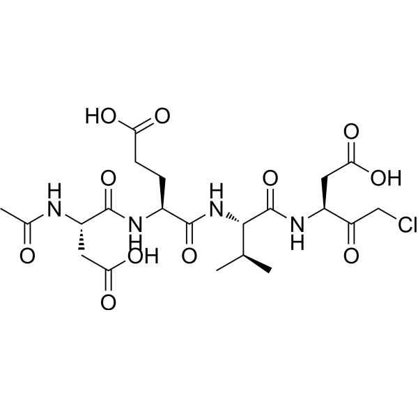
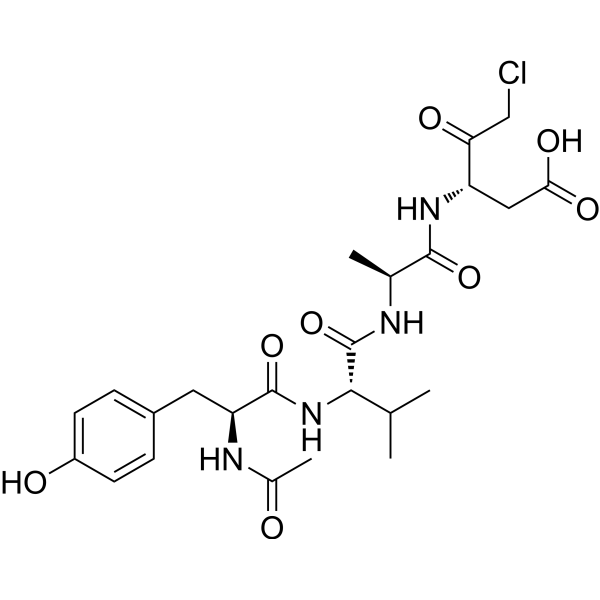
Ac-DEVD-CMK
Based on 3 publication(s) in Google Scholar
Ac-DEVD-CMK (Caspase-3 Inhibitor III) is a selective and irreversible caspase-3 inhibitor. Ac-DEVD-CMK significantly inhibits apoptosis induced by high levels of glucose or Ingenol 3,20-dibenzoate?(HY-137295). Ac-DEVD-CMK can be used in a variety of experimental approaches to inhibit apoptosis.
Nos produits utilisent uniquement pour la recherche. Nous ne vendons pas aux patients.
- Pureté : 98.81%
- CAS No.: 285570-60-7
- Formule: C21H31ClN4O11
- Masse moléculaire:550.94
-
Stockage:
Sealed storage, away from moisture.
Powder -80°C, 2 years , -20°C, 1 year* In solvent : -80°C, 6 months; -20°C, 1 month (sealed storage, away from moisture)

To place orders, for customer services and technical support, please contact: MedChemExpress USA
Tel: 609-228-6898 E-mail: [email protected] [email protected]


-
 Activité biologique
-
Chemical Information
- Protocole
- Pureté et documentation
- Références
-
 Help & FAQs


-
HY-16658B
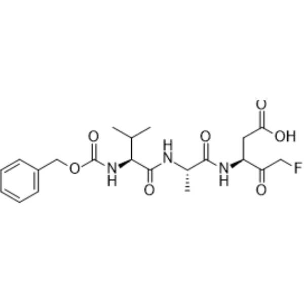
Z-VAD-FMKPan-Caspase Inhibitor -
HY-13205
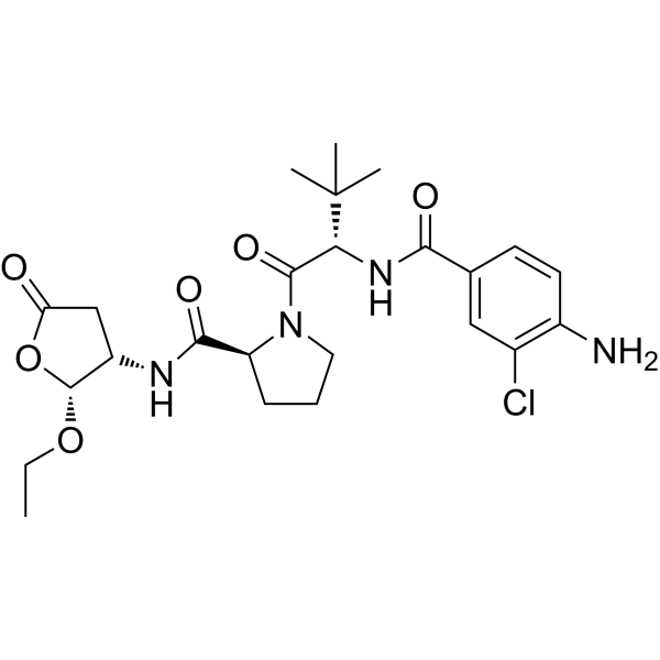
BelnacasanCaspase-1 Inhibitor -
HY-P1001
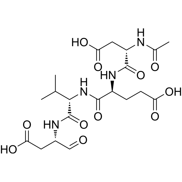
Ac-DEVD-CHOCaspase-3 Inhibitor -
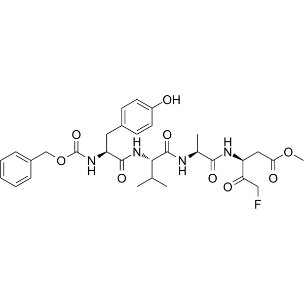
HY-P1009
Z-YVAD-FMKCaspase 1/4 Inhibitor -
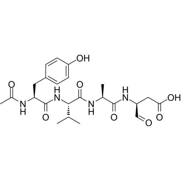
HY-120019
Ac-YVAD-CHOICE Inhibitor -
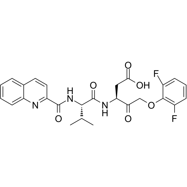
HY-12305
Q-VD-OPhPan-caspase Inhibitor -
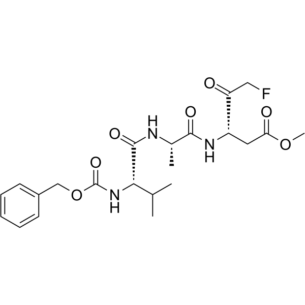
HY-16658
Z-VAD(OMe)-FMKPan-caspase Inhibitor -
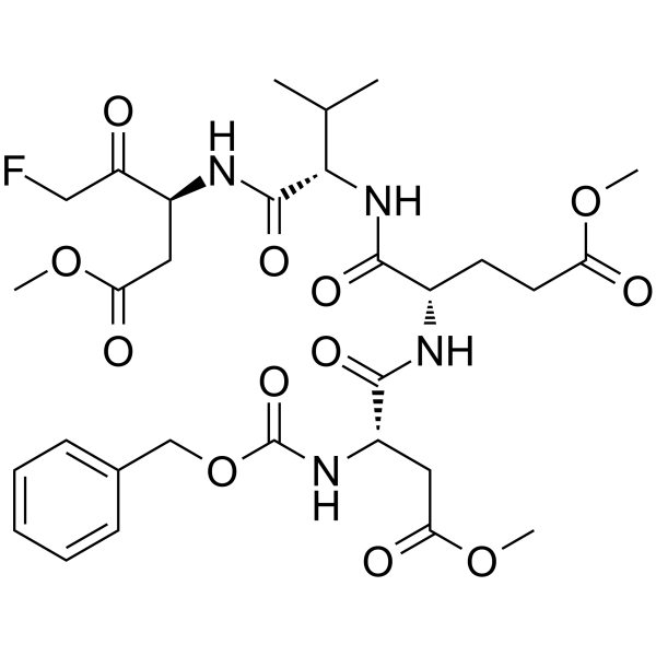
HY-12466
Z-DEVD-FMKCaspase-3 Inhibitor -
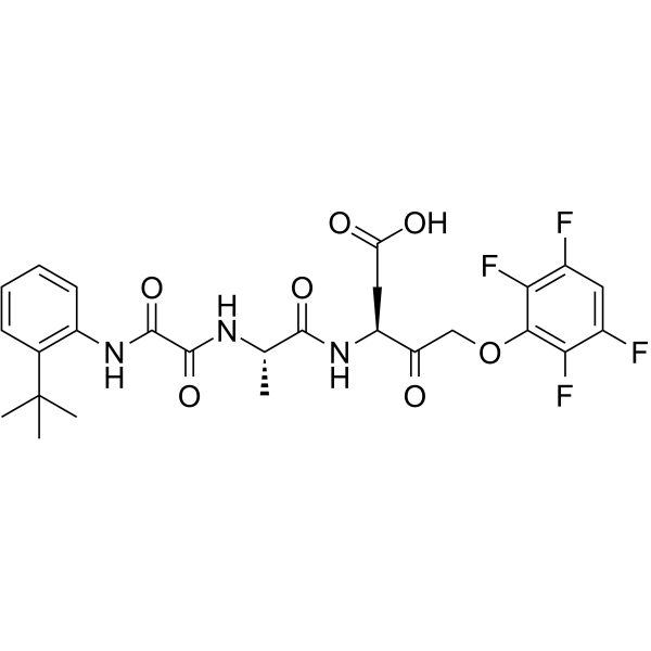
HY-10396
Emricasanpan-Caspase Inhibitor -
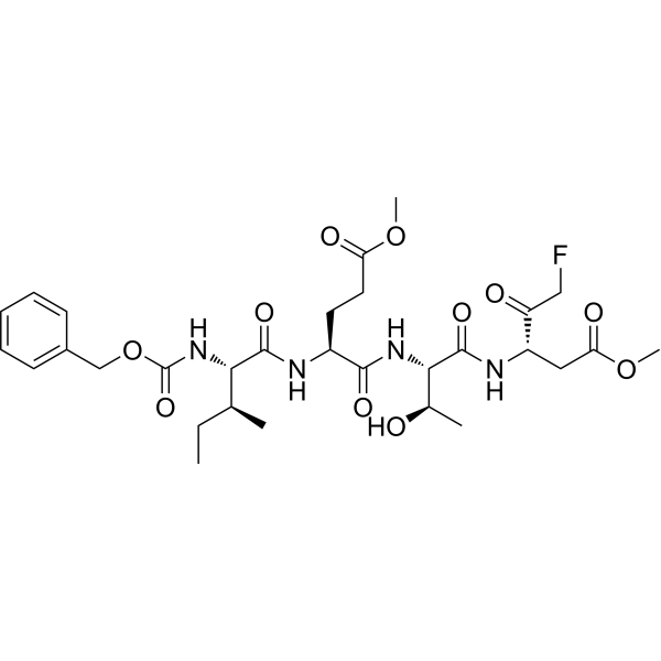
HY-101297
Z-IETD-FMKCaspase-8 Inhibitor -
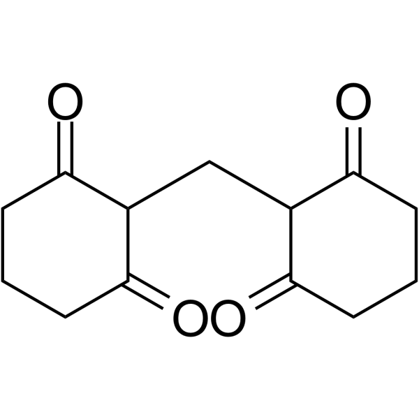
HY-103347
M50054Apoptosis Inhibitor -
HY-16990
Ac-YVAD-cmkCaspase-1 Inhibitor
Publications Citing Use of MedChemExpress (MCE) Ac-DEVD-CMK
MoreVoir tous les produits spécifiques à Isoform Caspase
More

Activité biologique
Description
|
Caspase-3 |
In Vitro
Ac-DEVD-CMK (100 μM; 24 h) inhibits IDB-induced apoptosis[3].
Ac-DEVD-CMK inhibits (10 μM; 36 h) inhibits citrate (10 mM)-induced p21 cleavage and G2/M accumulation in human pharyngeal squamous carcinoma FaDu and Detroit 562 cell lines[4].
MedChemExpress (MCE) has not independently confirmed the accuracy of these methods. They are for reference only.
-
Cell Line:Jurkat cells
-
Concentration:100 μM
-
Incubation Time:24 h
-
Result:Inhibited ingenol IDB (10 μM)-induced apoptosis.
In Vivo
Ac-DEVD-CMK (25 mg/kg; ip; single dose) significantly attenuates APAP-induced liver injury (AILI) in susceptible Sdc1−/− mice[6].
MedChemExpress (MCE) has not independently confirmed the accuracy of these methods. They are for reference only.
-
Animal Model:6- to 8-week-old females and males Sdc1-/- mice[4]
-
Dosage:25 mg/kg
-
Administration:IP; single dose; 3 hours post-APAP
-
Result:Significantly attenuated APAP (ip; 500 or 625 mg/kg)-induced liver injury (AILI), indicating that inhibition of GSK-3β or caspase-3 activity mitigates liver damage.


Chemical Information
-
CAS No. 285570-60-7
-
Appearance Solid
-
Masse moléculaire 550.94
-
Formule C21H31ClN4O11
-
Color White to off-white
-
Synonyms
Caspase-3 Inhibitor III
-
Livraison
Room temperature in continental US; may vary elsewhere.
-
Stockage
Sealed storage, away from moisture
Powder -80°C 2 years -20°C 1 year * In solvent : -80°C, 6 months; -20°C, 1 month (sealed storage, away from moisture)


Publications (3)
-
Journal Impact Factor
-
Most Recent
-
J Agric Food Chem
β-Hydroxybutyric Acid-Induced Bovine Endometrial Epithelial Cell Pyroptosis via Activating P2X7R. [Abstract]2025 Aug 27;73(34):21296-21308. PMID: 40742321 -
PLoS Pathog
2024 Jul 22;20(7):e1012408. PMID: 39038037 -
Exp Neurol
Muscone alleviates neuronal injury via increasing stress granules formation and reducing apoptosis in acute ischemic stroke. [Abstract]2024 Mar:373:114678. PMID: 38185313


Pureté et documentation
-
Fiche technique (276 KB)
-
SDS (252 KB)
- English - EN (252 KB)
- Français - FR (252 KB)
- Deutsch - DE (252 KB)
- Norwegian - NO (252 KB)
- Español - ES (252 KB)
- Swedish - SV (252 KB)
- Italian - IT (252 KB)
- Korean - KR (252 KB)
- Portuguese - PT (252 KB)
-
Instruction de manipulation (2659 KB)
Références
[1]. Eon Jeong Nam, et al. Syndecan-1 limits the progression of liver injury and promotes liver repair in acetaminophen-induced liver injury in mice. Hepatology. 2017 Nov;66(5):1601-1615. [Content Brief]
[2]. Lu Cai, et al. Hyperglycemia-induced apoptosis in mouse myocardium: mitochondrial cytochrome C-mediated caspase-3 activation pathway. Diabetes. 2002 Jun;51(6):1938-48. [Content Brief]
[3]. M M Mocanu, et al. Caspase inhibition and limitation of myocardial infarct size: protection against lethal reperfusion injury. Br J Pharmacol. 2000 May;130(2):197-200. [Content Brief]
[4]. M Blanco-Molina, et al. Ingenol esters induce apoptosis in Jurkat cells through an AP-1 and NF-kappaB independent pathway. Chem Biol. 2001 Aug;8(8):767-78. [Content Brief]
[6]. Hung KC, et al. Citrate-Induced p85α⁻PTEN Complex Formation Causes G2/M Phase Arrest in Human Pharyngeal Squamous Carcinoma Cell Lines. Int J Mol Sci. 2019 Apr 29;20(9):2105. [Content Brief]
[7]. Nam EJ, et al. Syndecan-1 limits the progression of liver injury and promotes liver repair in acetaminophen-induced liver injury in mice. Hepatology. 2017 Nov;66(5):1601-1615. [Content Brief]

Calculators
Concentration (start) × Volume (start) = Concentration (final) × Volume (final)


Ac-DEVD-CMK Related Classifications
HY-P0034 Related Classifications
 Powered by Bioz