



Rutaevin

Based on 1 Customer Validation
Rutaevin is an antifungal toxin and CYP3A4 inactivator. Rutaevin requires NADPH-dependent bioactivation to generate cis-but-2-ene-1,4-dial, which then covalently modifies and mechanism-based inactivates human CYP3A4 (IC50=4.48 μM, Ki=15.98 μM), and interacts with substances such as glutathione. Rutaevin disrupts the cellular structure of Sirococcus conigenus, accumulates linearly in resistant larch after pathogen exposure, and induces liver toxicity in mice via oral administration. Rutaevin can be applied to research in fields related to larch shoot blight and other associated areas.
Nur für Forschungszwecke. Wir verkaufen nicht an Patienten.
- Reinheit : 99.77%
- CAS. Nr.: 33237-37-5
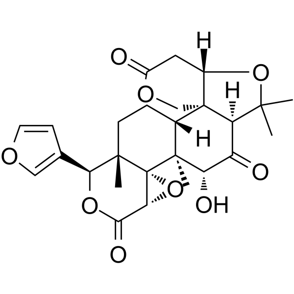
- Formel: C26H30O9
- Molecular Weight:486.51
-
Speicherung:
4°C, protect from light
* In solvent : -80°C, 6 months; -20°C, 1 month (protect from light)

To place orders, for customer services and technical support, please contact: MedChemExpress USA
Tel: 609-228-6898 E-mail: [email protected] [email protected]


-
 Biologische Aktivität
-
Chemical Information
- Protokoll
- Reinheit & Dokumentation
- Verweise
-
 Help & FAQs




Biologische Aktivität
Beschreibung
IC50 & Target
[1]|
CYP3A4 4.48 μM (IC50) |
In Vitro
Rutaevin (20-100 μM; 1-2 h) undergoes NADPH-dependent bioactivation to form a reactive BDA intermediate in rat and human liver microsomes, which reacts with nucleophilic trapping agents GSH, NAL, and MOA to form 10 distinct conjugates, including the cyclic mono(GSH) conjugate M1[1].
Rutaevin (0.1-30 μM; 30 min) acts as a potential mechanism-based inactivator of CYP3A4 in human liver microsomes, as shown by the 5.03-fold decrease in IC50 (to 4.48 μM) following NADPH-dependent preincubation[1].
Rutaevin (0.0625-1.0000 mg/mL; up to 5 days) potently inhibits the growth of Neofusicoccum laricinum with an IC50 of 0.27 mg/mL and achieves complete inhibition at 0.5 mg/mL[2].
MedChemExpress (MCE) has not independently confirmed the accuracy of these methods. They are for reference only.
In Vivo
Rutaevin (oral) induces hepatotoxicity in mice, as indicated by elevated serum ALT and AST activities[1].
Rutaevin (p.o.) induces hepatotoxicity in mice via bioactivation processes[2].
MedChemExpress (MCE) has not independently confirmed the accuracy of these methods. They are for reference only.
-
Animal Model:Sprague-Dawley (male, 220-250 g, bile duct cannulated)[1]
-
Dosage:5 mg/kg
-
Administration:i.v.; single dose
-
Result:Detected a cyclic mono(GSH) conjugate of rutaevin's bioactivated cis-butene-1,4-dial (BDA) intermediate (designated M1) as the most abundant metabolite in rat bile samples.


Chemical Information
-
CAS. Nr. 33237-37-5
-
Appearance Solid
-
Molecular Weight 486.51
-
Formel C26H30O9
-
Color White to off-white
-
SMILES
C[C@]1([C@H]2O)[C@@]3([C@@H]4O3)[C@]([C@H](C5=COC=C5)OC4=O)(CC[C@]1([H])[C@]67[C@](C(C)(C)O[C@@]6([H])CC(OC7)=O)([H])C2=O)C
-
Structure Classification
-
Initial Source
-
Versand
Room temperature in continental US; may vary elsewhere.
-
Speicherung
4°C, protect from light
* In solvent : -80°C, 6 months; -20°C, 1 month (protect from light)


Reinheit & Dokumentation
-
Data Sheet (280 KB)
-
SDS (252 KB)
- English - EN (252 KB)
- Français - FR (252 KB)
- Deutsch - DE (252 KB)
- Norwegian - NO (252 KB)
- Español - ES (252 KB)
- Swedish - SV (252 KB)
- Italian - IT (252 KB)
- Korean - KR (252 KB)
- Portuguese - PT (252 KB)
-
Handling Instructions (2659 KB)
Verweise

Calculators
Konzentration (Stammlösung) × Volumen (Stammlösung) = Konzentration (Ziellösung) × Volumen (Ziellösung)


Rutaevin Related Classifications
HY-N2620 Related Classifications
 Powered by Bioz