



Conbercept

Based on 1 Customer Validation
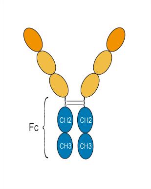
Conbercept (KH902) is a recombinant fusion protein composed of VEGFR-1 (second domain) and VEGFR-2 (third and fourth domains) regions fused to human IgG1 Fc. Conbercept is a VEGF inhibitor (IC50 = 8.8 pM) and is a soluble receptor decoy that blocks all isoforms of VEGF-A (Kd = 0.5 pM), VEGF-B (Kd = 8 pM), VEGF-C, and PlGF (Kd = 5 pM). Conbercept has anti-inflammatory effects, can lower the levels of VEGF, TNF-α and IL-6, and reduce the infiltration of inflammatory cells. Conbercept decreases tumor growth in several oncology studies. Conbercept can be used for various eye diseases such as polypoidal choroidal vasculopathy (PCV), diabetic macular edema (DME) and pathologic myopia choroidal neovascularization (pmCNV).
Nur für Forschungszwecke. Wir verkaufen nicht an Patienten.
- Reinheit : 98.42%
- CAS. Nr.: 1227158-72-6
- Molecular Weight:117.45 kDa
-
Speicherung:
Please store the product under the recommended conditions in the Certificate of Analysis.


To place orders, for customer services and technical support, please contact: MedChemExpress USA
Tel: 609-228-6898 E-mail: [email protected] [email protected]


-
 Biologische Aktivität
-
Chemical Information
- Protokoll
- Reinheit & Dokumentation
- Verweise
-
 Help & FAQs


Alle VEGFR Isoform-spezifische Produkte anzeigen
MoreAlle TNF Receptor Isoform-spezifische Produkte anzeigen
More

Biologische Aktivität
Beschreibung
IC50 & Target
[3]|
VEGFR1 |
VEGFR2 |
IL-6 |
In Vitro
Conbercept (0-586.9 pM, 4 d) shows an inhibition effect on VEGF-induced HUVEC proliferation with an EC50 value of 28 pM[3][5].
Conbercept inhibits VEGF-induced HUVEC migration and tube formation and suppressed the high glucose-induced migration and sprouting of human retinal endothelial cells[3].
MedChemExpress (MCE) has not independently confirmed the accuracy of these methods. They are for reference only.
In Vivo
Conbercept (10-50 mg/kg, i,v., single dose) significantly inhibits xylene-induced ear edema in mice[1].
Conbercept (0.64 mg, 40 μL, intravitreally injection, 4 times a week or 2 times every 2 weeks, for 2 weeks) has an inhibitory effect against VEGF over the course of 81 days in rabbit[3].
Conbercept (0.5 mg, intravitreally injection, single dose) is effective to prevent the formation of experimental choroidal neovascularization (CNV) and also to treat preexisted CNV without evidence of toxicity in monkey model[3][5].
Conbercept inhibits tumor growth in human hepatocellular carcinoma (HepG2), breast cancer (MCF-7), gastric carcinoma (A549), and colorectal cancer (LoVo) tumor xenograft models[3].
MedChemExpress (MCE) has not independently confirmed the accuracy of these methods. They are for reference only.
-
Animal Model:CIA model established in male Wistar rats (200 g, 4-6 weeks)[1]
-
Dosage:10 mg/kg
-
Administration:Intravenous injection (i.v.), once every other day for 2 weeks
-
Result:Significantly reduced paw edema volume and significantly reduced ankle joint swelling in CIA rats.
Significantly reduced Arthritis Index (AI) scores.
Reduced serum VEGF levels, TNF-α, and IL-6 in rats.
-
Animal Model:Xylene-induced ear edema model established in male Kunming mice (20-25 g)[1]
-
Dosage:10, 25 and 50 mg/kg
-
Administration:Intravenous injection (i.v.), single dose
-
Result:Significantly inhibited ear edema in the medium and high dose groups.
-
Animal Model:Experimental choroidal neovascularization (CNV) model established in rhesus monkeys[5]
-
Dosage:0.64 mg, 40 μL
-
Administration:Intravitreally injection, 4 times a week or 2 times every 2 weeks for 2 weeks
-
Result:Significantly reduced the incidence of CNV and had a significant therapeutic effect on established CNV.
No significant difference in the effects between weekly and biweekly administration.
Clinical Trial
| NCT Number | Sponsor | Condition | Start Date |
Phase

|
|---|---|---|---|---|
| NCT01329991 | 2011-05 | PHASE1 |
Conjugated
Unconjugated
Reconsititution
The product can be reconstituted/diluted with sterile PBS or saline.
Format
Anwendung
ELISA, FACS, Functional assay
Verified Bioactivity
-
 Immobilized VEGF165 Protein, Human can bind Conbercept. The EC50 for this effect is 4.31 ng/mL.


Chemical Information
-
CAS. Nr. 1227158-72-6
-
Appearance Liquid
-
Molecular Weight 117.45 kDa
-
Color Colorless to light yellow
-
SMILES
[Conbercept]
-
Synonyms
KH902
-
Versand
Shipping with dry ice.
-
Formulation
Please refer to the lot-specific COA for specific buffer information.
-
Speicherung
Please store the product under the recommended conditions in the Certificate of Analysis.


Reinheit & Dokumentation
-
Data Sheet (263 KB)
-
SDS (251 KB)
- English - EN (251 KB)
- Français - FR (251 KB)
- Deutsch - DE (251 KB)
- Norwegian - NO (251 KB)
- Español - ES (251 KB)
- Swedish - SV (251 KB)
- Italian - IT (251 KB)
- Korean - KR (251 KB)
- Portuguese - PT (251 KB)
-
Inhibitory Antibodies User Guide (603 KB)
Verweise
[1]. Liu Y, Fu H, Zuo L. Anti-inflammatory activities of a new VEGF blocker, Conbercept. Immunopharmacol Immunotoxicol. 2021 Oct;43(5):594-598. [Content Brief]
[2]. Liu H, et al. Updates on the Management of Ocular Vasculopathies with VEGF Inhibitor Conbercept. Curr Eye Res. 2020 Dec;45(12):1467-1476. [Content Brief]
[3]. Lu X, Sun X. Profile of conbercept in the treatment of neovascular age-related macular degeneration. Drug Des Devel Ther. 2015 Apr 22;9:2311-20. [Content Brief]
[4]. de Oliveira Dias JR, et al. Fusion proteins for treatment of retinal diseases: aflibercept, ziv-aflibercept, and conbercept. Int J Retina Vitreous. 2016 Feb 1;2:3. [Content Brief]
[5]. Zhang M, et al. The pharmacology study of a new recombinant human VEGF receptor-fc fusion protein on experimental choroidal neovascularization. Pharm Res. 2009 Jan;26(1):204-10. [Content Brief]

Calculators
Konzentration (Stammlösung) × Volumen (Stammlösung) = Konzentration (Ziellösung) × Volumen (Ziellösung)


Conbercept Related Classifications
HY-P99830 Related Classifications
 Powered by Bioz