



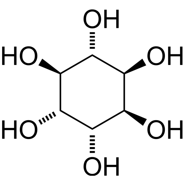
D-chiro-Inositol

Based on 1 Customer Validation
D-chiro-Inositol is a stereoisomer of inositol that exhibits activities such as improving glucose metabolism, anti-tumor effects, anti-inflammatory properties, and antioxidant activity. D-chiro-Inositol effectively alleviates cholestasis by enhancing bile acid secretion and reducing oxidative stress. D-chiro-Inositol improves insulin resistance, lowers hyperglycemia and circulating insulin levels, reduces serum androgen levels, and ameliorates some metabolic abnormalities associated with X syndrome by mimicking the action of insulin. Additionally, D-chiro-Inositol can induce a reduction in pro-inflammatory factors (such as Nf-κB) and cytokines (such as TNF-α), thereby exerting anti-inflammatory effects. D-chiro-Inositol may be used in the study of liver cirrhosis, breast cancer, type 2 diabetes, and polycystic ovary syndrome.
For research use only. We do not sell to patients.
- Purity : 98.0%
- CAS No.: 643-12-9
- Formula: C6H12O6
- Molecular Weight:180.16
-
Storage:
4°C, protect from light
* In solvent : -80°C, 6 months; -20°C, 1 month (protect from light)

To place orders, for customer services and technical support, please contact: MedChemExpress USA
Tel: 609-228-6898 E-mail: [email protected] [email protected]


-
 Biological Activity
-
Chemical Information
-
Solvent & Solubility
- Protocols
- Purity & Documentation
- References
-
 Help & FAQs


-
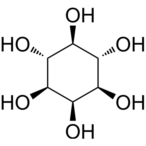
HY-B1411
i-InositolEndogenous Metabolite -
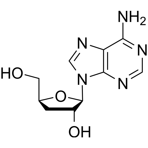
HY-N0262
CordycepinBacterial Inhibitor -
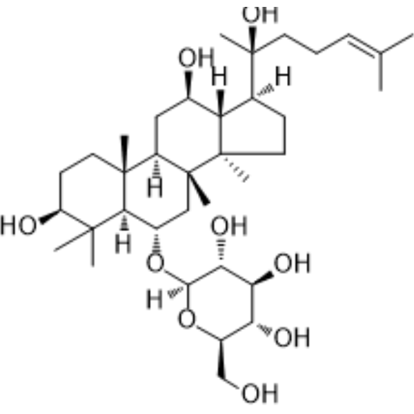
HY-N0604
Ginsenoside Rh1Endogenous Metabolite -

HY-Y1322
Triphenyl phosphateEndocrine Disruptor -
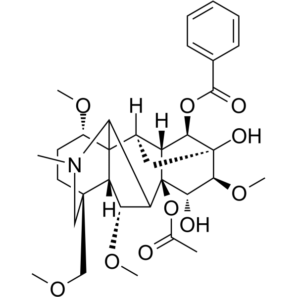
HY-N0267
HypaconitineTNF Receptor Inhibitor -

HY-N0262R
Cordycepin (Standard)Bacterial Inhibitor -
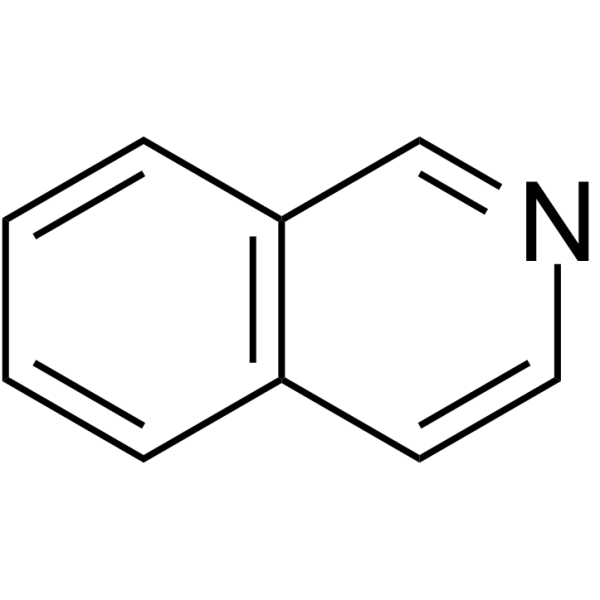
HY-W012732
IsoquinolineAlkaloid -
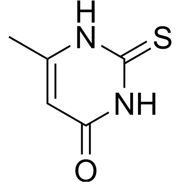
HY-B0513
MethylthiouracilTNF Receptor Inhibitor -

HY-N3021R
D-chiro-Inositol (Standard)Endogenous Metabolite -
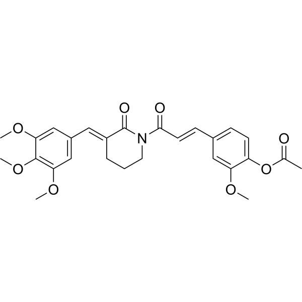
HY-148552
Anti-inflammatory agent 35Anti-inflammatory Agent -
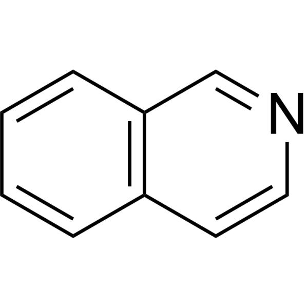
HY-W012732R
Isoquinoline (Standard)Anti-Cancer Agent -
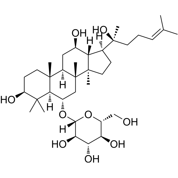
HY-N0604R
Ginsenoside Rh1 (Standard)Endogenous Metabolite
All Endogenous Metabolite Isoforms
More

Biological Activity
Description
IC50 & Target
|
Human Endogenous Metabolite |
In Vitro
D-chiro-Inositol (0.1-10 μmol, 3 h) improves glucose metabolism in HepG2 cells treated with Palmitoleic acid (HY-W011873) and inhibited the activity of FOXO1[3]. D-chiro-Inositol (1-40 mM, 48 h) inhibites the growth of breast cancer cells MDA-MB-231[4]. D-chiro-Inositol (0.125-1 mM, 24 and 48 h) regulates the GSH/GSSG ratio of 3T3-L1 adipocytes and reduces the pro-inflammatory state of cells[9].
MedChemExpress (MCE) has not independently confirmed the accuracy of these methods. They are for reference only. Further protocols information, click here.
-
Cell Line:PA-treated HepG2 cells
-
Concentration:0.1-10 μmol
-
Incubation Time:3 h
-
Result:Increased the mRNA expression levels of PEPCK and G6 Pase in HepG2 cells.
-
Cell Line:PA-treated HepG2 cells
-
Concentration:0.1-10 μmol
-
Incubation Time:3 h
-
Result:Inhibited the downregulation of IR β and p85 subunit expression levels and restored the phosphorylation levels of IRS-2, AKT and FOXO1.
-
Cell Line:MDA-MB-231 cells
-
Concentration:1, 5, 10, 20 and 40 mM
-
Incubation Time:48 h
-
Result:The cell proliferation abilities decreased by 18.3%, 17.2%, 17.5%, 18.4% and 24.9%, respectively.
In Vivo
MedChemExpress (MCE) has not independently confirmed the accuracy of these methods. They are for reference only.
-
Animal Model:Rat model of cholestasis induced by bile duct ligation [2]
-
Dosage:150 mg/kg
-
Administration:Oral gavage (p.o.) Once a day for 2 weeks
-
Result:Reduced serum alanine aminotransferase (ALT) and aspartate aminotransferase (AST) levels in rats, and alleviates bile duct hyperplasia, parenchymal necrosis and fibrosis. Increased total bile acid (TBA) levels in serum and bile and decreased total bile acid (TBA) levels in the liver. Increased the expression of genes encoding bile acid transporters BSEP (Abcb11) and MRP2 (Abcc2) in liver tissue. Decreased liver CD68 and NF-κB levels, serum and liver MDA levels, and significantly increased serum and liver superoxide dismutase activity.
-
Animal Model:High-fat diet mice [3]
-
Dosage:50 mg/kg
-
Administration:i.g.; once a day for 8 weeks
-
Result:Reduces hepatic DAG production and prevents PKCε ectopic translocation to the membrane, thereby inhibiting PKCε activation. Increased the low levels of IRβ and PI3Kp85 and reversed the downregulation of IRS-2 and AKT phosphorylation levels.
-
Animal Model:Female nude mice xenograft tumor model[4]
-
Dosage:500 和 1000 mg/kg
-
Administration:p.o.; once a day for 45 days
-
Result:The tumor growth inhibition rates were 22.1% and 67.6%, respectively.
Clinical Trial
| NCT Number | Sponsor | Condition | Start Date |
Phase

|
|---|---|---|---|---|
| NCT01329991 | Plexxikon| | 2011-05 | PHASE1 |


Chemical Information
-
CAS No. 643-12-9
-
Appearance Solid
-
Molecular Weight 180.16
-
Formula C6H12O6
-
Color White to off-white
-
SMILES
O[C@H]1[C@@H]([C@@H]([C@H]([C@@H]([C@H]1O)O)O)O)O
-
Structure Classification
-
Initial Source
-
Shipping
Room temperature in continental US; may vary elsewhere.
-
Storage
4°C, protect from light
* In solvent : -80°C, 6 months; -20°C, 1 month (protect from light)


Solvent & Solubility
In Vitro:
H2O : 75 mg/mL (416.30 mM; Need ultrasonic)
Please refer to the solubility information to select the appropriate solvent. Once prepared, please aliquot and store the solution to prevent product inactivation from repeated freeze-thaw cycles.
Storage method and period of stock solution: -80°C, 6 months; -20°C, 1 month (protect from light). When stored at -80°C, please use it within 6 months. When stored at -20°C, please use it within 1 month.
* Note: If you choose water as the stock solution, please dilute it to the working solution, then filter and sterilize it with a 0.22 μm filter before use.
Please refer to the solubility information to select the appropriate solvent. Once prepared, please aliquot and store the solution to prevent product inactivation from repeated freeze-thaw cycles.
Storage method and period of stock solution: -80°C, 6 months; -20°C, 1 month (protect from light). When stored at -80°C, please use it within 6 months. When stored at -20°C, please use it within 1 month.
* Note: If you choose water as the stock solution, please dilute it to the working solution, then filter and sterilize it with a 0.22 μm filter before use.
Concentration (start) × Volume (start) = Concentration (final) × Volume (final)
In Vivo:
For the following dissolution methods, please prepare the working solution directly:
It is recommended to prepare fresh solutions and use them promptly within a short period of time.
The percentages shown for the solvents indicate their volumetric ratio in the final prepared solution. If precipitation or phase separation occurs during preparation, heat and/or sonication can be used to aid dissolution.
Add each solvent one by one: PBS
Solubility: 100 mg/mL (555.06 mM); Clear solution; Need ultrasonic

Protocols
-
ROS/oxidative-stress fluorescent staining
ROS/oxidative-stress fluorescent staining uses cell-permeant fluorogenic probes that become fluorescent after oxidation inside cells or tissues; commonly used examples include DCFH-DA/DCFDA for broad cellular oxidant detection, DHE for superoxide-related signal detection, MitoSOX for mitochondrial superoxide-related signal detection, and CellROX probes for oxidative-stress-associated fluorescence readouts. The assay detects probe oxidation rather than a single ROS species unless the probe and analysis method have been chemically validated for that species. DCFH-DA enters cells, is deacetylated by intracellular esterases to DCFH, and produces fluorescent DCF after oxidation, so the readout is used as an operational measure of total cellular oxidative stress rather than a species-specific ROS measurement. DHE and MitoSOX can report superoxide-related oxidation, but red fluorescence alone can include non-specific ethidium-like oxidation products; HPLC or optimized spectral approaches are
-
Research Protocol for Endocrine Diseases
Endocrine diseases often arise from disrupted hormone production, hormone signaling, or target-tissue responsiveness; for diabetes-focused endocrine disease models, insulin signaling regulates glucose uptake, hepatic glucose output, lipid metabolism, and β-cell compensation. Type 2 diabetes develops through interacting defects in insulin resistance, β-cell dysfunction, adipose inflammation, hepatic glucose overproduction, altered incretin signaling, and ectopic lipid metabolism. A major unresolved question is whether endocrine dysfunction is driven primarily by target-tissue insulin resistance, intrinsic β-cell failure, immune/inflammatory stress, or combined multi-organ failure that differs by disease stage.
-
Research Protocol for Inflammation-related Diseases
The NLRP3 inflammasome is a cytosolic innate immune signaling platform that integrates priming signals and danger-signal activation to promote caspase-1 activation, maturation of IL-1β and IL-18, and gasdermin D-mediated pyroptotic cell death. The core experimental logic is to determine whether inflammatory disease phenotypes are driven by increased NLRP3 expression, ASC-containing inflammasome assembly, caspase-1 cleavage, GSDMD cleavage, and extracellular release of IL-1β/IL-18 rather than by nonspecific cell injury alone. The pathway is strongly linked to inflammation-related disease phenotypes because monosodium urate crystals activate NALP3/NLRP3 inflammasome signaling in gout-like crystal inflammation, cholesterol crystals activate NLRP3 inflammasomes in atherogenesis models, and DSS-induced intestinal inflammation has been reported to involve NLRP3 inflammasome activity. However, experimental colitis studies also show context-dependent protective effects of NLRP3 inflammasome co
-
Research Protocol for Metabolic Diseases
AMP-activated protein kinase, AMPK, is a conserved cellular energy sensor that responds to reduced cellular energy status and coordinates metabolism by increasing ATP-generating catabolic pathways while suppressing ATP-consuming anabolic processes. In metabolic disease research, the AMPK pathway is experimentally relevant because it regulates hepatic lipid synthesis, fatty acid oxidation, glucose production, skeletal-muscle glucose disposal, mTORC1-linked biosynthesis, autophagy, mitochondrial homeostasis, and whole-body energy balance. The central pathway logic is that energy stress, metformin, exercise-like stimulation, or direct AMPK activators increase AMPKα Thr172 phosphorylation and downstream substrate phosphorylation, including ACC and RAPTOR. Phosphorylation of ACC suppresses lipogenesis and supports fatty acid oxidation, whereas phosphorylation of RAPTOR suppresses mTORC1 signaling and links cellular energy status to growth and protein synthesis control. The pathway is linked
-
Breast Cancer Modeling
Breast cancer is a heterogeneous cancer, and it has been distinguished into four subtypes: luminal A, luminal B, HER2-positive and basal-like. Molecular mutations, epigenetic alterations, hormone exposure and immune microenvironment are related to the progression of breast cancer.


Purity & Documentation
-
Data Sheet (285 KB)
-
SDS (393 KB)
- English - EN (393 KB)
- Français - FR (393 KB)
- Deutsch - DE (393 KB)
- Norwegian - NO (393 KB)
- Español - ES (393 KB)
- Swedish - SV (393 KB)
- Italian - IT (393 KB)
- Korean - KR (393 KB)
- Portuguese - PT (393 KB)
-
Handling Instructions (2659 KB)
References
[1]. Ostlund RE Jr, et al. A stereospecific myo-inositol/D-chiro-inositol transporter in HepG2 liver cells. Identification with D-chiro-[3-3H]inositol. J Biol Chem. 1996 Apr 26;271(17):10073-8. [Content Brief]
[2]. Zhao SS, et al. D-chiro-inositol effectively attenuates cholestasis in bile duct ligated rats by improving bile acidsecretion and attenuating oxidative stress. Acta Pharmacol Sin. 2018 Feb;39(2):213-221. [Content Brief]
[3]. Cheng F, et al. chiro-Inositol Ameliorates High Fat Diet-Induced Hepatic Steatosis and Insulin Resistance viaPKCε-PI3K/AKT Pathway. J Agric Food Chem. 2019 May 29;67(21):5957-5967. [Content Brief]
[5]. Gambioli R, et al. New insights into the activities of D-chiro-inositol: a narrative review[J]. Biomedicines, 2021, 9(10): 1378. [Content Brief]
[6]. Iuorno M J, et al. Effects of d-chiro-inositol in lean women with the polycystic ovary syndrome[J]. Endocrine Practice, 2002, 8(6): 417-423. [Content Brief]
[7]. Pintaudi B, et al. The effectiveness of Myo‐inositol and D‐chiro inositol treatment in type 2 diabetes[J]. International journal of endocrinology, 2016, 2016(1): 9132052. [Content Brief]
[8]. Larner J, et al. D‐chiro‐inositol–its functional role in insulin action and its deficit in insulin resistance[J]. Journal of Diabetes Research, 2002, 3(1): 47-60. [Content Brief]
[9]. Fortis-Barrera Á, et al. C ucurbita ficifolia Bouché (Cucurbitaceae) and D-chiro-inositol modulate the redox state and inflammation in 3T3-L1 adipocytes[J]. Journal of Pharmacy and Pharmacology, 2013, 65(10): 1563-1576. [Content Brief]


Complete Stock Solution Preparation Table
Please refer to the solubility information to select the appropriate solvent. Once prepared, please aliquot and store the solution to prevent product inactivation from repeated freeze-thaw cycles.
Storage method and period of stock solution: -80°C, 6 months; -20°C, 1 month (protect from light). When stored at -80°C, please use it within 6 months. When stored at -20°C, please use it within 1 month.
| Optional Solvent | Concentration Solvent Mass | 1 mg | 5 mg | 10 mg | 25 mg |
|---|---|---|---|---|---|
| H2O | 1 mM | 5.5506 mL | 27.7531 mL | 55.5062 mL | 138.7655 mL |
| 5 mM | 1.1101 mL | 5.5506 mL | 11.1012 mL | 27.7531 mL | |
| 10 mM | 0.5551 mL | 2.7753 mL | 5.5506 mL | 13.8766 mL | |
| 15 mM | 0.3700 mL | 1.8502 mL | 3.7004 mL | 9.2510 mL | |
| 20 mM | 0.2775 mL | 1.3877 mL | 2.7753 mL | 6.9383 mL | |
| 25 mM | 0.2220 mL | 1.1101 mL | 2.2202 mL | 5.5506 mL | |
| 30 mM | 0.1850 mL | 0.9251 mL | 1.8502 mL | 4.6255 mL | |
| 40 mM | 0.1388 mL | 0.6938 mL | 1.3877 mL | 3.4691 mL | |
| 50 mM | 0.1110 mL | 0.5551 mL | 1.1101 mL | 2.7753 mL | |
| 60 mM | 0.0925 mL | 0.4626 mL | 0.9251 mL | 2.3128 mL | |
| 80 mM | 0.0694 mL | 0.3469 mL | 0.6938 mL | 1.7346 mL | |
| 100 mM | 0.0555 mL | 0.2775 mL | 0.5551 mL | 1.3877 mL |
* Note: If you choose water as the stock solution, please dilute it to the working solution, then filter and sterilize it with a 0.22 μm filter before use.


D-chiro-Inositol Related Classifications
HY-N3021 Related Classifications
 Powered by Bioz