



Polyphyllin I
Based on 5 publication(s) in Google Scholar
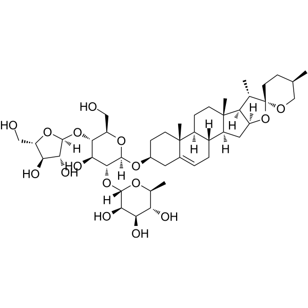
Polyphyllin I is a bioactive constituent extracted from Paris polyphylla, has strong anti-tumor activity. Polyphyllin I is an activator of the JNK signaling pathway and is an inhibitor of PDK1/Akt/mTOR signaling. Polyphyllin I induces autophagy, G2/M phase arrest and apoptosis.
For research use only. We do not sell to patients.
- Purity : 99.91%
- CAS No.: 50773-41-6
- Formula: C44H70O16
- Molecular Weight:855.02
-
Storage:Powder -20°C, 3 years , 4°C, 2 years ; In solvent -80°C, 6 months , -20°C, 1 month

To place orders, for customer services and technical support, please contact: MedChemExpress USA
Tel: 609-228-6898 E-mail: [email protected] [email protected]


-
 Biological Activity
-
Chemical Information
-
Solvent & Solubility
- Protocols
- Purity & Documentation
- References
-
 Help & FAQs


-
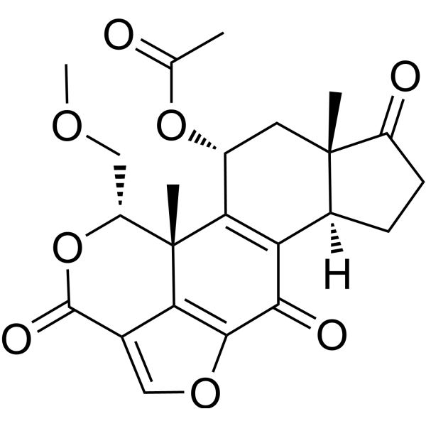
HY-10197
WortmanninPI3K Inhibitor -
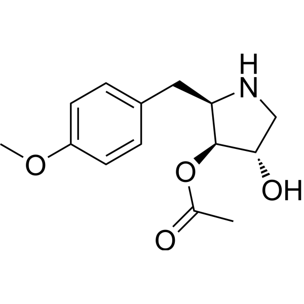
HY-18982
AnisomycinProtein Synthesis Inhibitor -
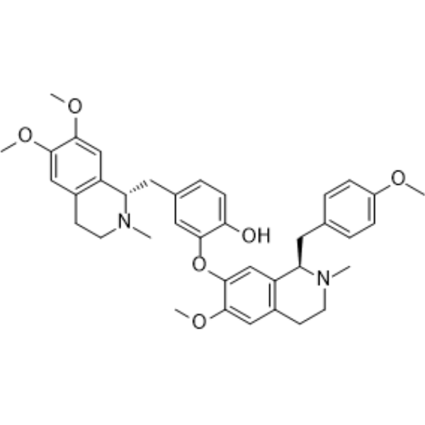
HY-N0441
NeferineAutophagy Modulator -

HY-10219
RapamycinmTOR Inhibitor -
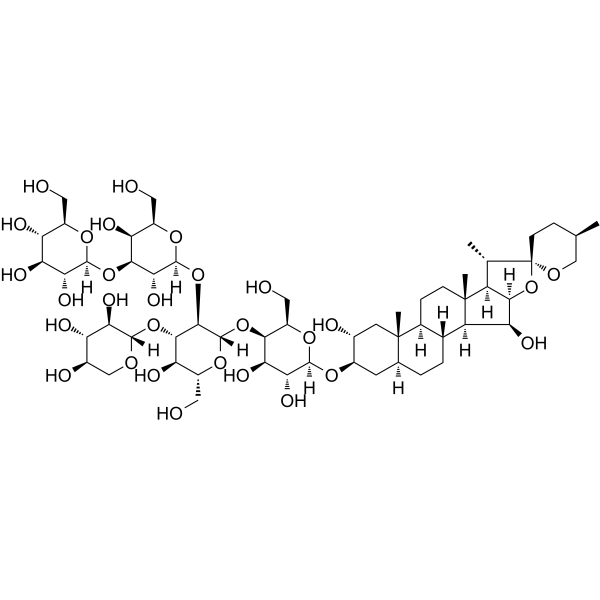
HY-N4000
DigitoninDetergent -
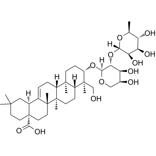
HY-N0255
alpha-HederinApoptosis Inducer -
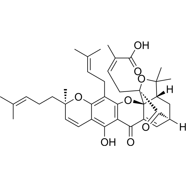
HY-N0087
Gambogic AcidBcl-2 Family Inhibitor -
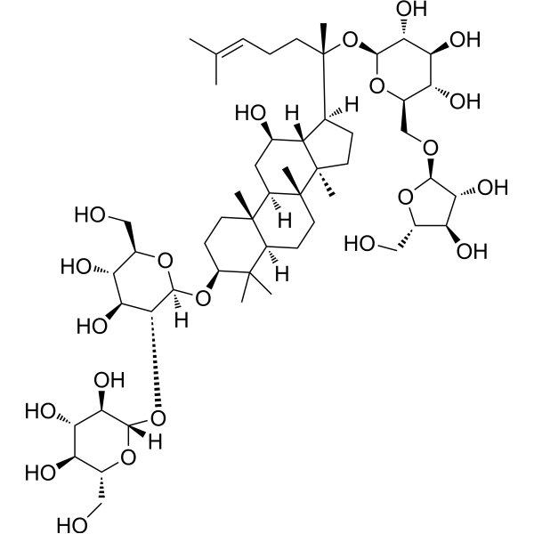
HY-N0042
Ginsenoside RcTNF Receptor Inhibitor -
HY-14664
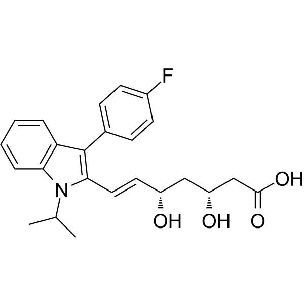
FluvastatinHMG-CoA Reductase Inhibitor -
HY-K2014
PEI Transfection Reagent -
HY-N3584
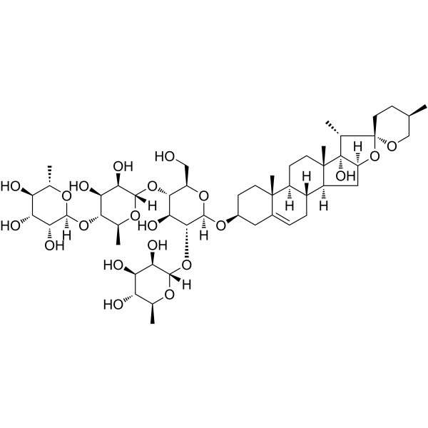
Paris saponin VIICaspase Inhibitor -
HY-N0124
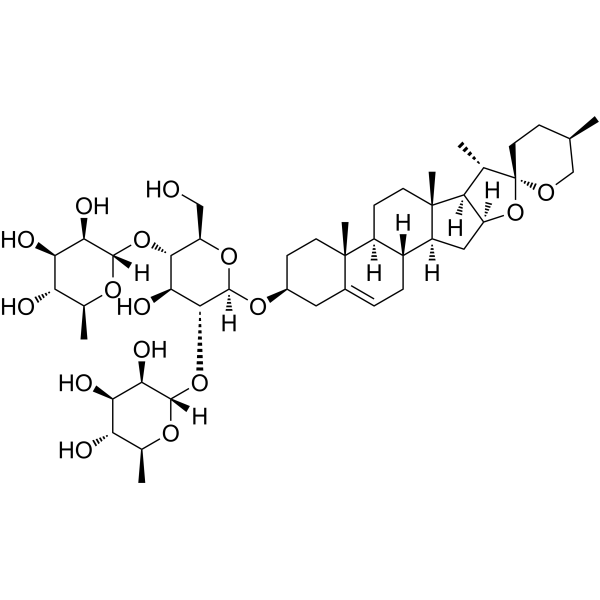
DioscinAnticancer Agent
Publications Citing Use of MedChemExpress (MCE) Polyphyllin I
More Customer Validation & Images

-
WB

-
Bio/Physico-chemical Assay

-
Cell Imaging/Staining

-
RT-PCR

-
WB



Biological Activity
Description
IC50 & Target
Cellular Effect
|
Cell Line

|
Type | Value | Description | References |
|---|---|---|---|---|
| Bel-7402 | IC50 |
4.74 μM
Compound: PPI
|
Cytotoxicity against human Bel7402 cells after 24 hrs by MTT assay
Cytotoxicity against human Bel7402 cells after 24 hrs by MTT assay
|
[PMID: 27623551] |
| HepG2 | IC50 |
4.01 μM
Compound: PPI
|
Cytotoxicity against human HepG2 cells after 24 hrs by MTT assay
Cytotoxicity against human HepG2 cells after 24 hrs by MTT assay
|
[PMID: 27623551] |
| NCI-H1299 | IC50 |
<1 μM
Compound: PSI
|
Cytotoxicity against human NCI-H1299 cells assessed as reduction in cell viability after 48 hrs by MTT assay
Cytotoxicity against human NCI-H1299 cells assessed as reduction in cell viability after 48 hrs by MTT assay
|
[PMID: 27721159] |
| NCI-H446 | IC50 |
<1 μM
Compound: PSI
|
Cytotoxicity against human NCI-H446 cells assessed as reduction in cell viability after 48 hrs by MTT assay
Cytotoxicity against human NCI-H446 cells assessed as reduction in cell viability after 48 hrs by MTT assay
|
[PMID: 27721159] |
| NCI-H460 | IC50 |
<1 μM
Compound: PSI
|
Cytotoxicity against human NCI-H460 cells assessed as reduction in cell viability after 48 hrs by MTT assay
Cytotoxicity against human NCI-H460 cells assessed as reduction in cell viability after 48 hrs by MTT assay
|
[PMID: 27721159] |
| NCI-H520 | IC50 |
<1 μM
Compound: PSI
|
Cytotoxicity against human NCI-H520 cells assessed as reduction in cell viability after 48 hrs by MTT assay
Cytotoxicity against human NCI-H520 cells assessed as reduction in cell viability after 48 hrs by MTT assay
|
[PMID: 27721159] |
In Vitro
Polyphyllin I (0.625-10 mg/mL; 24-72 h) inhibits the proliferation of three non-small cell lung cancer (NSCLC) cell lines, with the IC50s of 1.24, 2.40, and 2.33 μg/ml for A549, H460, and SK-MES-1 cells, respectively[1].
Polyphyllin I (2.5 mg/mL; 6-24 h) induces apoptosis of A549 cells[1].
Polyphyllin I (0.25-2 μM; 24 h) increase in LC3-II expression and decrease in P62 expression in HGC-27 cells[3].
MedChemExpress (MCE) has not independently confirmed the accuracy of these methods. They are for reference only.
-
Cell Line:A549, H460, and SK-MES-1 cells[1]
-
Concentration:24, 48, 72 hours
-
Incubation Time:24, 48, 72 hours
-
Result:Markedly reduced proliferation capacity in vitro and in a concentration-dependent manner.
In Vivo
MedChemExpress (MCE) has not independently confirmed the accuracy of these methods. They are for reference only.
-
Animal Model:BALB/c nude mice (5 weeks old) were s.c. inoculated with A549 cells[1]
-
Dosage:1.5 mg/kg
-
Administration:I.p. twice daily from day 2 to day 11
-
Result:The tumor growth rate was much lower than that in cisplatin group and PBS group.
- Detection of Apoptosis
- TUNEL staining for apoptotic DNA fragmentation
- Flow cytometric DNA-content cell-cycle staining
- Annexin V plus membrane-impermeant dye apoptosis staining
- Autophagy
- Apoptosis Solutions
- Protocol for Cell Cycle
- Lysosome and acidic-vesicle live-cell staining
- Macroautophagy Solutions


Chemical Information
-
CAS No. 50773-41-6
-
Appearance Solid
-
Molecular Weight 855.02
-
Formula C44H70O16
-
Color White to off-white
-
SMILES
C[C@H](CC1)CO[C@]21O[C@@]3([H])C[C@@]4([H])[C@@]5([H])[C@]([C@](CC[C@H](O[C@]6([H])O[C@H](CO)[C@@H](O[C@]7([H])O[C@@H](CO)[C@H](O)[C@H]7O)[C@H](O)[C@H]6O[C@@]8([H])[C@H](O)[C@H](O)[C@@H](O)[C@H](C)O8)C9)(C)C9=CC5)([H])CC[C@]4(C)[C@@]3([H])[C@@H]2C
-
Structure Classification
-
Initial Source
-
Shipping
Room temperature in continental US; may vary elsewhere.
-
Storage
Powder -20°C 3 years 4°C 2 years In solvent -80°C 6 months -20°C 1 month


Publications (5)
-
Journal Impact Factor
-
Most Recent
-
Int J Mol Sci
Polyphyllin I Inhibits the Metastasis of Cervical Cancer Through the Regulation of the β-Catenin Signaling Pathway. [Abstract]2025 May 12;26(10):4630. PMID: 40429774
Polyphyllin I purchased from MedChemExpress. Usage Cited in: Int J Mol Sci. 2025 May 12;26(10):4630. [Abstract]
The thermostability of Polyphyllin I (PPI) on the β-catenin protein was confirmed through CETSA.
Polyphyllin I purchased from MedChemExpress. Usage Cited in: Int J Mol Sci. 2025 May 12;26(10):4630. [Abstract]
SPR assay for the identification of Polyphyllin I (PPI) targeting the β-catenin protein.
Polyphyllin I purchased from MedChemExpress. Usage Cited in: Int J Mol Sci. 2025 May 12;26(10):4630. [Abstract]
An immunofluorescence assay was conducted to detect the effect of different concentrations of Polyphyllin I (PPI, 0-1 μM) on the changes in the nucleoplasmic distribution of β-catenin protein after a 6-h treatment of HO-8910PM cells.
Polyphyllin I purchased from MedChemExpress. Usage Cited in: Int J Mol Sci. 2025 May 12;26(10):4630. [Abstract]
qRT-PCR analysis of β-catenin mRNA levels in Polyphyllin I (PPI, 0-1 μM)-treated cells.
Polyphyllin I purchased from MedChemExpress. Usage Cited in: Int J Mol Sci. 2025 May 12;26(10):4630. [Abstract]
Assessment of the impact of PPI on β-catenin protein degradation; HO-8910PM cells were exposed to Polyphyllin I (PPI) (1 μM) either alone or in combination with either MG132 (20 μM), which is a proteasome inhibitor, or BAF (200 nM), which is an autophagy inhibitor, for 6 h. Subsequently, the cells were subjected to CHX (100 µg/mL) treatment for the indicated durations.
-
Biochim Biophys Acta Mol Basis Dis
Polyphyllin I inhibits glioblastoma progression by initiating ferroptosis via the Sirt1/Nrf2/HO-1/GPX4 signaling cascade. [Abstract]2025 Aug 26;1872(1):168028. PMID: 40876787 -
Med Oncol
Polyphyllin I inhibits ovarian cancer growth by inducing G0/G1 phase arrest and inhibiting the c-Myc signaling pathway. [Abstract]2025 Jun 12;42(7):254. PMID: 40506557 -
FASEB J
Polyphyllin I attenuates cognitive impairments and reduces AD-like pathology through CIP2A-PP2A signaling pathway in 3XTg-AD mice. [Abstract]2020 Dec;34(12):16414-16431. PMID: 33070372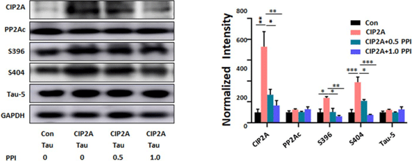
Polyphyllin I purchased from MedChemExpress. Usage Cited in: FASEB J. 2020 Dec;34(12):16414-16431. [Abstract]
HEK293-T cells are co-transfected with pFUW vector and tau plasmids, or pFUW-CIP2A and tau plasmids, with or without Polyphyllin I (PPI; 0.5 or 1.0 μM) incubation for 48 h.
-


Solvent & Solubility
In Vitro:
DMSO : 100 mg/mL (116.96 mM; Need ultrasonic; Hygroscopic DMSO has a significant impact on the solubility of product, please use newly opened DMSO)
H2O : < 0.1 mg/mL (insoluble)
Please refer to the solubility information to select the appropriate solvent. Once prepared, please aliquot and store the solution to prevent product inactivation from repeated freeze-thaw cycles.
Storage method and period of stock solution: -80°C, 6 months; -20°C, 1 month. When stored at -80°C, please use it within 6 months. When stored at -20°C, please use it within 1 month.
Please refer to the solubility information to select the appropriate solvent. Once prepared, please aliquot and store the solution to prevent product inactivation from repeated freeze-thaw cycles.
Storage method and period of stock solution: -80°C, 6 months; -20°C, 1 month. When stored at -80°C, please use it within 6 months. When stored at -20°C, please use it within 1 month.
Concentration (start) × Volume (start) = Concentration (final) × Volume (final)
In Vivo:
Select the appropriate dissolution method based on your experimental animal and administration route.
- For the following dissolution methods, please ensure to first prepare a clear stock solution using an In Vitro approach and then sequentially add co-solvents:
- To ensure reliable experimental results, the clarified stock solution can be appropriately stored based on storage conditions. As for the working solution for In Vivo experiments, it is recommended to prepare freshly and use it on the same day.
- The percentages shown for the solvents indicate their volumetric ratio in the final prepared solution. If precipitation or phase separation occurs during preparation, heat and/or sonication can be used to aid dissolution.
Add each solvent one by one: 10% DMSO 90% (20% SBE-β-CD in Saline)
Solubility: ≥ 2.5 mg/mL (2.92 mM); Clear solution
This protocol yields a clear solution of ≥ 2.5 mg/mL (saturation unknown).
Taking 1 mL working solution as an example, add 100 μL DMSO stock solution (25.0 mg/mL) to 900 μL 20% SBE-β-CD in Saline, and mix evenly.
Preparation of 20% SBE-β-CD in Saline (4°C, storage for one week): 2 g SBE-β-CD powder is dissolved in 10 mL Saline, completely dissolve until clear.
Add each solvent one by one: 10% DMSO 40% PEG300 5% Tween-80 45% Saline
Solubility: ≥ 0.83 mg/mL (0.97 mM); Clear solution
This protocol yields a clear solution of ≥ 0.83 mg/mL (saturation unknown).
Taking 1 mL working solution as an example, add 100 μL DMSO stock solution (8.3 mg/mL) to 400 μL PEG300, and mix evenly; then add 50 μL Tween-80 and mix evenly; then add 450 μL Saline to adjust the volume to 1 mL.
Preparation of Saline: Dissolve 0.9 g sodium chloride in ddH₂O and dilute to 100 mL to obtain a clear Saline solution.
In Vivo Dissolution Calculator
Please enter the basic information of animal experiments:
-
-
-
-
Recommended: Prepare an additional quantity of animals to account for potential losses during experiments.
Please enter your animal formula composition:
-
%DMSO +
Recommended: Keep the proportion of DMSO in working solution below 2% if your animal is weak.
-
%+
-
+%Tween-80 + +
-
%Saline +
The co-solvents required include: DMSO, . All of co-solvents are available by MedChemExpress (MCE). , Tween 80. All of co-solvents are available by MedChemExpress (MCE).
Working solution concentration: 0.22 mg/mL
Method for preparing stock solution: mg drug dissolved in μL DMSO. Stock solution concentration: mg/mL.
1. Take μL DMSO stock solution;
2. Add μL .
μL , mix evenly;
3. Then add μL Tween 80, mix evenly;
4. Then add μL
Please ensure that the stock solution in the first step is dissolved to a clear state, and add co-solvents in sequence. You can use ultrasonic heating (ultrasonic cleaner, recommended frequency 20-40 kHz), vortexing, etc. to assist dissolution.


Purity & Documentation
-
Data Sheet (281 KB)
-
SDS (252 KB)
- English - EN (252 KB)
- Français - FR (252 KB)
- Deutsch - DE (252 KB)
- Norwegian - NO (252 KB)
- Español - ES (252 KB)
- Swedish - SV (252 KB)
- Italian - IT (252 KB)
- Korean - KR (252 KB)
- Portuguese - PT (252 KB)
-
Handling Instructions (2659 KB)
References
[1]. Kong M, et al. Effects of polyphyllin I on growth inhibition of human non-small lung cancer cells and in xenograft. Acta Biochim Biophys Sin (Shanghai). 2010 Nov;42(11):827-33. [Content Brief]
[2]. Liu J, et al. Polyphyllin I induces G2/M phase arrest and apoptosis in U251 human glioma cells via mitochondrial dysfunction and the JNK signaling pathway. Acta Biochim Biophys Sin (Shanghai). 2017 Jun 1;49(6):479-486. [Content Brief]
[3]. He J, et al. Polyphyllin I induces autophagy and cell cycle arrest via inhibiting PDK1/Akt/mTOR signal and downregulating cyclin B1 in human gastric carcinoma HGC-27 cells. Biomed Pharmacother. 2019 Sep;117:109189. [Content Brief]


Complete Stock Solution Preparation Table
Please refer to the solubility information to select the appropriate solvent. Once prepared, please aliquot and store the solution to prevent product inactivation from repeated freeze-thaw cycles.
Storage method and period of stock solution: -80°C, 6 months; -20°C, 1 month. When stored at -80°C, please use it within 6 months. When stored at -20°C, please use it within 1 month.
| Optional Solvent | Concentration Solvent Mass | 1 mg | 5 mg | 10 mg | 25 mg |
|---|---|---|---|---|---|
| DMSO | 1 mM | 1.1696 mL | 5.8478 mL | 11.6956 mL | 29.2391 mL |
| 5 mM | 0.2339 mL | 1.1696 mL | 2.3391 mL | 5.8478 mL | |
| 10 mM | 0.1170 mL | 0.5848 mL | 1.1696 mL | 2.9239 mL | |
| 15 mM | 0.0780 mL | 0.3899 mL | 0.7797 mL | 1.9493 mL | |
| 20 mM | 0.0585 mL | 0.2924 mL | 0.5848 mL | 1.4620 mL | |
| 25 mM | 0.0468 mL | 0.2339 mL | 0.4678 mL | 1.1696 mL | |
| 30 mM | 0.0390 mL | 0.1949 mL | 0.3899 mL | 0.9746 mL | |
| 40 mM | 0.0292 mL | 0.1462 mL | 0.2924 mL | 0.7310 mL | |
| 50 mM | 0.0234 mL | 0.1170 mL | 0.2339 mL | 0.5848 mL | |
| 60 mM | 0.0195 mL | 0.0975 mL | 0.1949 mL | 0.4873 mL | |
| 80 mM | 0.0146 mL | 0.0731 mL | 0.1462 mL | 0.3655 mL | |
| 100 mM | 0.0117 mL | 0.0585 mL | 0.1170 mL | 0.2924 mL |


Polyphyllin I Related Classifications
HY-N0047 Related Classifications
 Powered by Bioz