



Duligotuzumab

Based on 1 Customer Validation
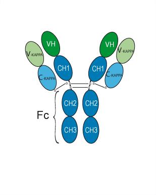
Duligotuzumab (MEHD-7945A; RG 7597) is a humanized IgG-κ monoclonal antibody targeting EGFR. Duligotuzumab blocks the binding of ligands to these two receptors, inhibits downstream HER/ErbB, AKT and MAPK signaling pathways, induces antibody-dependent cellular cytotoxicity, reduces the proliferation and migration abilities of cancer cells, promotes apoptosis, exerts radiosensitizing effects, and reverses EGFR resistance in cancer cells. Duligotuzumab can be used in tumor-related research.
For research use only. We do not sell to patients.
- Purity : 98.70%
- CAS No.: 1314238-96-4
- Molecular Weight:144.78 kDa
-
Storage:
Please store the product under the recommended conditions in the Certificate of Analysis.


To place orders, for customer services and technical support, please contact: MedChemExpress USA
Tel: 609-228-6898 E-mail: [email protected] [email protected]


-
 Biological Activity
-
Chemical Information
- Protocols
- Purity & Documentation
- References
-
 Help & FAQs



All EGFR Isoforms
More

Biological Activity
Description
Isotype
Human IgG1 kappa
Recommend Isotype Controls
Species Reactivity
Human
IC50 & Target
[1]|
HER3 |
HER2 |
EGFR |
Akt |
p38 MAPK |
In Vitro
Duligotuzumab (0.01-25 μM; 72 h) inhibits proliferation of HER2-positive OE33, OE19, and NCI-N87 gastric cancer cell lines with IC50 values of ≈0.1 to ≈0.5 μM over 72 h, but has no effect on HER2-negative MKN28 gastric cancer cells[1].
Duligotuzumab (0.5 μM; 7 days) reduces colony formation in HER2-positive OE33, OE19, and NCI-N87 gastric cancer cell lines over 7 days[1].
Duligotuzumab (0.5 μM; 72 h) reduces migration of HER2-positive OE33, OE19, and NCI-N87 gastric cancer cell lines over 72 h[1].
Duligotuzumab (0.5 μM; 72 h) induces apoptosis in HER2-positive OE33, OE19, and NCI-N87 gastric cancer cell lines over 72 h, with an apoptotic rate of ≈35% in OE33 cells[1].
Duligotuzumab (0.5 μM; 48 h) reduces phosphorylated HER2 and HER3 levels but does not alter total or phosphorylated AKT, MAPK, or total HER2 or HER3 protein levels in HER2-positive OE33 and OE19 gastric cancer cell lines[1].
Duligotuzumab induces antibody-dependent cell-mediated cytotoxicity in EGFR-overexpressing cell lines, with activity comparable to cetuximab[2].
Duligotuzumab binds to EGFR with a dissociation constant of 0.4 nM[2].
Duligotuzumab enhances antiproliferative activity in cancer cells, overcomes EGFR resistance, and enhances radiation effects compared to monospecific HER antibodies, including in cetuximab-resistant cells[3].
Duligotuzumab potently blocks EGFR and HER3 receptors and inhibits downstream HER/ErbB signaling pathways in an in vitro system[3].
MedChemExpress (MCE) has not independently confirmed the accuracy of these methods. They are for reference only.
-
Cell Line:HER2-positive human gastric cancer cell lines (OE33, OE19, NCI-N87), HER2-negative human gastric cancer cell line (MKN28)
-
Concentration:0.01, 0.05, 0.1, 0.5, 1, 5, 10, 25 μM
-
Incubation Time:72 h
-
Result:Inhibited proliferation of HER2-positive OE33, OE19, and NCI-N87 cells, with IC50 values ranging from ≈0.1 to ≈0.5 μM.
Exhibited no antiproliferative effect in HER2-negative MKN28 cells.
-
Cell Line:HER2-positive human gastric cancer cell lines (OE33, OE19, NCI-N87)
-
Concentration:0.5 μM
-
Incubation Time:72 h
-
Result:Reduced migration of OE33, OE19, and NCI-N87 cells, though to a lesser extent than the combination of Duligotuzumab and trastuzumab.
-
Cell Line:HER2-positive human gastric cancer cell lines (OE33, OE19, NCI-N87)
-
Concentration:0.5 μM
-
Incubation Time:72 h
-
Result:Induced apoptosis in OE33, OE19, and NCI-N87 cells, with OE33 cells showing an apoptotic rate of ≈35%.
-
Cell Line:HER2-positive human gastric cancer cell lines (OE33, OE19)
-
Concentration:0.5 μM
-
Incubation Time:48 h
-
Result:Had no effect on the expression of total or phosphorylated AKT and MAPK proteins in OE33 and OE19 cells.
Reduced phosphorylated HER2 and HER3 receptor levels in these cell lines, though not to the extent seen with the combination of Duligotuzumab and trastuzumab.
Did not alter total HER2 or HER3 receptor levels.
Gene ID
Accession
Conjugated
Unconjugated
Reconsititution
The product can be reconstituted/diluted with sterile PBS or saline.
Format
-
Human IgG1 kappa
Application
ELISA, FACS, Functional assay
Verified Bioactivity
-
 Immobilized EGFR Protein, Human can bind Duligotuzumab. The EC50 for this effect is 4.244 ng/mL.
 -
 Immobilized HER3 Protein, Human can bind Duligotuzumab. The EC50 for this effect is 1.423 ng/mL.
 -
 Flow Cytometry analysis of Hela cells labelling EGFR (red) with Duligotuzumab (HY-P99866). Cells were fixed with 4% paraformaldehyde and permeabilised with 90% methanol. Then cells were stained with the primary antibody at 1/200 dilution for an hour at 4℃. Goat Anti-Human IgG H&L (AF488) (HY-P83776) was used as the secondary antibody at 1/1,000 dilution for 30 minutes at 4℃. Human IgG1 kappa (HY-P99001, blue) was used as the isotype control, cells without incubation with primary antibody were used as the unlabeled control (black).


Chemical Information
-
CAS No. 1314238-96-4
-
Appearance Liquid
-
Molecular Weight 144.78 kDa
-
Color Colorless to light yellow
-
SMILES
[Duligotuzumab]
-
Synonyms
MEHD-7945A; RG 7597
-
Shipping
Shipping with dry ice.
-
Formulation
Please refer to the lot-specific COA for specific buffer information.
-
Storage
Please store the product under the recommended conditions in the Certificate of Analysis.


Purity & Documentation
-
Data Sheet (265 KB)
-
SDS (252 KB)
- English - EN (252 KB)
- Français - FR (252 KB)
- Deutsch - DE (252 KB)
- Norwegian - NO (252 KB)
- Español - ES (252 KB)
- Swedish - SV (252 KB)
- Italian - IT (252 KB)
- Korean - KR (252 KB)
- Portuguese - PT (252 KB)
-
Inhibitory Antibodies User Guide (603 KB)
References
[1]. Laterza MM, et al. Enhanced Antitumor Effect of Trastuzumab and Duligotuzumab or Ipatasertib Combination in HER-2 Positive Gastric Cancer Cells. Cancers (Basel). 2021;13(10):2339. [Content Brief]
[2]. Fayette J, et al. Randomized Phase II Study of Duligotuzumab (MEHD7945A) vs. Cetuximab in Squamous Cell Carcinoma of the Head and Neck (MEHGAN Study). Front Oncol. 2016;6:232. [Content Brief]
[3]. Cole, P. DULIGOTUZUMAB. Drugs of the Future 2015, 40(3): 167-172.

Calculators
Concentration (start) × Volume (start) = Concentration (final) × Volume (final)


Duligotuzumab Related Classifications
HY-P99866 Related Classifications
 Powered by Bioz
Keywords