



Lumisterol
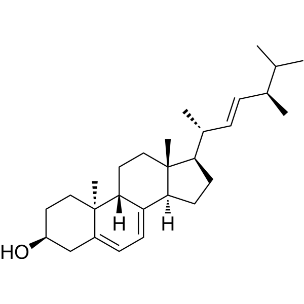
Lumisterol (9β,10α-Ergosterol) is a photoproduct of 7-dehydrocholesterol, present in the skin, and acts as an orally active VDR non-genomic modulator and ROR inverse agonist. Lumisterol binds to the SARS-CoV-2 Mpro substrate-binding pocket and the RdRP active site, inhibiting enzyme activity. Lumisterol induces NRF2-regulated antioxidant responses, p53 phosphorylation and nuclear translocation, and intracellular free radical scavenging. Lumisterol inhibits the proliferation of epidermal keratinocytes and melanoma cells, modulates cell cycle progression, and suppresses basal and TNFα-induced NFκB transcriptional activity. Lumisterol inhibits RORγ transcriptional activity and IL-17 production. Lumisterol is used in research on UVB-induced skin damage, melanoma, psoriasis, vitamin D deficiency, and COVID-19.
For research use only. We do not sell to patients.
- CAS No.: 474-69-1
- Formula: C28H44O
- Molecular Weight:396.65
-
Storage:
-80°C, protect from light, stored under nitrogen

To place orders, for customer services and technical support, please contact: MedChemExpress USA
Tel: 609-228-6898 E-mail: [email protected] [email protected]


-
 Biological Activity
-
Chemical Information
-
Solvent & Solubility
- Protocols
- Purity & Documentation
- References
-
 Help & FAQs


All Endogenous Metabolite Isoforms
More

Biological Activity
Description
|
RORγ |
VDR |
Nrf2 |
NFκB |
IL-17 |
p53 |
TNFα |
In Vitro
Lumisterol shows favorable docking scores and binding poses in the LBDs of RORα and RORγ, consistent with its role as a ligand for these receptors[2].
In human malignant melanoma A375 and SK-MEL-28 cell lines, treatment with 100 nM Lumisterol (100 nM; 72 h) enhances the migration of A375 cells and SK-MEL-28 cells[4].
In human malignant melanoma A375 cell line, 100 nM Lumisterol (100 nM; 48 h) increases the proportion of cells in the G0/G1 phase and decreases the percentage in G2/M, but has no effect on SK-MEL-28 cells[4].
In human malignant melanoma SK-MEL-28 cell line, 100 nM Lumisterol (100 nM; 24 h) causes a small but significant increase in CYP24A1 mRNA levels, but has no effect on CYP24A1 or other vitamin D metabolizing genes in A375 cells[4].
Lumisterol (1150 µW cm-2 UV-B pre-exposure; 30 min pre-exposure, 8 h thermal) is converted to Vitamin D2 (HY-76542) in a cell-free system, with the strongest conversion occurring when lumisterol is pre-exposed to UV-B light[5].
Lumisterol (24-72 h) incubation of a UV-B-exposed yeast diet at 37 °C results in a time-dependent conversion of Lumisterol to Vitamin D2, with a 63% increase in Vitamin D2 content after 72 h[5].
In human malignant melanoma A375 and SK-MEL-28 cell lines, Lumisterol (1 μM; 72 h) fails to inhibit cell proliferation, and instead increases proliferation by 10% in SK-MEL-28 cells[4].
MedChemExpress (MCE) has not independently confirmed the accuracy of these methods. They are for reference only. Further protocols information, click here.
-
Cell Line:A375 and SK-MEL-28
-
Concentration:1 μM
-
Incubation Time:72 h
-
Result:Failed to inhibit proliferation of either cell type.
Increased proliferation by 10% in SK-MEL-28 cells.
-
Cell Line:A375 and SK-MEL-28
-
Concentration:100 nM
-
Incubation Time:72 h
-
Result:Enhanced the migration of A375 cells.
Increased migration of SK-MEL-28 cells to a lesser extent than 22(OH)L3.
-
Cell Line:A375 and SK-MEL-28
-
Concentration:100 nM
-
Incubation Time:48 h
-
Result:Increased the percentage of cells in the G0/G1 phase and decreased the percentage in G2/M in A375 cells.
Had no effect on the cell cycle of SK-MEL-28 cells.
-
Cell Line:A375 and SK-MEL-28
-
Concentration:100 nM
-
Incubation Time:24 h
-
Result:Caused a small but significant increase in CYP24A1 mRNA levels in SK-MEL-28 cells.
Had no effect on CYP24A1 or other vitamin D metabolizing genes in A375 cells.


Chemical Information
-
CAS No. 474-69-1
-
Appearance Solid
-
Molecular Weight 396.65
-
Formula C28H44O
-
Color White to off-white
-
SMILES
C[C@H](C(C)C)/C=C/[C@@H](C)[C@H]1CC[C@@]2([H])C3=CC=C4C[C@@H](O)CC[C@@]4(C)[C@]3([H])CC[C@]12C
-
Synonyms
9β,10α-Ergosterol
-
Structure Classification
-
Initial Source
-
Shipping
Shipping with dry ice.
-
Storage
-80°C, protect from light, stored under nitrogen


Solvent & Solubility
In Vitro:
DMSO : 100 mg/mL (252.11 mM; ultrasonic and warming and heat to 60°C; Hygroscopic DMSO has a significant impact on the solubility of product, please use newly opened DMSO)
Please refer to the solubility information to select the appropriate solvent. Once prepared, please aliquot and store the solution to prevent product inactivation from repeated freeze-thaw cycles.
Storage method and period of stock solution: -80°C, 6 months (protect from light, stored under nitrogen). When stored at -80°C, please use it within 6 months.
Please refer to the solubility information to select the appropriate solvent. Once prepared, please aliquot and store the solution to prevent product inactivation from repeated freeze-thaw cycles.
Storage method and period of stock solution: -80°C, 6 months (protect from light, stored under nitrogen). When stored at -80°C, please use it within 6 months.
Concentration (start) × Volume (start) = Concentration (final) × Volume (final)
In Vivo:
Select the appropriate dissolution method based on your experimental animal and administration route.
- For the following dissolution methods, please ensure to first prepare a clear stock solution using an In Vitro approach and then sequentially add co-solvents:
- To ensure reliable experimental results, the clarified stock solution can be appropriately stored based on storage conditions. As for the working solution for In Vivo experiments, it is recommended to prepare freshly and use it on the same day.
- The percentages shown for the solvents indicate their volumetric ratio in the final prepared solution. If precipitation or phase separation occurs during preparation, heat and/or sonication can be used to aid dissolution.
Add each solvent one by one: 10% DMSO 40% PEG300 5% Tween-80 45% Saline
Solubility: ≥ 2.5 mg/mL (6.30 mM); Clear solution
This protocol yields a clear solution of ≥ 2.5 mg/mL (saturation unknown).
Taking 1 mL working solution as an example, add 100 μL DMSO stock solution (25.0 mg/mL) to 400 μL PEG300, and mix evenly; then add 50 μL Tween-80 and mix evenly; then add 450 μL Saline to adjust the volume to 1 mL.
Preparation of Saline: Dissolve 0.9 g sodium chloride in ddH₂O and dilute to 100 mL to obtain a clear Saline solution.
In Vivo Dissolution Calculator
Please enter the basic information of animal experiments:
-
-
-
-
Recommended: Prepare an additional quantity of animals to account for potential losses during experiments.
Please enter your animal formula composition:
-
%DMSO +
Recommended: Keep the proportion of DMSO in working solution below 2% if your animal is weak.
-
%+
-
+%Tween-80 + +
-
%Saline +
The co-solvents required include: DMSO, . All of co-solvents are available by MedChemExpress (MCE). , Tween 80. All of co-solvents are available by MedChemExpress (MCE).
Working solution concentration: 0.22 mg/mL
Method for preparing stock solution: mg drug dissolved in μL DMSO. Stock solution concentration: mg/mL.
1. Take μL DMSO stock solution;
2. Add μL .
μL , mix evenly;
3. Then add μL Tween 80, mix evenly;
4. Then add μL
Please ensure that the stock solution in the first step is dissolved to a clear state, and add co-solvents in sequence. You can use ultrasonic heating (ultrasonic cleaner, recommended frequency 20-40 kHz), vortexing, etc. to assist dissolution.

Protocols
-
Kinase activity and phosphorylation assays
Kinase activity assays measure the ability of kinases to transfer phosphate groups from ATP to specific substrates, while phosphorylation assays detect the presence and levels of phosphorylated proteins. Common methods include radiolabeled ATP incorporation (e. g. ,), ADP release detection via bioluminescence (e. g. ,[3]), enzyme-linked immunosorbent assays (ELISA) for phospho-specific epitopes (e. g. ,[6]), and microtiter-based formats for high-throughput screening (e. g. ,[8]). The ADP-Glo assay quantifies kinase activity by measuring ADP produced during phosphorylation using a luciferase-based system. Radiometric assays involve autoradiography or scintillation counting after incorporation of 32P-labeled ATP into substrate proteins. ELISA-based approaches rely on phospho-specific antibodies to detect activated kinases in cell lysates or purified samples.
-
Western Blot
Western blotting (WB) is a commonly used experimental method in molecular biology, biochemistry, and immunogenetics for identifying and quantifying target proteins. It combines gel electrophoresis with immunoassay, enabling researchers to analyze protein expression, post-translational modifications, and molecular weight.
-
Cytoplasmic-Nuclear Fractionated Protein Extraction
Cytoplasmic-nuclear fractionated protein extraction separates soluble cytoplasmic proteins from nuclear-enriched proteins by mild plasma-membrane permeabilization, differential centrifugation, washing of nuclei, and extraction of nuclear proteins for downstream immunoblotting or related molecular analysis. The readout is the relative abundance of a protein in cytoplasmic and nuclear fractions, commonly assessed by western blotting together with compartment markers such as tubulin or pyruvate kinase for cytoplasm and lamin, nucleoporin, hnRNP, H2AX, or Lamin B for nuclear fractions.
-
Dual Luciferin reporter gene assay
Luciferin reporter gene assay is a reporting system to detect the activity of Firefly Luciferase using luciferin as a substrate, which is often used in the research of miRNA target gene verification and promoter transcriptive activity regulation. Dual luciferase usually refers to Firefly luciferase and Renilla luciferase.
-
Flow cytometric DNA-content cell-cycle staining
Flow cytometric DNA-content cell-cycle staining measures the fluorescence intensity of DNA-bound fluorochromes in single cells or nuclei to estimate DNA content distributions, allowing assignment of populations to G0/G1, S, and G2/M phases by DNA histogram deconvolution. Propidium iodide (PI) intercalates into DNA, and PI fluorescence is proportional to cellular DNA content when staining is performed under conditions that make DNA accessible and minimize non-DNA signal. Cells with G2/M DNA content are expected to show approximately twice the fluorescence intensity of G0/G1 cells, while S-phase cells occupy intermediate fluorescence values. PI-based DNA-content analysis can also detect cells with fractional DNA content, often reported as sub-G1, when DNA fragmentation and extraction during staining reduce retained DNA signal in apoptotic cells. DAPI is an alternative DNA fluorochrome for univariate DNA-content analysis, while bivariate approaches combining DNA content with proliferation
-
Imiquimod-Induced Psoriasiform Dermatitis
Imiquimod (IMQ)-induced psoriasiform dermatitis is a widely used murine model in which topical application of IMQ, a Toll-like receptor 7 (TLR7) agonist, triggers innate immune activation in the skin and induces a psoriasis-like inflammatory cascade characterized by epidermal hyperplasia, immune cell infiltration, and cytokine production dominated by the IL-23/IL-17 axis. This inflammatory response is mediated through activation of dendritic cells and downstream induction of IL-23, IL-17A, IL-22, and related pro-inflammatory mediators, recapitulating key features of human plaque psoriasis and enabling mechanistic and therapeutic studies. The model is commonly induced using Aldara (5% IMQ cream) applied topically to murine skin, resulting in rapid onset of erythema, scaling, and thickening that can be quantified as disease severity indices and validated histologically.
-
BrdU Incorporation Assay
Bromodeoxyuridine (BrdU) incorporation assay is based on the principle that BrdU, a thymidine analog, is incorporated into newly synthesized DNA during the S phase of the cell cycle, thereby serving as a marker of DNA replication and cellular proliferation. Incorporated BrdU can be detected using anti-BrdU antibodies following DNA denaturation, enabling visualization or quantification of proliferating cells through immunochemical detection methods such as immunofluorescence or immunohistochemistry.
-
Protocol for Cell Cycle
Cell-cycle analysis by flow cytometry measures DNA content in single cells to estimate the fraction of cells in G0/G1, S, and G2/M phases. Propidium iodide intercalates into DNA, and after RNA removal with RNase, fluorescence intensity reflects cellular DNA content: 2N cells are assigned to G0/G1, cells between 2N and 4N to S phase, and 4N cells to G2/M. DNA-content analysis alone cannot reliably separate G0 from G1 or G2 from M. Ki-67 can distinguish quiescent G0 cells from cycling cells, EdU or BrdU incorporation marks active DNA synthesis in S phase, and phospho-histone H3 staining identifies mitotic cells within the 4N population.
-
Protocol for Kinase activity and phosphorylation assays
Kinase activity assays measure transfer of phosphate from ATP to a protein or peptide substrate, generating phosphorylated substrate, ADP, or incorporated radiolabeled phosphate as the readout; phosphorylation assays measure site-specific phosphorylation in cells or tissues as a proxy for kinase-pathway activation, inhibition, or substrate regulation. Phosphorylation can be detected by phospho-specific Western blot, immunoprecipitation kinase assay, phospho-immunofluorescence, phospho-flow cytometry, luminescent ADP detection, radiolabeled ATP incorporation, or reporter-based pathway assays, and these readouts can be applied to cancer cells, primary neurons, mouse tumors, organoids, inflammatory macrophages, ferroptosis studies, and mitophagy studies when the kinase target is biologically relevant.
-
Cell Viability Determination by MTT Colorimetric Assay
The following protocol uses the MTT colorimetric assay as a classic literature-established method for assessing cell viability/metabolic activity in cultured mammalian cells. MTT[3-(4,5-dimethylthiazol-2-yl)-2,5-diphenyltetrazolium bromide] is reduced by metabolically active cells to a colored formazan product; the amount of formazan is quantified spectrophotometrically and provides an indirect measure of metabolically active viable cells. Importantly, MTT reduction reflects cellular oxidoreductase/metabolic activity rather than an absolute direct count of living cells, so changes in cellular metabolism can alter the signal independently of cell number.


Purity & Documentation
-
Data Sheet (290 KB)
-
SDS (394 KB)
- English - EN (394 KB)
- Français - FR (394 KB)
- Deutsch - DE (394 KB)
- Norwegian - NO (394 KB)
- Español - ES (394 KB)
- Swedish - SV (394 KB)
- Italian - IT (394 KB)
- Korean - KR (394 KB)
- Portuguese - PT (394 KB)
-
Handling Instructions (2659 KB)
References
[1]. Slominski AT, et al. Photoprotective Properties of Vitamin D and Lumisterol Hydroxyderivatives. Cell biochemistry and biophysics. 2020 Jun;78(2):165-180. [Content Brief]
[2]. Slominski AT, et al. Characterization of a new pathway that activates lumisterol in vivo to biologically active hydroxylumisterols. Scientific reports. 2017 Sep 12;7(1):11434. [Content Brief]
[3]. Slominski AT, et al. On the role of cholesterol, vitamin D, lumisterol, and tachysterol signaling in psoriasis. J Invest Dermatol. 2026 Apr;146(4):887-889. [Content Brief]
[4]. Domżalski P, et al. Anticancer Activity of Vitamin D, Lumisterol and Selected Derivatives against Human Malignant Melanoma Cell Lines. International journal of molecular sciences. 2024 Oct 10;25(20):10914. [Content Brief]
[5]. Kotwan J, et al. Oral Intake of Lumisterol Affects the Metabolism of Vitamin D. Molecular nutrition & food research. 2021 Jul;65(14):e2001165. [Content Brief]
[6]. Tuckey RC, et al. Selective ability of rat 7-Dehydrocholesterol reductase (DHCR7) to act on some 7-Dehydrocholesterol metabolites but not on lumisterol metabolites. The Journal of steroid biochemistry and molecular biology. 2021 Sep;212:105929. [Content Brief]
[7]. Qayyum S, et al. Vitamin D and lumisterol novel metabolites can inhibit SARS-CoV-2 replication machinery enzymes. American journal of physiology. Endocrinology and metabolism. 2021 Aug 01;321(2):E246-E251. [Content Brief]


Complete Stock Solution Preparation Table
Please refer to the solubility information to select the appropriate solvent. Once prepared, please aliquot and store the solution to prevent product inactivation from repeated freeze-thaw cycles.
Storage method and period of stock solution: -80°C, 6 months (protect from light, stored under nitrogen). When stored at -80°C, please use it within 6 months.
| Optional Solvent | Concentration Solvent Mass | 1 mg | 5 mg | 10 mg | 25 mg |
|---|---|---|---|---|---|
| DMSO | 1 mM | 2.5211 mL | 12.6056 mL | 25.2111 mL | 63.0279 mL |
| 5 mM | 0.5042 mL | 2.5211 mL | 5.0422 mL | 12.6056 mL | |
| 10 mM | 0.2521 mL | 1.2606 mL | 2.5211 mL | 6.3028 mL | |
| 15 mM | 0.1681 mL | 0.8404 mL | 1.6807 mL | 4.2019 mL | |
| 20 mM | 0.1261 mL | 0.6303 mL | 1.2606 mL | 3.1514 mL | |
| 25 mM | 0.1008 mL | 0.5042 mL | 1.0084 mL | 2.5211 mL | |
| 30 mM | 0.0840 mL | 0.4202 mL | 0.8404 mL | 2.1009 mL | |
| 40 mM | 0.0630 mL | 0.3151 mL | 0.6303 mL | 1.5757 mL | |
| 50 mM | 0.0504 mL | 0.2521 mL | 0.5042 mL | 1.2606 mL | |
| 60 mM | 0.0420 mL | 0.2101 mL | 0.4202 mL | 1.0505 mL | |
| 80 mM | 0.0315 mL | 0.1576 mL | 0.3151 mL | 0.7878 mL | |
| 100 mM | 0.0252 mL | 0.1261 mL | 0.2521 mL | 0.6303 mL |


Lumisterol Related Classifications
HY-N0181A Related Classifications
 Powered by Bioz