



Crocin
Based on 17 publication(s) in Google Scholar
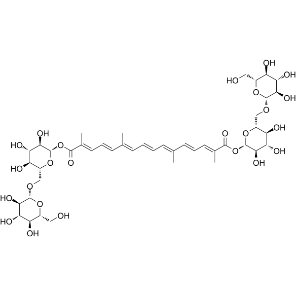
Crocin (Crocin I) is an orally active natural product that can be isolated from the stigma of Crocus sativus. Crocin inhibits tumor cell proliferation and promotes apoptosis through JAK pathway. Crocin has anti-inflammatory, antioxidant and antitumor activities .
For research use only. We do not sell to patients.
- Purity : 99.30%
- CAS No.: 42553-65-1
- Formula: C44H64O24
- Molecular Weight:976.96
-
Storage:
4°C, protect from light
* In solvent : -80°C, 6 months; -20°C, 1 month (protect from light)

To place orders, for customer services and technical support, please contact: MedChemExpress USA
Tel: 609-228-6898 E-mail: [email protected] [email protected]


-
 Biological Activity
-
Chemical Information
-
Solvent & Solubility
- Protocols
- Purity & Documentation
- References
-
 Help & FAQs


-
HY-N2072
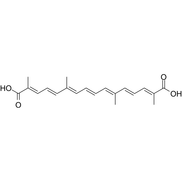
CrocetinAnticancer/Neuroprotective Agent -
HY-N4114
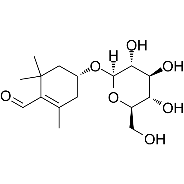
PicrocrocinAnticancer Agent -
HY-N0698
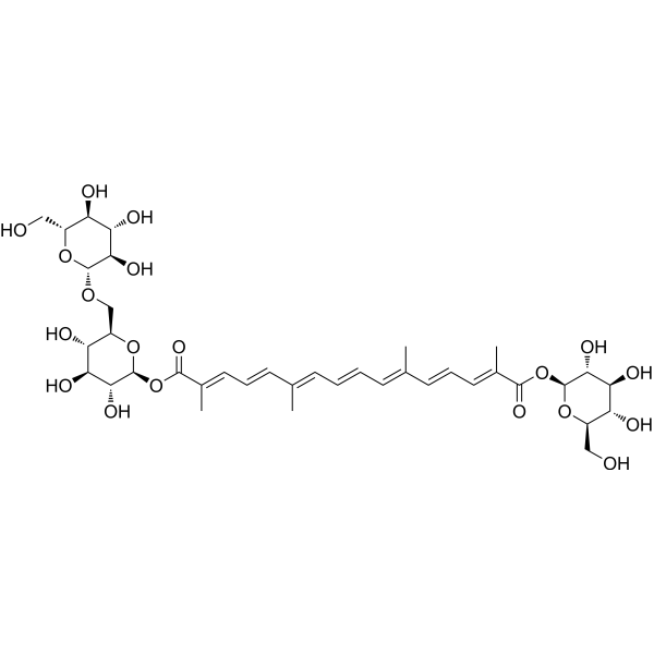
Crocin IIα-synuclein Inhibitor -

HY-N0697R
Crocin (Standard)Endogenous Metabolite -
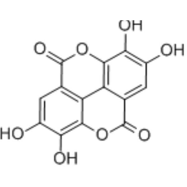
HY-B0183
Ellagic acidSHP2 Inhibitor -
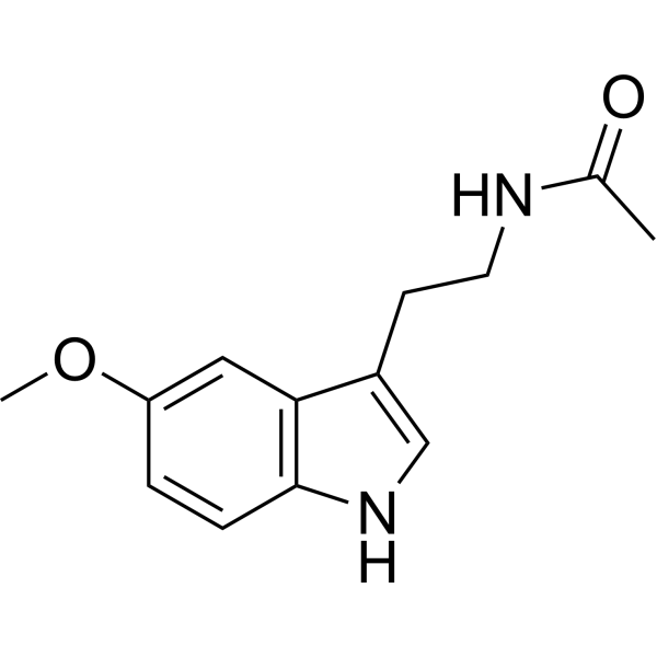
HY-B0075
MelatoninMelatonin Receptor Activator -
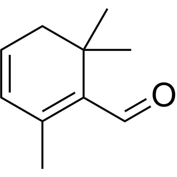
HY-N7560
SafranalPerfume Analogue -

HY-B0015
PaclitaxelAntitumor Agent -
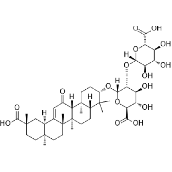
HY-N0184
Glycyrrhizic acidHMGB1 Antagonist -
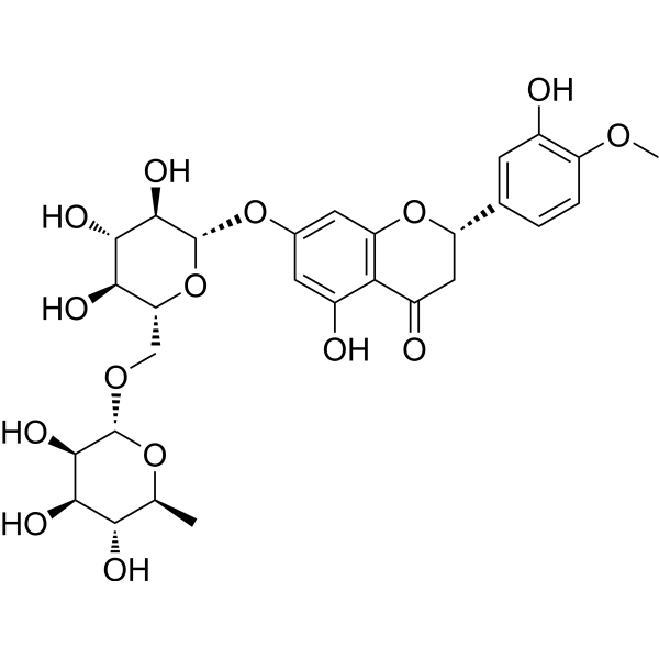
HY-15337
HesperidinAntioxidant -
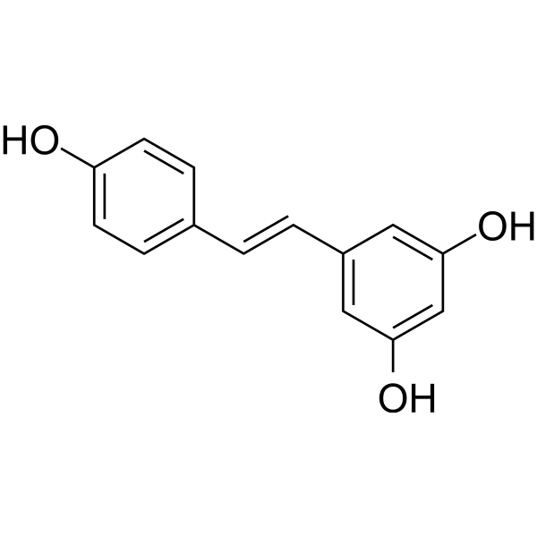
HY-16561
ResveratrolPolyphenolic Phytoalexin -
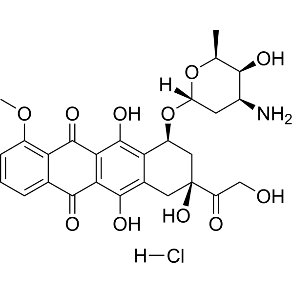
HY-15142
Doxorubicin hydrochlorideTopoisomerase Inhibitor
Publications Citing Use of MedChemExpress (MCE) Crocin
More- Int Immunopharmacol. 2026 Mar 1:172:116231. [Abstract]
- Int Immunopharmacol. 2025 Sep 5:165:115437. [Abstract]
- Int J Mol Sci. 2025 Feb 6;26(3):1381. [Abstract]
- Int J Mol Sci. 2024 Sep 20;25(18):10124. [Abstract]
- Biomolecules. 2022 Dec 5;12(12):1813. [Abstract]
- J Mol Struct. 2025 Dec 15.
- Cell Cycle. 2022 Jan;21(2):202-218. [Abstract]
- Ren Fail. 2023;45(2):2253924. [Abstract]
- Cytokine. 2022 Jun;154:155888. [Abstract]
- Chem Biol Drug Des. 2024 Feb;103(2):e14467. [Abstract]
- J Phys Chem B. 2026 Jan 8;130(1):205-214. [Abstract]
- Mol Cell Toxicol. 2025 Dec 10.
- Pharm Chem J. 2024 May 15.
- Res Sq. 2026 Apr 12.
- Biomed Pharmacother. 2025 May:186:118043. [Abstract]
- SSRN. 2023 Dec 19.
- Bioengineered. 2021 Dec;12(1):4569-4580. [Abstract]
 Customer Validation & Images

-
Histological Imaging/Staining
-
IF
-
Histological Imaging/Staining
-
WB
-
IHC
All Endogenous Metabolite Isoforms
More

Biological Activity
Description
Cellular Effect
|
Cell Line

|
Type | Value | Description | References |
|---|---|---|---|---|
| RAW264.7 | IC50 |
>100 μM
Compound: 16, crocin
|
Inhibition of nitric oxide production in lipopolysaccharide-activated mouse RAW264.7 cells by Griess reaction based method
Inhibition of nitric oxide production in lipopolysaccharide-activated mouse RAW264.7 cells by Griess reaction based method
|
[PMID: 23305920] |
In Vitro
Crocin (150, 200 μM, 24 h) plays an antitumor role in colon cancer cells by inhibiting the JAK pathway[1].
MedChemExpress (MCE) has not independently confirmed the accuracy of these methods. They are for reference only.
-
Cell Line:HCT116
-
Concentration:0, 50, 100, 150, 200, 250, 300, 350, 400, and 450 μM
-
Incubation Time:24 h
-
Result:Weakened the cell vitality in a concentration-dependent manner.
-
Cell Line:HCT116
-
Concentration:150, 200 μM
-
Incubation Time:24 h
-
Result:Facilitated apoptosis of HCT-116 cells in a concentration-dependent manner.
Abated the colony number and downregulated the positive expression of Ki-67.
-
Cell Line:HCT116
-
Concentration:150, 200 μM
-
Incubation Time:24 h
-
Result:Decreased the levels of p-JAK2, p-STAT3, and p-ERK, as well as the ratios of p-JAK2/JAK2, p-STAT3/STAT3, and pERK/ERK.
In Vivo
MedChemExpress (MCE) has not independently confirmed the accuracy of these methods. They are for reference only.
-
Animal Model:Bleomycin-induced pulmonary fibrosis in rat[2]
-
Dosage:20 mg/kg
-
Administration:p.o.
-
Result:Reduced the expression of TLR4 and IL-10.
Reduced the expression of TNF-α and TGF-β1.
- Annexin V plus membrane-impermeant dye apoptosis staining
- BrdU Incorporation Assay
- Somatic Cell Culture
- CFSE Dye Dilution Proliferation Assay
- Dye-dilution cell tracking and proliferation staining
- CCK-8/WST-8 Cell Proliferation Assay
- Protocol for Cell Cycle
- Cell Counting-Based Growth Curve Assay
- Research Protocol for Inflammation-related Diseases
- Colony Formation (Clonogenic) Assay
- Cell Viability Determination by MTT Colorimetric Assay
- EdU Incorporation Assay (Click Chemistry-Based DNA Synthesis Measurement)
- Ki-67 Immunostaining Proliferation Assay
- Detection of Apoptosis
- TUNEL staining for apoptotic DNA fragmentation
- PCNA Immunodetection Proliferation Assay
- Protocol for Cell Counting and Cell Density Analysis
- Apoptosis Solutions
- MTT Cell Proliferation Assay
Clinical Trial
| NCT Number | Sponsor | Condition | Start Date |
Phase
|
|---|---|---|---|---|
| NCT01329991 | 2011-05 | PHASE1 |


Chemical Information
-
CAS No. 42553-65-1
-
Appearance Solid
-
Molecular Weight 976.96
-
Formula C44H64O24
-
Color Brown to reddish brown
-
SMILES
O[C@@H]([C@H](O)[C@H]1O)[C@H](O[C@H]1OC(/C(C)=C/C=C/C(C)=C/C=C/C=C(C)/C=C/C=C(C)/C(O[C@@H]([C@@H]([C@@H](O)[C@@H]2O)O)O[C@@H]2CO[C@@H]([C@@H]([C@@H](O)[C@@H]3O)O)O[C@@H]3CO)=O)=O)CO[C@@H]([C@@H]([C@@H](O)[C@@H]4O)O)O[C@@H]4CO
-
Synonyms
Crocin I
-
Structure Classification
-
Initial Source
-
Shipping
Room temperature in continental US; may vary elsewhere.
-
Storage
4°C, protect from light
* In solvent : -80°C, 6 months; -20°C, 1 month (protect from light)


Publications (17)
-
Journal Impact Factor
-
Most Recent
-
Int Immunopharmacol
Rutin ameliorates hepatic ischemia-reperfusion injury by targeting CD36 to suppress hepatocyte ferroptosis. [Abstract]2026 Mar 1:172:116231. PMID: 41576568 -
Int Immunopharmacol
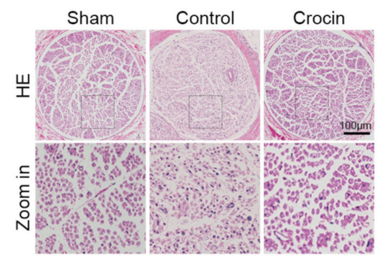
Crocin facilitates peripheral nerve regeneration through modulation of the STAT3/Bcl-2/Beclin-1 signaling axis-mediated autophagic pathway. [Abstract]2025 Sep 5:165:115437. PMID: 40913861
Crocin purchased from MedChemExpress. Usage Cited in: Int Immunopharmacol. 2025 Sep 5:165:115437. [Abstract]
Hematoxylin and eosin (H&E) staining of transverse sections 28 days after surgery showed well-preserved tissue structure with no necrotic or scar tissue in the Crocin (50 mg/kg; i.p.; once daily; rats) group, whereas the control group exhibited sparse and disorganized axonal regeneration. (scale bar = 100 μm).
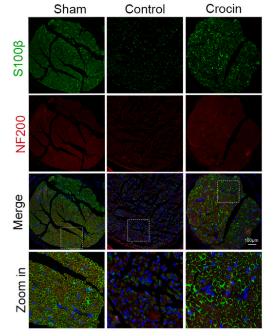
Crocin purchased from MedChemExpress. Usage Cited in: Int Immunopharmacol. 2025 Sep 5:165:115437. [Abstract]
Immunofluorescence analysis showed that Schwann cell regeneration markers (S100β+) and axon regeneration markers (/NF200+) were significantly up-regulated in the Crocin (50 mg/kg; i.p.; once daily; rats) group compared with the control group.
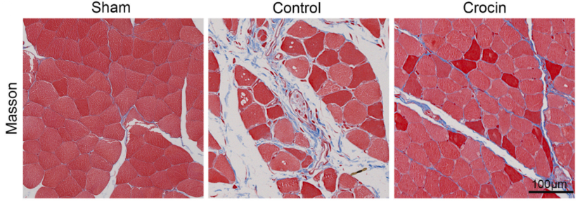
Crocin purchased from MedChemExpress. Usage Cited in: Int Immunopharmacol. 2025 Sep 5:165:115437. [Abstract]
Morphological analysis of the target muscle via Masson's trichrome staining revealed a significant reduction in collagen fiber area in the Crocin (50 mg/kg; i.p.; once daily; rats)-treated group compared to the crush injury group (scale bar = 100 μm).
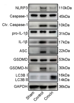
Crocin purchased from MedChemExpress. Usage Cited in: Int Immunopharmacol. 2025 Sep 5:165:115437. [Abstract]
Western blot analysis of NLRP3 inflammasome components, LC3B, and GSDMD under compound posttreatment. Western blot analysis demonstrated that the Crocin (50 mg/kg; i.p.; once daily; rats)-treated group exhibited significant suppression of NLRP3-mediated pyroptosis-related proteins (including NLRP3, IL-1β, caspase-1, and GSDMD) alongside elevated LC3B expression.
-
Int J Mol Sci
Discovery of TRPV4-Targeting Small Molecules with Anti-Influenza Effects Through Machine Learning and Experimental Validation. [Abstract]2025 Feb 6;26(3):1381. PMID: 39941149 -
Int J Mol Sci
Crocin Protects the 661W Murine Photoreceptor Cell Line against the Toxic Effects of All- Trans-Retinal. [Abstract]2024 Sep 20;25(18):10124. PMID: 39337609 -
Biomolecules
Establishment of a Cell Line Stably Expressing the Growth Hormone Secretagogue Receptor to Identify Crocin as a Ghrelin Agonist. [Abstract]2022 Dec 5;12(12):1813. PMID: 36551241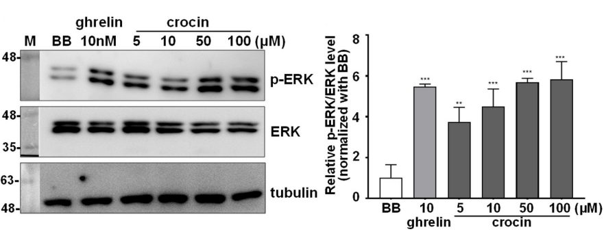
Crocin purchased from MedChemExpress. Usage Cited in: Biomolecules. 2022 Dec 5;12(12):1813. [Abstract]
Crocin (5, 10, 50,100 μM) significantly increases the level of ERK1/2 phosphorylation in GHSR1a-mCherry HEK293T cells.
-
-
Cell Cycle
Crocin ameliorates atherosclerosis by promoting the reverse cholesterol transport and inhibiting the foam cell formation via regulating PPARγ/LXR-α. [Abstract]2022 Jan;21(2):202-218. PMID: 34978526 -
Ren Fail
Crocin improves the renal autophagy in rat experimental membranous nephropathy via regulating the SIRT1/Nrf2/HO-1 signaling pathway. [Abstract]2023;45(2):2253924. PMID: 37724538 -
Cytokine
Crocin attenuates NF-κB-mediated inflammation and proliferation in breast cancer cells by down-regulating PRKCQ. [Abstract]2022 Jun;154:155888. PMID: 35447530 -
Chem Biol Drug Des
Crocin enhances the sensitivity to paclitaxel in human breast cancer cells by reducing BIRC5 expression. [Abstract]2024 Feb;103(2):e14467. PMID: 38661582 -
J Phys Chem B
2026 Jan 8;130(1):205-214. PMID: 41404815 -
-
-
-
Biomed Pharmacother
Crocin-I mitigates diquat-induced pulmonary fibrosis via activation of the SIRT3/FOXO3a pathway. [Abstract]2025 May:186:118043. PMID: 40194334
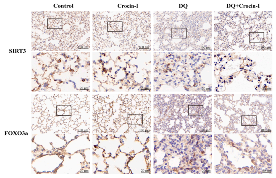
Crocin purchased from MedChemExpress. Usage Cited in: Biomed Pharmacother. 2025 May:186:118043. [Abstract]
Representative immunohistochemical images of SIRT3 and FOXO3a expression intensity in each group. Immunohistochemical analysis indicated that Crocin (Crocin-I) (40 mg/kg; gavage; once daily for 3 weeks) treatment enhanced the positive expression of SIRT3 and FOXO3a in the lung tissues of DQ-treated mice.
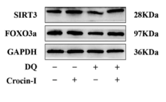
Crocin purchased from MedChemExpress. Usage Cited in: Biomed Pharmacother. 2025 May:186:118043. [Abstract]
Crocin (Crocin-I) (40 mg/kg; gavage; once daily for 3 weeks) markedly reversed the downregulated expression of SIRT3 and FOXO3a induced by fibrosis in DQ-triggered pulmonary fibrosis mice.
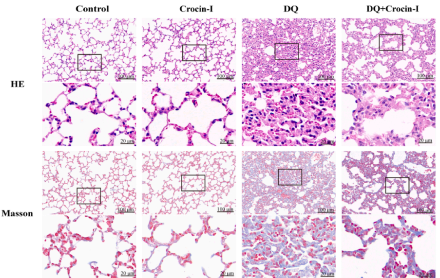
Crocin purchased from MedChemExpress. Usage Cited in: Biomed Pharmacother. 2025 May:186:118043. [Abstract]
Representative hematoxylin and eosin and Masson-stained images illustrating the effect of crocin-I on the lung tissues of DQ-induced PF mice. HE staining and Masson staining confirmed that Crocin (Crocin-I) (40 mg/kg; gavage; once daily for 3 weeks) significantly reduced the severity of PF in DQ-intoxicated mice. Scale bars: 100 µm (upper) and 50 µm (lower).
-
-
Bioengineered
Crocin exerts anti-proliferative and apoptotic effects on cutaneous squamous cell carcinoma via miR-320a/ATG2B. [Abstract]2021 Dec;12(1):4569-4580. PMID: 34320900


Solvent & Solubility
In Vitro:
DMSO : 125 mg/mL (127.95 mM; Need ultrasonic; Hygroscopic DMSO has a significant impact on the solubility of product, please use newly opened DMSO)
Please refer to the solubility information to select the appropriate solvent. Once prepared, please aliquot and store the solution to prevent product inactivation from repeated freeze-thaw cycles.
Storage method and period of stock solution: -80°C, 6 months; -20°C, 1 month (protect from light). When stored at -80°C, please use it within 6 months. When stored at -20°C, please use it within 1 month.
Please refer to the solubility information to select the appropriate solvent. Once prepared, please aliquot and store the solution to prevent product inactivation from repeated freeze-thaw cycles.
Storage method and period of stock solution: -80°C, 6 months; -20°C, 1 month (protect from light). When stored at -80°C, please use it within 6 months. When stored at -20°C, please use it within 1 month.
Concentration (start) × Volume (start) = Concentration (final) × Volume (final)
In Vivo:
Select the appropriate dissolution method based on your experimental animal and administration route.
- For the following dissolution methods, please ensure to first prepare a clear stock solution using an In Vitro approach and then sequentially add co-solvents:
- To ensure reliable experimental results, the clarified stock solution can be appropriately stored based on storage conditions. As for the working solution for In Vivo experiments, it is recommended to prepare freshly and use it on the same day.
- The percentages shown for the solvents indicate their volumetric ratio in the final prepared solution. If precipitation or phase separation occurs during preparation, heat and/or sonication can be used to aid dissolution.
Add each solvent one by one: 10% DMSO 40% PEG300 5% Tween-80 45% Saline
Solubility: ≥ 2.08 mg/mL (2.13 mM); Clear solution
This protocol yields a clear solution of ≥ 2.08 mg/mL (saturation unknown).
Taking 1 mL working solution as an example, add 100 μL DMSO stock solution (20.8 mg/mL) to 400 μL PEG300, and mix evenly; then add 50 μL Tween-80 and mix evenly; then add 450 μL Saline to adjust the volume to 1 mL.
Preparation of Saline: Dissolve 0.9 g sodium chloride in ddH₂O and dilute to 100 mL to obtain a clear Saline solution.
Add each solvent one by one: 10% DMSO 90% (20% SBE-β-CD in Saline)
Solubility: ≥ 2.08 mg/mL (2.13 mM); Clear solution
This protocol yields a clear solution of ≥ 2.08 mg/mL (saturation unknown).
Taking 1 mL working solution as an example, add 100 μL DMSO stock solution (20.8 mg/mL) to 900 μL 20% SBE-β-CD in Saline, and mix evenly.
Preparation of 20% SBE-β-CD in Saline (4°C, storage for one week): 2 g SBE-β-CD powder is dissolved in 10 mL Saline, completely dissolve until clear.
In Vivo Dissolution Calculator
Please enter the basic information of animal experiments:
-
-
-
-
Recommended: Prepare an additional quantity of animals to account for potential losses during experiments.
Please enter your animal formula composition:
-
%DMSO +
Recommended: Keep the proportion of DMSO in working solution below 2% if your animal is weak.
-
%+
-
+%Tween-80 + +
-
%Saline +
The co-solvents required include: DMSO, . All of co-solvents are available by MedChemExpress (MCE). , Tween 80. All of co-solvents are available by MedChemExpress (MCE).
Working solution concentration: 0.22 mg/mL
Method for preparing stock solution: mg drug dissolved in μL DMSO. Stock solution concentration: mg/mL. * In solvent : -80°C, 6 months; -20°C, 1 month (protect from light)
1. Take μL DMSO stock solution;
2. Add μL .
μL , mix evenly;
3. Then add μL Tween 80, mix evenly;
4. Then add μL
Please ensure that the stock solution in the first step is dissolved to a clear state, and add co-solvents in sequence. You can use ultrasonic heating (ultrasonic cleaner, recommended frequency 20-40 kHz), vortexing, etc. to assist dissolution.


Purity & Documentation
-
Data Sheet (277 KB)
-
SDS (396 KB)
- English - EN (396 KB)
- Français - FR (396 KB)
- Deutsch - DE (396 KB)
- Norwegian - NO (396 KB)
- Español - ES (396 KB)
- Swedish - SV (396 KB)
- Italian - IT (396 KB)
- Korean - KR (396 KB)
- Portuguese - PT (396 KB)
-
Handling Instructions (2659 KB)
References
[1]. Zaghloul MS, et al. Crocin attenuates lung inflammation and pulmonary vascular dysfunction in a rat model of bleomycin-induced pulmonary fibrosis. Life Sci. 2019 Aug 26:116794. [Content Brief]
[2]. Yang H, et al. Crocin exerts anti-tumor effect in colon cancer cells via repressing the JAK pathway. Eur J Histochem. 2023 Sep 12;67(3):3697. [Content Brief]


Complete Stock Solution Preparation Table
Please refer to the solubility information to select the appropriate solvent. Once prepared, please aliquot and store the solution to prevent product inactivation from repeated freeze-thaw cycles.
Storage method and period of stock solution: -80°C, 6 months; -20°C, 1 month (protect from light). When stored at -80°C, please use it within 6 months. When stored at -20°C, please use it within 1 month.
| Optional Solvent | Concentration Solvent Mass | 1 mg | 5 mg | 10 mg | 25 mg |
|---|---|---|---|---|---|
| DMSO | 1 mM | 1.0236 mL | 5.1179 mL | 10.2358 mL | 25.5896 mL |
| 5 mM | 0.2047 mL | 1.0236 mL | 2.0472 mL | 5.1179 mL | |
| 10 mM | 0.1024 mL | 0.5118 mL | 1.0236 mL | 2.5590 mL | |
| 15 mM | 0.0682 mL | 0.3412 mL | 0.6824 mL | 1.7060 mL | |
| 20 mM | 0.0512 mL | 0.2559 mL | 0.5118 mL | 1.2795 mL | |
| 25 mM | 0.0409 mL | 0.2047 mL | 0.4094 mL | 1.0236 mL | |
| 30 mM | 0.0341 mL | 0.1706 mL | 0.3412 mL | 0.8530 mL | |
| 40 mM | 0.0256 mL | 0.1279 mL | 0.2559 mL | 0.6397 mL | |
| 50 mM | 0.0205 mL | 0.1024 mL | 0.2047 mL | 0.5118 mL | |
| 60 mM | 0.0171 mL | 0.0853 mL | 0.1706 mL | 0.4265 mL | |
| 80 mM | 0.0128 mL | 0.0640 mL | 0.1279 mL | 0.3199 mL | |
| 100 mM | 0.0102 mL | 0.0512 mL | 0.1024 mL | 0.2559 mL |


Crocin Related Classifications
HY-N0697 Related Classifications
 Powered by Bioz