



FeTMPyP
Based on 1 publication(s) in Google Scholar
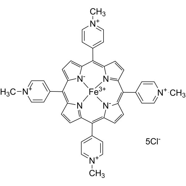
FeTMPyP is an orally active poly (ADP-ribose) polymerase (PARP) inhibitor. FeTMPyP inhibits cell death, nitrotyrosine formation, and depolarization of mitochondrial transmembrane potential. FeTMPyP reduces homocysteine-induced nitrosative stress and partially restores TFEB protein and mRNA levels. FeTMPyP improves functional and behavioral deficits caused by chronic constriction injury in rats. FeTMPyP alleviates acute cerebral infarction in a rat model of middle cerebral artery occlusion with mild hyperglycemia. FeTMPyP can be used in studies related to neuropathic pain, renal aging, ischemic penumbra, and hyperglycemic stroke.
For research use only. We do not sell to patients.
- Purity : 99.98%
- CAS No.: 133314-07-5
- Formula: C44H36Cl5FeN8
- Molecular Weight:909.92
-
Storage:
-20°C, sealed storage, away from moisture
* In solvent : -80°C, 6 months; -20°C, 1 month (sealed storage, away from moisture)

To place orders, for customer services and technical support, please contact: MedChemExpress USA
Tel: 609-228-6898 E-mail: [email protected] [email protected]


-
 Biological Activity
-
Chemical Information
-
Solvent & Solubility
- Protocols
- Purity & Documentation
- References
-
 Help & FAQs




Publications Citing Use of MedChemExpress (MCE) FeTMPyP
More

Biological Activity
Description
In Vitro
FeTMPyP partially rescues the reduction in TFEB protein expression caused by Hcy stimulation in MPC-5 mouse podocyte cells[2].
FeTMPyP (0.01-100 μM; 6 h) concentration-dependently reduces LDH release, a marker of cell death, in glucose-deprived immunostimulated rat prefrontal cortex astrocytes after 6 h of treatment[3].
FeTMPyP (0.01-100 μM; 4 h) reduces LDH release in SIN-1-treated, glucose-deprived rat prefrontal cortex astrocytes after 4 h of treatment[3].
FeTMPyP (10 μM; 6 h) inhibits the increase in nitrotyrosine immunoreactivity, a marker of peroxynitrite, in glucose-deprived immunostimulated rat prefrontal cortex astrocytes[3].
FeTMPyP (10 μM; 3 h) inhibits the increase in nitrotyrosine immunoreactivity, a marker of peroxynitrite, in SIN-1-treated, glucose-deprived rat prefrontal cortex astrocytes[3].
FeTMPyP (0.33-50 μM; 15 min) concentration-dependently inhibits peroxynitrite-mediated tyrosine nitration of bovine serum albumin in a cell-free assay after 15 min of incubation[3].
FeTMPyP (10 μM) inhibits peroxynitrite-mediated oxidation of DCF-H to DCF in SIN-1-treated, glucose-deprived rat prefrontal cortex astrocytes[3].
FeTMPyP (10 μM; 3 h) prevents mitochondrial transmembrane potential depolarization in SIN-1-treated, glucose-deprived rat prefrontal cortex astrocytes over 3 h of co-treatment[3].
FeTMPyP (50 μM; full experimental protocol) reverses HG MCAO plasma-induced increases in myogenic tone and lumen narrowing in isolated nonischemic rat MCA, likely via inhibiting the vasoconstricting properties of peroxynitrite[4].
FeTMPyP (50 μM; full experimental protocol) does not alter HG Sham plasma-induced increases in myogenic tone in isolated nonischemic rat MCA, indicating peroxynitrite is not involved in this response[4].
MedChemExpress (MCE) has not independently confirmed the accuracy of these methods. They are for reference only. Further protocols information, click here.
-
Cell Line:rat prefrontal cortex astrocytes
-
Concentration:0.01-100 μM
-
Incubation Time:6 h
-
Result:Attenuated astrocyte death in a concentration-dependent manner, and reduced LDH release in glucose-deprived immunostimulated astrocytes.
-
Cell Line:rat prefrontal cortex astrocytes
-
Concentration:0.01-100 μM
-
Incubation Time:4 h
-
Result:Blocked the augmented astrocyte death induced by glucose deprivation plus SIN-1 treatment.
-
Cell Line:rat prefrontal cortex astrocytes
-
Concentration:10 μM
-
Incubation Time:6 h
-
Result:Completely blocked the elevated nitrotyrosine immunoreactivity observed in glucose-deprived immunostimulated astrocytes.
-
Cell Line:rat prefrontal cortex astrocytes
-
Concentration:10 μM
-
Incubation Time:3 h
-
Result:Completely blocked the elevated nitrotyrosine immunoreactivity observed in SIN-1-treated, glucose-deprived astrocytes.
In Vivo
FeTMPyP (10 mg/kg; i.v.; single dose 10 minutes before reperfusion) acutely reduces corrected acute infarct volume by ~72% in hyperglycemic rats with mild ischemic stroke (<68% CBF decrease) but provides no neuroprotection in rats with severe ischemic stroke (>68% CBF decrease), with no effect on reperfusion CBF[4].
MedChemExpress (MCE) has not independently confirmed the accuracy of these methods. They are for reference only.
-
Animal Model:Sprague Dawley (male, 180-230 g, chronic constriction injury of left sciatic nerve)[1]
-
Dosage:1 mg/kg; 3 mg/kg
-
Administration:p.o.; daily; 14 days
-
Result:Significantly improved sciatic functional index (SFI) at 3 mg/kg.
Increased paw withdrawal latencies to cold (4°C) and hot (52.5°C) thermal stimuli, increased paw withdrawal thresholds to mechanical von Frey filament stimuli , and reduced cold chemical allodynia scores from acetone spray tests relative to CCI controls on day 14 at 3 mg/kg.
Reversed CCI-induced foot deformity at both 1 mg/kg and 3 mg/kg.
Significantly reduced sciatic nerve malondialdehyde (MDA) levels and nitrite levels.
Reversed CCI-induced elevations in iNOS, COX-2, and phospho-NF-κB protein expression and restored reduced Nrf2 and HO-1 protein expression.
-
Animal Model:Wistar rats (male, ≈300 g, hyperglycemic model via streptozotocin injection, transient middle cerebral artery occlusion reperfusion)[4]
-
Dosage:10 mg/kg
-
Administration:i.v.; single dose 10 minutes before reperfusion
-
Result:Reduced corrected acute infarct volume in rats with mild ischemia.
Showed no significant effect on corrected acute infarct volume in rats with severe ischemia.
Did not alter reperfusion CBF in either mild or severe ischemia groups.


Chemical Information
-
CAS No. 133314-07-5
-
Appearance Solid
-
Molecular Weight 909.92
-
Formula C44H36Cl5FeN8
-
Color Brown to black
-
SMILES
C[N+](C=C1)=CC=C1C2=C3C=CC(C(C4=CC=[N+](C)C=C4)=C5C=CC([N-]56)=C7C8=CC=[N+](C)C=C8)=[N]3[Fe+3]96[N]%10=C7C=CC%10=C(C%11=CC=[N+](C)C=C%11)C%12=CC=C2[N-]%129.[5Cl-]
-
Shipping
Room temperature in continental US; may vary elsewhere.
-
Storage
-20°C, sealed storage, away from moisture
* In solvent : -80°C, 6 months; -20°C, 1 month (sealed storage, away from moisture)


Publications (1)
-
Journal Impact Factor
-
Most Recent


Solvent & Solubility
In Vitro:
H2O : 3.57 mg/mL (3.92 mM; ultrasonic and warming and heat to 60°C)
Please refer to the solubility information to select the appropriate solvent. Once prepared, please aliquot and store the solution to prevent product inactivation from repeated freeze-thaw cycles.
Storage method and period of stock solution: -80°C, 6 months; -20°C, 1 month (sealed storage, away from moisture). When stored at -80°C, please use it within 6 months. When stored at -20°C, please use it within 1 month.
* Note: If you choose water as the stock solution, please dilute it to the working solution, then filter and sterilize it with a 0.22 μm filter before use.
Please refer to the solubility information to select the appropriate solvent. Once prepared, please aliquot and store the solution to prevent product inactivation from repeated freeze-thaw cycles.
Storage method and period of stock solution: -80°C, 6 months; -20°C, 1 month (sealed storage, away from moisture). When stored at -80°C, please use it within 6 months. When stored at -20°C, please use it within 1 month.
* Note: If you choose water as the stock solution, please dilute it to the working solution, then filter and sterilize it with a 0.22 μm filter before use.
Concentration (start) × Volume (start) = Concentration (final) × Volume (final)

Protocols
-
RNA extraction experimental
By lysing cells, releasing RNA, and removing impurities such as proteins and DNA, high-purity RNA products are finally obtained. The commonly used traditional method is the guanidine isothiocyanate/phenol/chloroform method (Trizol), which is suitable for a variety of animal materials including animal tissues, microorganisms, cultured cells, etc., and most plant materials.
-
Cell Cytotoxicity Assay
Cytotoxicity assays are usually based on the assessment of cell membrane damage, which can also be indirectly detected by measuring cell viability. Detection methods include MTT assay, CKK-8 assay, LDH assay and ATP assay, etc.
-
Mammalian live/dead viability and cytotoxicity staining
Live/dead viability and cytotoxicity staining assays are based on the simultaneous detection of intracellular esterase activity in metabolically active (viable) cells and membrane integrity loss in non-viable cells. In commonly used dual-staining approaches, membrane-permeant fluorogenic substrates are converted by intracellular esterases into fluorescent products in live cells, while impermeant DNA-binding dyes selectively enter cells with compromised plasma membranes and label nucleic acids in dead or dying cells, enabling discrimination between viable and non-viable populations by fluorescence microscopy or flow cytometry.
-
How to Select a Suitable Non-Mouse Animal Model
Selecting a suitable non-mouse animal model is a structured decision based on the research question, required anatomy or physiology, disease mechanism, endpoint feasibility, translational relevance, and ethical justification. Non-mouse models are preferred when mice cannot reproduce key human-relevant features, such as organ size, surgical anatomy, cardiovascular physiology, neuroanatomy, immune features, pharmacology, toxicology, or long-term clinical procedures. Candidate species may include rats, rabbits, guinea pigs, ferrets, zebrafish, pigs, sheep, goats, dogs, cats, horses, and non-human primates, but each species must be justified by its specific scientific advantage rather than convenience or tradition. Unresolved questions include how to quantify translational superiority across species, how to balance increased biological relevance against higher ethical burden, and when human-derived systems or new approach methodologies should replace animal use.
-
Apoptosis Solutions
Apoptosis is a regulated, generally non-lytic cell-death pathway that removes unwanted, damaged, infected, or abnormal cells through coordinated morphological changes, caspase activation, DNA fragmentation, and membrane remodeling. The intrinsic apoptosis pathway is controlled mainly by mitochondrial outer membrane permeabilization, BCL-2 family proteins, cytochrome c release, apoptosome formation, caspase-9 activation, and downstream executioner caspase-3/7 activation. The extrinsic apoptosis pathway is initiated by death receptors such as Fas, TNFR, and TRAIL receptors, which recruit adaptor proteins and activate caspase-8 before engaging executioner caspases or mitochondrial amplification through BID cleavage. Apoptosis is linked to many phenotypes, including cancer cell killing, tissue homeostasis, immune regulation, neurodegeneration, infection response, and treatment-induced cytotoxicity; unresolved questions include how apoptosis interacts with necroptosis, pyroptosis, ferroptos
-
How to Choose the Right Model Animal
Choosing the right model animal is a validity-driven decision in which the species, strain, sex, age, genetic background, disease-induction method, outcome measures, and welfare burden must match the scientific question rather than laboratory tradition or convenience. A model should be selected by judging face validity, construct validity, and predictive validity: whether it resembles the human phenotype, whether it reproduces relevant mechanisms, and whether results are likely to predict human biology or treatment response. Animal studies often fail to translate because of species differences, weak disease resemblance, poor experimental design, inadequate reporting, publication bias, and underuse of randomization, blinding, and sample-size justification. Unresolved questions include how to rank competing models objectively, how much human-disease complexity must be reproduced for a given objective, and when non-animal systems such as organoids, ex vivo tissue, or computational models


Purity & Documentation
-
Data Sheet (289 KB)
-
SDS (252 KB)
- English - EN (252 KB)
- Français - FR (252 KB)
- Deutsch - DE (252 KB)
- Norwegian - NO (252 KB)
- Español - ES (252 KB)
- Swedish - SV (252 KB)
- Italian - IT (252 KB)
- Korean - KR (252 KB)
- Portuguese - PT (252 KB)
-
Handling Instructions (2659 KB)
References
[1]. Komirishetty P, et al. FeTMPyP a peroxynitrite decomposition catalyst ameliorated functional and behavioral deficits in chronic constriction injury induced neuropathic pain in rats. Free Radic Res. 2021;55(9-10):1005-1017. [Content Brief]
[2]. Zhang S, et al. Nitrative Stress-Related Autophagic Insufficiency Participates in Hyperhomocysteinemia-Induced Renal Aging. Oxid Med Cell Longev. 2020;2020:4252047. Published 2020 Jan 25. [Content Brief]
[3]. Choi IY, et al. Augmented death in immunostimulated astrocytes deprived of glucose: inhibition by an iron porphyrin FeTMPyP. J Neuroimmunol. 2001;112(1-2):55-62. [Content Brief]
[4]. Palomares SM, et al. Peroxynitrite decomposition with FeTMPyP improves plasma-induced vascular dysfunction and infarction during mild but not severe hyperglycemic stroke. J Cereb Blood Flow Metab. 2012;32(6):1035-1045. [Content Brief]


Complete Stock Solution Preparation Table
Please refer to the solubility information to select the appropriate solvent. Once prepared, please aliquot and store the solution to prevent product inactivation from repeated freeze-thaw cycles.
Storage method and period of stock solution: -80°C, 6 months; -20°C, 1 month (sealed storage, away from moisture). When stored at -80°C, please use it within 6 months. When stored at -20°C, please use it within 1 month.
| Optional Solvent | Concentration Solvent Mass | 1 mg | 5 mg | 10 mg | 25 mg |
|---|---|---|---|---|---|
| H2O | 1 mM | 1.0990 mL | 5.4950 mL | 10.9900 mL | 27.4749 mL |
* Note: If you choose water as the stock solution, please dilute it to the working solution, then filter and sterilize it with a 0.22 μm filter before use.


FeTMPyP Related Classifications
HY-120380 Related Classifications
 Powered by Bioz