



Bilobetin
Based on 2 publication(s) in Google Scholar
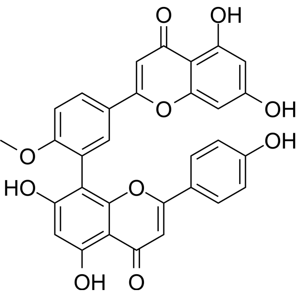
Bilobetin, an active component of Ginkgo biloba, can reduce blood lipids and improve the effects of insulin. Bilobetin ameliorated insulin resistance, increased the hepatic uptake and oxidation of lipids, reduced very-low-density lipoprotein triglyceride secretion and blood triglyceride levels, enhanced the expression and activity of enzymes involved in β-oxidation and attenuated the accumulation of triglycerides and their metabolites in tissues. Bilobetin also increased the phosphorylation, nuclear translocation and activity of PPARα accompanied by elevated cAMP level and PKA activity.
For research use only. We do not sell to patients.
- Purity : 99.71%
- CAS No.: 521-32-4
- Formula: C31H20O10
- Molecular Weight:552.48
-
Storage:
4°C, protect from light
* In solvent : -80°C, 6 months; -20°C, 1 month (protect from light)

To place orders, for customer services and technical support, please contact: MedChemExpress USA
Tel: 609-228-6898 E-mail: [email protected] [email protected]


-
 Biological Activity
-
Chemical Information
-
Solvent & Solubility
- Protocols
- Purity & Documentation
- References
-
 Help & FAQs


-
HY-N0662
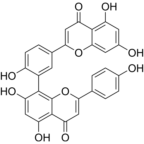
AmentoflavoneGABA(A) Negative Modulator -
HY-N2117
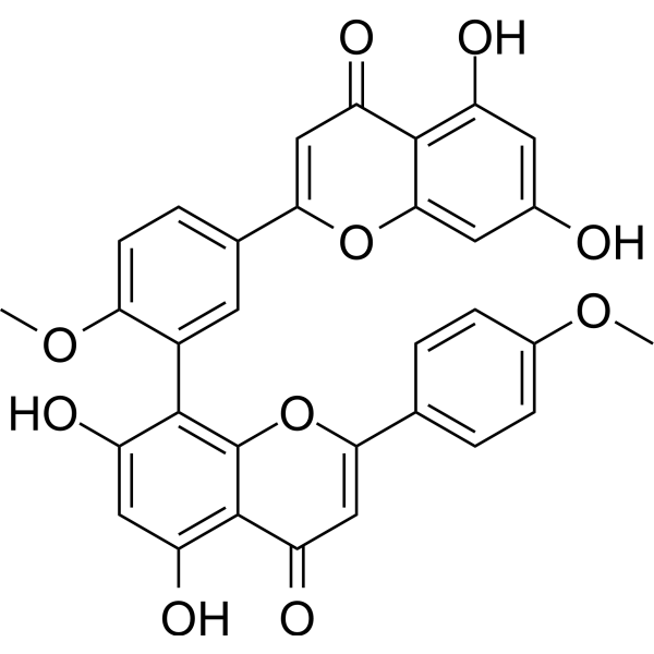
Isoginkgetinpre-mRNA Splicing Inhibitor -
HY-N0889
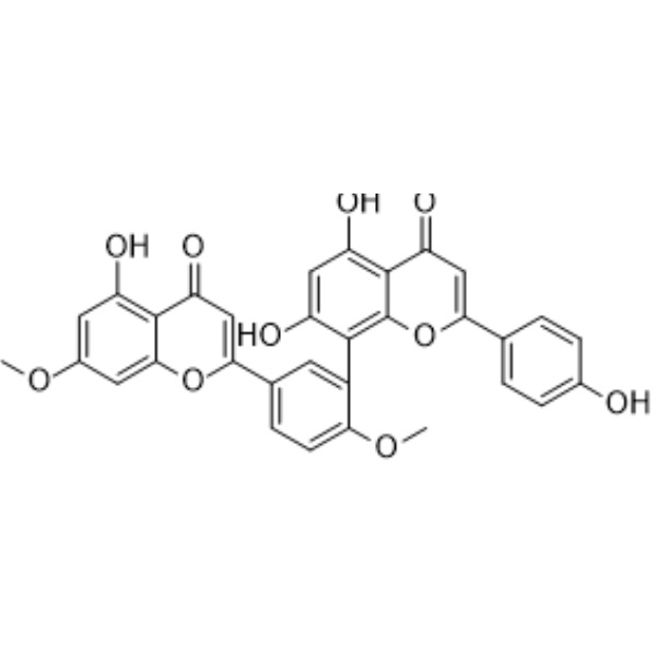
GinkgetinAutophagy Inducer -
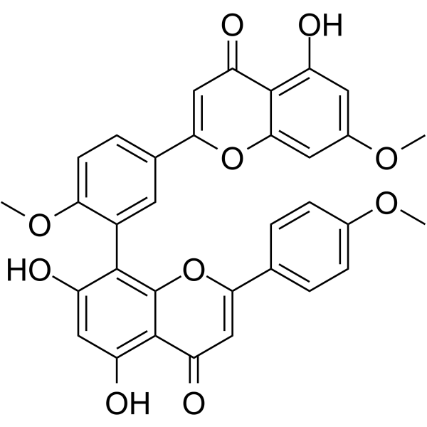
HY-N2119
SciadopitysinNF-κB Inhibitor -
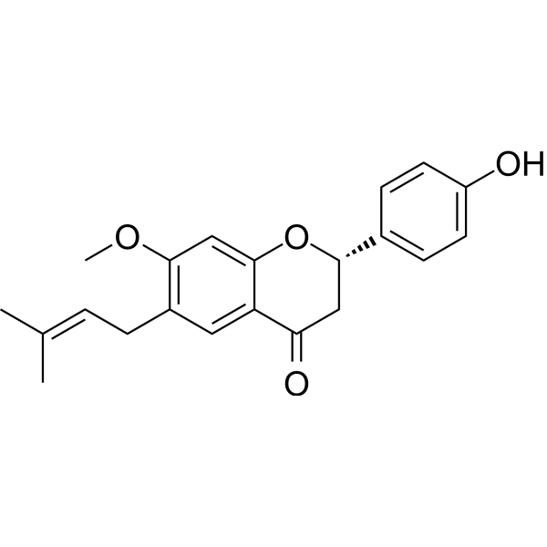
HY-N0234
BavachininPan-PPAR Agonist, HIF-1α Inhibitor -

HY-N2118R
Bilobetin (Standard)PPAR Activator -
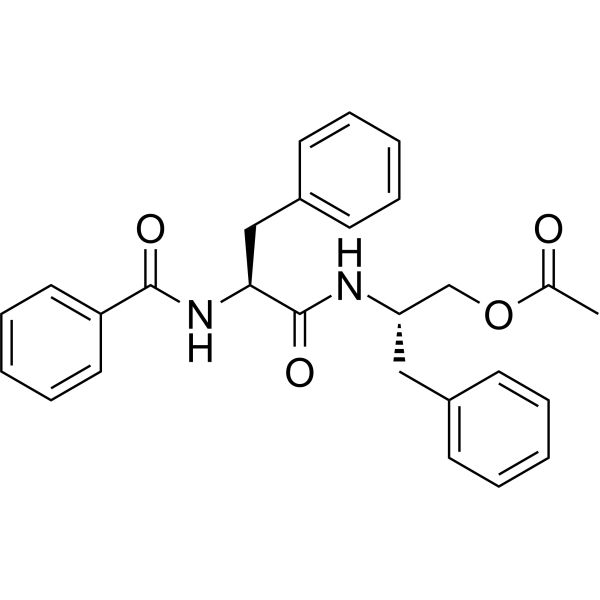
HY-N2905
Aurantiamide acetateCathepsin Inhibitor -
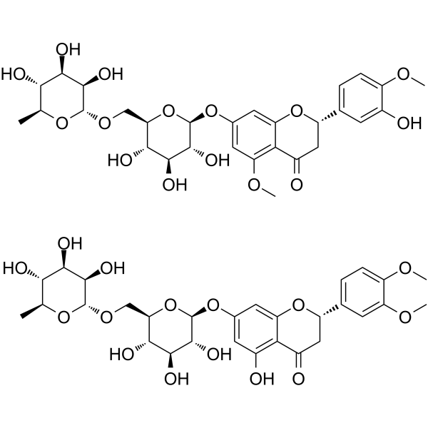
HY-N0165
Methyl-HesperidinVasodilating Agent -
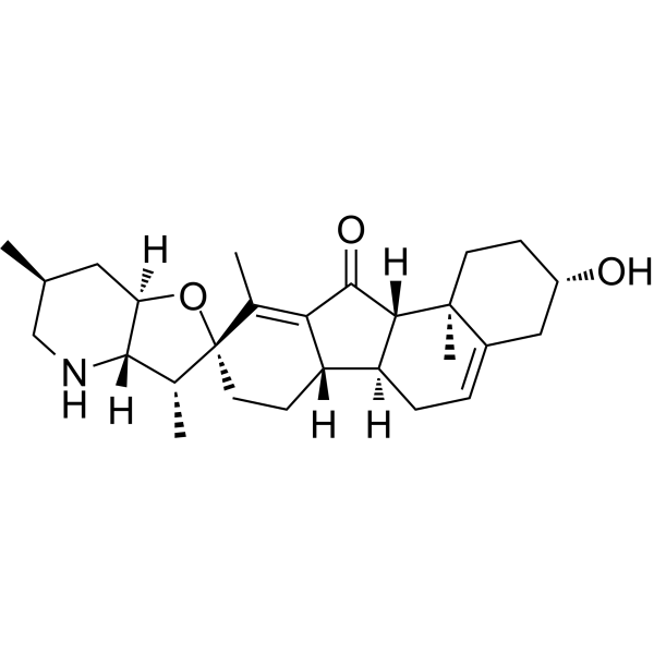
HY-N0836
JervineAnti-cancer Agent -
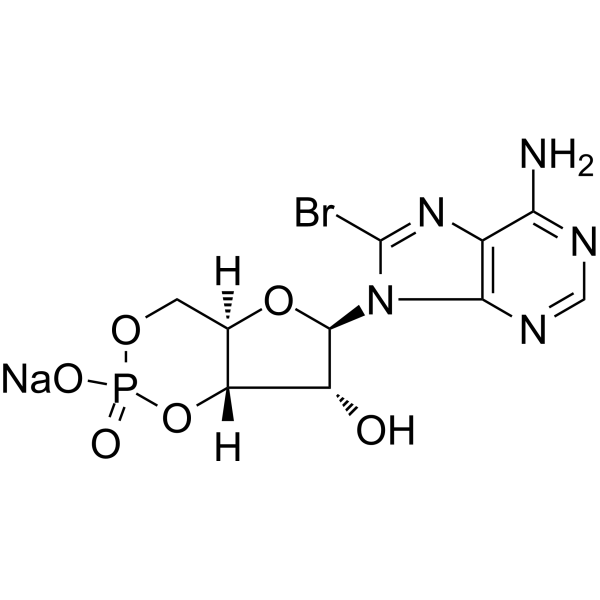
HY-12306
8-Bromo-cAMP sodium saltPKA Activator -
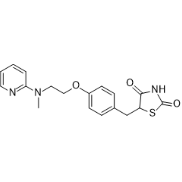
HY-17386
RosiglitazonePPARγ Agonist -
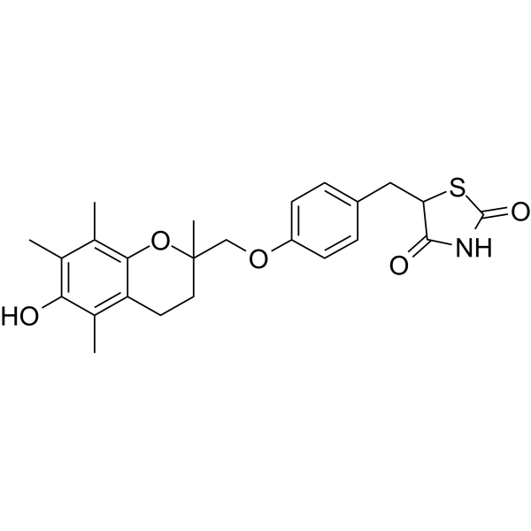
HY-50935
TroglitazonePPARγ Agonist
Publications Citing Use of MedChemExpress (MCE) Bilobetin
More

Biological Activity
Description
IC50 & Target
[1]|
PPARα |
PKA |
Cellular Effect
|
Cell Line

|
Type | Value | Description | References |
|---|---|---|---|---|
| A549 | IC50 |
>50 μM
Compound: 12
|
Cytotoxicity against human A549 cells assessed as reduction in cell viability after 48 hrs by MTT assay
Cytotoxicity against human A549 cells assessed as reduction in cell viability after 48 hrs by MTT assay
|
[PMID: 29921475] |
| HepG2 | IC50 |
>50 μM
Compound: 12
|
Cytotoxicity against human HepG2 cells assessed as reduction in cell viability after 48 hrs by MTT assay
Cytotoxicity against human HepG2 cells assessed as reduction in cell viability after 48 hrs by MTT assay
|
[PMID: 29921475] |
| MCF7 | IC50 |
26.5 μM
Compound: 7
|
Cytotoxicity against human MCF7 cells measured after 24 hrs by MTT assay
Cytotoxicity against human MCF7 cells measured after 24 hrs by MTT assay
|
[PMID: 34875389] |
| MGC-803 | IC50 |
>50 μM
Compound: 12
|
Cytotoxicity against human MGC803 cells assessed as reduction in cell viability after 48 hrs by MTT assay
Cytotoxicity against human MGC803 cells assessed as reduction in cell viability after 48 hrs by MTT assay
|
[PMID: 29921475] |
| T-24 | IC50 |
>50 μM
Compound: 12
|
Cytotoxicity against human T24 cells assessed as reduction in cell viability after 48 hrs by MTT assay
Cytotoxicity against human T24 cells assessed as reduction in cell viability after 48 hrs by MTT assay
|
[PMID: 29921475] |
In Vitro
Bilobetin (0-40 μM, 24/48/72 h) inhibits the cell proliferation in Huh7 and HepG2 cells[4].
Bilobetin (0-20 μM, 24 and 48 h) induces apoptosis and increases sub G1 population in Huh7 and HepG2 cells[4].
Bilobetin (0-20 μM, 24 h) induces accumulation of ROS and DNA damage in Huh7 and HepG2 cells[4].
Bilobetin (1-2 μM, 1 days) inhibits the phosphorylation of AKT in sebocytes[5].
MedChemExpress (MCE) has not independently confirmed the accuracy of these methods. They are for reference only. Further protocols information, click here.
-
Cell Line:Huh7 and HepG2 cells
-
Concentration:0, 2.5, 5, 10, 20, and 40 μM
-
Incubation Time:24/48/72 h
-
Result:Inhibited cell proliferation in a dose dependent manner, with IC50s of 18.28 μM at 48 h in Huh7 cells, and 19 μM at 72 h in HepG2 cells.
-
Cell Line:Huh7 and HepG2 cells
-
Concentration:0, 2.5, 5, 10, 20 μM
-
Incubation Time:24/48 h
-
Result:Showed the total apoptotic cell rate of 2.2, 2.2, 1.9, and 10.6% for 24 h (Huh7 cells) at 0, 5, 10, and 20 μM, and 1.7, 1.5, 7.5, and 22.5% for 48 h (Huh7 cells) at 0, 5, 10, and 20 μM.
Showed the total apoptotic cell rate of 1.1, 1.2, 1.6, and 3.0% for 24 h (Huh7 cells) at 0, 5, 10, and 20 μM, and 1.5, 1.1, 3.2, and 14.2% for 48 h (Huh7 cells) at 0, 5, 10, and 20 μM, respectively.
In Vivo
Bilobetin (i.p., 50 mg/kg, for 7 days) induces kidney injury in rats, and promotes the trafficking of AQP-2 onto the plasma membrane in rats[2].
Bilobetin (6 and 12 mg/kg, i.p., daily for ten days.) shows protective effects against Cisplatin (HY-17394) (7 mg/kg, i.p., a single dose on the third day)-induced testicular toxicity in rats[3].
MedChemExpress (MCE) has not independently confirmed the accuracy of these methods. They are for reference only.
-
Animal Model:rats fed a high-fat diet[1]
-
Dosage:12 mg/kg/day
-
Administration:i.p., 4 or 14 days after mice were fed a HFD for 8 weeks.
-
Result:Increased glucose infusion rate (GIR, P < 0.05) and reduced EGP (P < 0.05) in basal and clamp states.
Reduced total TG (P < 0.01) and VLDL-TG (P < 0.01).
Enhances hepatic Intralipid-TG uptake.
Reduced the total amount of lipid level in the liver and muscle.
Promoted the phosphorylation of PPARα and the translocation of PPARα from the cytoplasm to the nucleus in rats liver.


Chemical Information
-
CAS No. 521-32-4
-
Appearance Solid
-
Molecular Weight 552.48
-
Formula C31H20O10
-
Color Light yellow to yellow
-
SMILES
O=C1C=C(C2=CC=C(O)C=C2)OC3=C(C4=CC(C5=CC(C6=C(O)C=C(O)C=C6O5)=O)=CC=C4OC)C(O)=CC(O)=C13
-
Structure Classification
-
Initial Source
-
Shipping
Room temperature in continental US; may vary elsewhere.
-
Storage
4°C, protect from light
* In solvent : -80°C, 6 months; -20°C, 1 month (protect from light)


Publications (2)
-
Journal Impact Factor
-
Most Recent


Solvent & Solubility
In Vitro:
DMSO : 125 mg/mL (226.25 mM; Need ultrasonic; Hygroscopic DMSO has a significant impact on the solubility of product, please use newly opened DMSO)
Please refer to the solubility information to select the appropriate solvent. Once prepared, please aliquot and store the solution to prevent product inactivation from repeated freeze-thaw cycles.
Storage method and period of stock solution: -80°C, 6 months; -20°C, 1 month (protect from light). When stored at -80°C, please use it within 6 months. When stored at -20°C, please use it within 1 month.
Please refer to the solubility information to select the appropriate solvent. Once prepared, please aliquot and store the solution to prevent product inactivation from repeated freeze-thaw cycles.
Storage method and period of stock solution: -80°C, 6 months; -20°C, 1 month (protect from light). When stored at -80°C, please use it within 6 months. When stored at -20°C, please use it within 1 month.
Concentration (start) × Volume (start) = Concentration (final) × Volume (final)
In Vivo:
Select the appropriate dissolution method based on your experimental animal and administration route.
- For the following dissolution methods, please ensure to first prepare a clear stock solution using an In Vitro approach and then sequentially add co-solvents:
- To ensure reliable experimental results, the clarified stock solution can be appropriately stored based on storage conditions. As for the working solution for In Vivo experiments, it is recommended to prepare freshly and use it on the same day.
- The percentages shown for the solvents indicate their volumetric ratio in the final prepared solution. If precipitation or phase separation occurs during preparation, heat and/or sonication can be used to aid dissolution.
Add each solvent one by one: 10% DMSO 90% (20% SBE-β-CD in Saline)
Solubility: 2.08 mg/mL (3.76 mM); Suspended solution; Need ultrasonic
This protocol yields a suspended solution of 2.08 mg/mL. Suspended solution can be used for oral and intraperitoneal injection.
Taking 1 mL working solution as an example, add 100 μL DMSO stock solution (20.8 mg/mL) to 900 μL 20% SBE-β-CD in Saline, and mix evenly.
Preparation of 20% SBE-β-CD in Saline (4°C, storage for one week): 2 g SBE-β-CD powder is dissolved in 10 mL Saline, completely dissolve until clear.
In Vivo Dissolution Calculator
Please enter the basic information of animal experiments:
-
-
-
-
Recommended: Prepare an additional quantity of animals to account for potential losses during experiments.
Please enter your animal formula composition:
-
%DMSO +
Recommended: Keep the proportion of DMSO in working solution below 2% if your animal is weak.
-
%+
-
+%Tween-80 + +
-
%Saline +
The co-solvents required include: DMSO, . All of co-solvents are available by MedChemExpress (MCE). , Tween 80. All of co-solvents are available by MedChemExpress (MCE).
Working solution concentration: 0.22 mg/mL
Method for preparing stock solution: mg drug dissolved in μL DMSO. Stock solution concentration: mg/mL. * In solvent : -80°C, 6 months; -20°C, 1 month (protect from light)
1. Take μL DMSO stock solution;
2. Add μL .
μL , mix evenly;
3. Then add μL Tween 80, mix evenly;
4. Then add μL
Please ensure that the stock solution in the first step is dissolved to a clear state, and add co-solvents in sequence. You can use ultrasonic heating (ultrasonic cleaner, recommended frequency 20-40 kHz), vortexing, etc. to assist dissolution.

Protocols
-
Kinase activity and phosphorylation assays
Kinase activity assays measure the ability of kinases to transfer phosphate groups from ATP to specific substrates, while phosphorylation assays detect the presence and levels of phosphorylated proteins. Common methods include radiolabeled ATP incorporation (e. g. ,), ADP release detection via bioluminescence (e. g. ,[3]), enzyme-linked immunosorbent assays (ELISA) for phospho-specific epitopes (e. g. ,[6]), and microtiter-based formats for high-throughput screening (e. g. ,[8]). The ADP-Glo assay quantifies kinase activity by measuring ADP produced during phosphorylation using a luciferase-based system. Radiometric assays involve autoradiography or scintillation counting after incorporation of 32P-labeled ATP into substrate proteins. ELISA-based approaches rely on phospho-specific antibodies to detect activated kinases in cell lysates or purified samples.
-
RNA extraction experimental
By lysing cells, releasing RNA, and removing impurities such as proteins and DNA, high-purity RNA products are finally obtained. The commonly used traditional method is the guanidine isothiocyanate/phenol/chloroform method (Trizol), which is suitable for a variety of animal materials including animal tissues, microorganisms, cultured cells, etc., and most plant materials.
-
Western Blot
Western blotting (WB) is a commonly used experimental method in molecular biology, biochemistry, and immunogenetics for identifying and quantifying target proteins. It combines gel electrophoresis with immunoassay, enabling researchers to analyze protein expression, post-translational modifications, and molecular weight.
-
Cytoplasmic-Nuclear Fractionated Protein Extraction
Cytoplasmic-nuclear fractionated protein extraction separates soluble cytoplasmic proteins from nuclear-enriched proteins by mild plasma-membrane permeabilization, differential centrifugation, washing of nuclei, and extraction of nuclear proteins for downstream immunoblotting or related molecular analysis. The readout is the relative abundance of a protein in cytoplasmic and nuclear fractions, commonly assessed by western blotting together with compartment markers such as tubulin or pyruvate kinase for cytoplasm and lamin, nucleoporin, hnRNP, H2AX, or Lamin B for nuclear fractions.
-
Research Protocol for Endocrine Diseases
Endocrine diseases often arise from disrupted hormone production, hormone signaling, or target-tissue responsiveness; for diabetes-focused endocrine disease models, insulin signaling regulates glucose uptake, hepatic glucose output, lipid metabolism, and β-cell compensation. Type 2 diabetes develops through interacting defects in insulin resistance, β-cell dysfunction, adipose inflammation, hepatic glucose overproduction, altered incretin signaling, and ectopic lipid metabolism. A major unresolved question is whether endocrine dysfunction is driven primarily by target-tissue insulin resistance, intrinsic β-cell failure, immune/inflammatory stress, or combined multi-organ failure that differs by disease stage.
-
Protocol for Kinase activity and phosphorylation assays
Kinase activity assays measure transfer of phosphate from ATP to a protein or peptide substrate, generating phosphorylated substrate, ADP, or incorporated radiolabeled phosphate as the readout; phosphorylation assays measure site-specific phosphorylation in cells or tissues as a proxy for kinase-pathway activation, inhibition, or substrate regulation. Phosphorylation can be detected by phospho-specific Western blot, immunoprecipitation kinase assay, phospho-immunofluorescence, phospho-flow cytometry, luminescent ADP detection, radiolabeled ATP incorporation, or reporter-based pathway assays, and these readouts can be applied to cancer cells, primary neurons, mouse tumors, organoids, inflammatory macrophages, ferroptosis studies, and mitophagy studies when the kinase target is biologically relevant.
-
Research Protocol for Metabolic Diseases
AMP-activated protein kinase, AMPK, is a conserved cellular energy sensor that responds to reduced cellular energy status and coordinates metabolism by increasing ATP-generating catabolic pathways while suppressing ATP-consuming anabolic processes. In metabolic disease research, the AMPK pathway is experimentally relevant because it regulates hepatic lipid synthesis, fatty acid oxidation, glucose production, skeletal-muscle glucose disposal, mTORC1-linked biosynthesis, autophagy, mitochondrial homeostasis, and whole-body energy balance. The central pathway logic is that energy stress, metformin, exercise-like stimulation, or direct AMPK activators increase AMPKα Thr172 phosphorylation and downstream substrate phosphorylation, including ACC and RAPTOR. Phosphorylation of ACC suppresses lipogenesis and supports fatty acid oxidation, whereas phosphorylation of RAPTOR suppresses mTORC1 signaling and links cellular energy status to growth and protein synthesis control. The pathway is linked


Purity & Documentation
-
Data Sheet (288 KB)
-
SDS (393 KB)
- English - EN (393 KB)
- Français - FR (393 KB)
- Deutsch - DE (393 KB)
- Norwegian - NO (393 KB)
- Español - ES (393 KB)
- Swedish - SV (393 KB)
- Italian - IT (393 KB)
- Korean - KR (393 KB)
- Portuguese - PT (393 KB)
-
Handling Instructions (2659 KB)
References
[1]. Kou XH, et al. Bilobetin ameliorates insulin resistance by PKA-mediated phosphorylation of PPARα in rats fed a high-fat diet. Br J Pharmacol. 2012 Apr;165(8):2692-706. [Content Brief]
[2]. Wang Q, et al. Bilobetin induces kidney injury by influencing cGMP-mediated AQP-2 trafficking and podocyte cell cycle arrest. Phytomedicine. 2019 Nov;64:153073. [Content Brief]
[3]. Negm WA, et al. The Mechanistic Perspective of Bilobetin Protective Effects against Cisplatin-Induced Testicular Toxicity: Role of Nrf-2/Keap-1 Signaling, Inflammation, and Apoptosis. Biomedicines. 2022 May 13;10(5):1134 [Content Brief]


Complete Stock Solution Preparation Table
Please refer to the solubility information to select the appropriate solvent. Once prepared, please aliquot and store the solution to prevent product inactivation from repeated freeze-thaw cycles.
Storage method and period of stock solution: -80°C, 6 months; -20°C, 1 month (protect from light). When stored at -80°C, please use it within 6 months. When stored at -20°C, please use it within 1 month.
| Optional Solvent | Concentration Solvent Mass | 1 mg | 5 mg | 10 mg | 25 mg |
|---|---|---|---|---|---|
| DMSO | 1 mM | 1.8100 mL | 9.0501 mL | 18.1002 mL | 45.2505 mL |
| 5 mM | 0.3620 mL | 1.8100 mL | 3.6200 mL | 9.0501 mL | |
| 10 mM | 0.1810 mL | 0.9050 mL | 1.8100 mL | 4.5251 mL | |
| 15 mM | 0.1207 mL | 0.6033 mL | 1.2067 mL | 3.0167 mL | |
| 20 mM | 0.0905 mL | 0.4525 mL | 0.9050 mL | 2.2625 mL | |
| 25 mM | 0.0724 mL | 0.3620 mL | 0.7240 mL | 1.8100 mL | |
| 30 mM | 0.0603 mL | 0.3017 mL | 0.6033 mL | 1.5084 mL | |
| 40 mM | 0.0453 mL | 0.2263 mL | 0.4525 mL | 1.1313 mL | |
| 50 mM | 0.0362 mL | 0.1810 mL | 0.3620 mL | 0.9050 mL | |
| 60 mM | 0.0302 mL | 0.1508 mL | 0.3017 mL | 0.7542 mL | |
| 80 mM | 0.0226 mL | 0.1131 mL | 0.2263 mL | 0.5656 mL | |
| 100 mM | 0.0181 mL | 0.0905 mL | 0.1810 mL | 0.4525 mL |


Bilobetin Related Classifications
HY-N2118 Related Classifications
 Powered by Bioz