



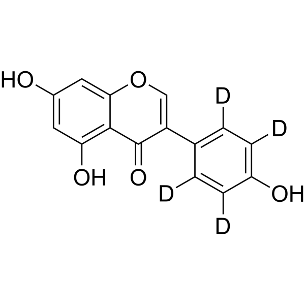
Genistein-d4
Based on 1 publication(s) in Google Scholar
Genistein-d4 is the deuterium labeled Genistein. Genistein, a soy isoflavone, is a multiple tyrosine kinases (e.g., EGFR) inhibitor which acts as a chemotherapeutic agent against different types of cancer, mainly by altering apoptosis, the cell cycle, and angiogenesis and inhibiting metastasis.
Nos produits utilisent uniquement pour la recherche. Nous ne vendons pas aux patients.
- Pureté : 98.81%
- CAS No.: 187960-08-3
- Formule: C15H6D4O5
- Masse moléculaire:274.26
-
Stockage:Powder -20°C, 3 years , 4°C, 2 years ; In solvent -80°C, 6 months , -20°C, 1 month

To place orders, for customer services and technical support, please contact: MedChemExpress USA
Tel: 609-228-6898 E-mail: [email protected] [email protected]


-
 Activité biologique
-
Chemical Information
-
Solvant et solubilité
- Protocole
- Pureté et documentation
- Références
-
 Help & FAQs


-
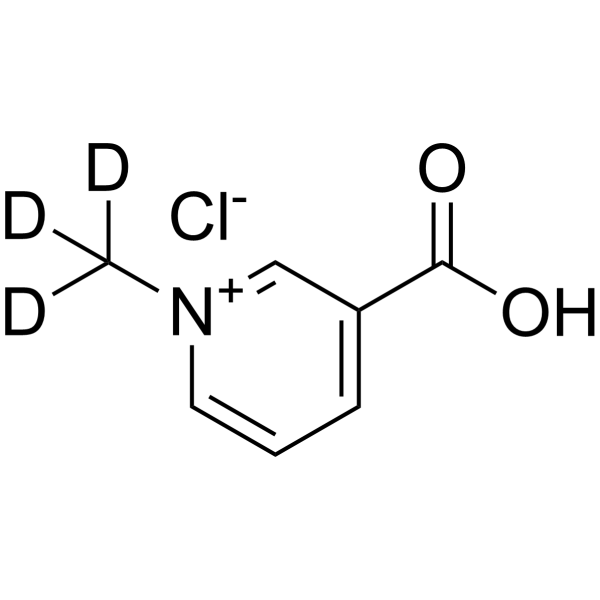
HY-N0415S
Trigonelline-d3 chlorideStable Isotope -
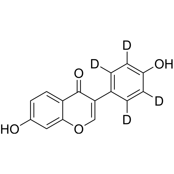
HY-N0019S
Daidzein-d4Stable Isotope -
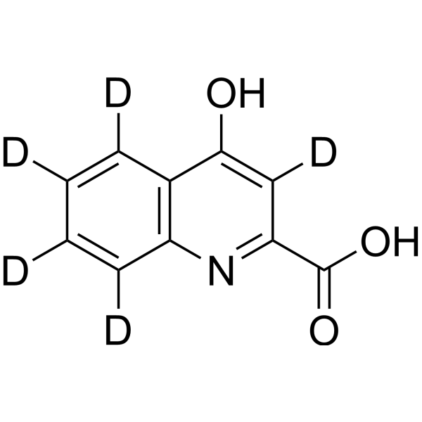
HY-100806S
Kynurenic acid-d5Stable Isotope -
HY-90001
RitonavirHIV Protease Inhibitor -
HY-N0039
Ginsenoside Rb1Autophagy Inducer -
HY-B0143S2
Niacin-d4Stable Isotope -
HY-N0229S3
L-Alanine-d4Stable Isotope -
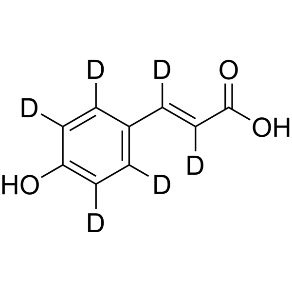
HY-N0351S1
p-Coumaric acid-d6Stable Isotope -
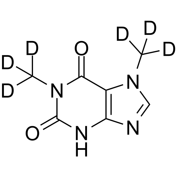
HY-W016498S
Paraxanthine-d6Stable Isotope -
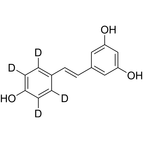
HY-16561S
Resveratrol-d4Stable Isotope -
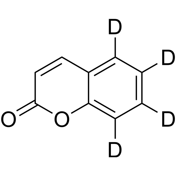
HY-N0709S
Coumarin-d4Stable Isotope -
HY-W355129S
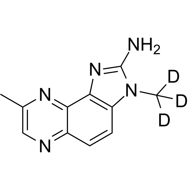
MeIQx-d3Stable Isotope
Publications Citing Use of MedChemExpress (MCE) Genistein-d4
MoreVoir tous les produits spécifiques à Isoform EGFR
MoreVoir tous les produits spécifiques à Isoform Endogenous Metabolite
More

Activité biologique
Description
In Vitro
Stable heavy isotopes of hydrogen, carbon, and other elements have been incorporated into drug molecules, largely as tracers for quantitation during the drug development process. Deuteration has gained attention because of its potential to affect the pharmacokinetic and metabolic profiles of drugs[1].
MedChemExpress (MCE) has not independently confirmed the accuracy of these methods. They are for reference only.
- Detection of Apoptosis
- TUNEL staining for apoptotic DNA fragmentation
- Human pluripotent stem cell endothelial-cell differentiation
- Flow cytometric DNA-content cell-cycle staining
- Vascular/Branching Fractal Analysis
- Annexin V plus membrane-impermeant dye apoptosis staining
- BrdU Incorporation Assay
- Endothelial Tube Formation Assay
- Apoptosis Solutions
- Protocol for Cell Cycle
- Endothelial Cell Migration/Angiogenic Sprouting Assay
- Orthotopic Cell-Line Xenograft
Application
1. This compound can be used as a tracer
2. This compound can be used as an internal standard for quantitative analysis by NMR, GC-MS, or LC-MS.


Chemical Information
-
CAS No. 187960-08-3
-
Unlabeled CAS 446-72-0
-
Appearance Solid
-
Masse moléculaire 274.26
-
Formule C15H6D4O5
-
Color White to off-white
-
SMILES
O=C1C(C2=C(C([2H])=C(C([2H])=C2[2H])O)[2H])=COC3=CC(O)=CC(O)=C31
-
Synonyms
NPI 031L-d4
-
Livraison
Room temperature in continental US; may vary elsewhere.
-
Stockage
Powder -20°C 3 years 4°C 2 years In solvent -80°C 6 months -20°C 1 month


Publications (1)
-
Journal Impact Factor
-
Most Recent


Solvant et solubilité
In Vitro:
DMSO : 50 mg/mL (182.31 mM; Need ultrasonic; Hygroscopic DMSO has a significant impact on the solubility of product, please use newly opened DMSO)
Please refer to the solubility information to select the appropriate solvent. Once prepared, please aliquot and store the solution to prevent product inactivation from repeated freeze-thaw cycles.
Storage method and period of stock solution: -80°C, 6 months; -20°C, 1 month. When stored at -80°C, please use it within 6 months. When stored at -20°C, please use it within 1 month.
Please refer to the solubility information to select the appropriate solvent. Once prepared, please aliquot and store the solution to prevent product inactivation from repeated freeze-thaw cycles.
Storage method and period of stock solution: -80°C, 6 months; -20°C, 1 month. When stored at -80°C, please use it within 6 months. When stored at -20°C, please use it within 1 month.
Concentration (start) × Volume (start) = Concentration (final) × Volume (final)
In Vivo:
Select the appropriate dissolution method based on your experimental animal and administration route.
- For the following dissolution methods, please ensure to first prepare a clear stock solution using an In Vitro approach and then sequentially add co-solvents:
- To ensure reliable experimental results, the clarified stock solution can be appropriately stored based on storage conditions. As for the working solution for In Vivo experiments, it is recommended to prepare freshly and use it on the same day.
- The percentages shown for the solvents indicate their volumetric ratio in the final prepared solution. If precipitation or phase separation occurs during preparation, heat and/or sonication can be used to aid dissolution.
Add each solvent one by one: 10% DMSO 40% PEG300 5% Tween-80 45% Saline
Solubility: ≥ 1.25 mg/mL (4.56 mM); Clear solution
This protocol yields a clear solution of ≥ 1.25 mg/mL (saturation unknown).
Taking 1 mL working solution as an example, add 100 μL DMSO stock solution (12.5 mg/mL) to 400 μL PEG300, and mix evenly; then add 50 μL Tween-80 and mix evenly; then add 450 μL Saline to adjust the volume to 1 mL.
Preparation of Saline: Dissolve 0.9 g sodium chloride in ddH₂O and dilute to 100 mL to obtain a clear Saline solution.
Add each solvent one by one: 10% DMSO 90% (20% SBE-β-CD in Saline)
Solubility: ≥ 1.25 mg/mL (4.56 mM); Clear solution
This protocol yields a clear solution of ≥ 1.25 mg/mL (saturation unknown).
Taking 1 mL working solution as an example, add 100 μL DMSO stock solution (12.5 mg/mL) to 900 μL 20% SBE-β-CD in Saline, and mix evenly.
Preparation of 20% SBE-β-CD in Saline (4°C, storage for one week): 2 g SBE-β-CD powder is dissolved in 10 mL Saline, completely dissolve until clear.
In Vivo Dissolution Calculator
Please enter the basic information of animal experiments:
-
-
-
-
Recommended: Prepare an additional quantity of animals to account for potential losses during experiments.
Please enter your animal formula composition:
-
%DMSO +
Recommended: Keep the proportion of DMSO in working solution below 2% if your animal is weak.
-
%+
-
+%Tween-80 + +
-
%Saline +
The co-solvents required include: DMSO, . All of co-solvents are available by MedChemExpress (MCE). , Tween 80. All of co-solvents are available by MedChemExpress (MCE).
Working solution concentration: 0.22 mg/mL
Method for preparing stock solution: mg drug dissolved in μL DMSO. Stock solution concentration: mg/mL.
1. Take μL DMSO stock solution;
2. Add μL .
μL , mix evenly;
3. Then add μL Tween 80, mix evenly;
4. Then add μL
Please ensure that the stock solution in the first step is dissolved to a clear state, and add co-solvents in sequence. You can use ultrasonic heating (ultrasonic cleaner, recommended frequency 20-40 kHz), vortexing, etc. to assist dissolution.


Pureté et documentation
-
Fiche technique (274 KB)
-
SDS (393 KB)
- English - EN (393 KB)
- Français - FR (393 KB)
- Deutsch - DE (393 KB)
- Norwegian - NO (393 KB)
- Español - ES (393 KB)
- Swedish - SV (393 KB)
- Italian - IT (393 KB)
- Korean - KR (393 KB)
- Portuguese - PT (393 KB)
-
Instruction de manipulation (2659 KB)
Références
[1]. Russak EM, et al. Impact of Deuterium Substitution on the Pharmacokinetics of Pharmaceuticals. Ann Pharmacother. 2019;53(2):211-216. [Content Brief]
[2]. Peterson G, et al. Genistein inhibits both estrogen and growth factor-stimulated proliferation of human breast cancer cells. Cell Growth Differ. 1996 Oct;7(10):1345-51. [Content Brief]
[3]. Ding S, et al. Environmentally Relevant Dose of Bisphenol A Does Not Affect Lipid Metabolism and Has No Synergetic or Antagonistic Effects on Genistein's Beneficial Roles on Lipid Metabolism. PLoS One. 2016 May 12;11(5):e0155352. [Content Brief]


Complete Stock Solution Preparation Table
Please refer to the solubility information to select the appropriate solvent. Once prepared, please aliquot and store the solution to prevent product inactivation from repeated freeze-thaw cycles.
Storage method and period of stock solution: -80°C, 6 months; -20°C, 1 month. When stored at -80°C, please use it within 6 months. When stored at -20°C, please use it within 1 month.
| Optional Solvent | Concentration Solvent Mass | 1 mg | 5 mg | 10 mg | 25 mg |
|---|---|---|---|---|---|
| DMSO | 1 mM | 3.6462 mL | 18.2309 mL | 36.4618 mL | 91.1544 mL |
| 5 mM | 0.7292 mL | 3.6462 mL | 7.2924 mL | 18.2309 mL | |
| 10 mM | 0.3646 mL | 1.8231 mL | 3.6462 mL | 9.1154 mL | |
| 15 mM | 0.2431 mL | 1.2154 mL | 2.4308 mL | 6.0770 mL | |
| 20 mM | 0.1823 mL | 0.9115 mL | 1.8231 mL | 4.5577 mL | |
| 25 mM | 0.1458 mL | 0.7292 mL | 1.4585 mL | 3.6462 mL | |
| 30 mM | 0.1215 mL | 0.6077 mL | 1.2154 mL | 3.0385 mL | |
| 40 mM | 0.0912 mL | 0.4558 mL | 0.9115 mL | 2.2789 mL | |
| 50 mM | 0.0729 mL | 0.3646 mL | 0.7292 mL | 1.8231 mL | |
| 60 mM | 0.0608 mL | 0.3038 mL | 0.6077 mL | 1.5192 mL | |
| 80 mM | 0.0456 mL | 0.2279 mL | 0.4558 mL | 1.1394 mL | |
| 100 mM | 0.0365 mL | 0.1823 mL | 0.3646 mL | 0.9115 mL |


Genistein-d4 Related Classifications
HY-14596S Related Classifications
 Powered by Bioz