



Glyoxal (40% w/w in water)

Based on 1 Customer Validation
Glyoxal (40% w/w in water) is an α-oxoaldehyde that inhibits Aldose Reductase, Glutathione Reductase, and NADPH synthase. Glyoxal (40% w/w in water) exhibits cytotoxicity, triggers oxidative stress, induces ROS accumulation, lipid peroxidation, mitochondrial membrane potential collapse, DNA damage, apoptosis, and massive production of advanced glycation end products (AGEs). Glyoxal (40% w/w in water) depletes glutathione and activates MAPK phosphorylation. It has lower toxicity as a fixative than paraformaldehyde (PFA) and serves as a precursor for the synthesis of oxalates and dietary carcinogens. Glyoxal (40% w/w in water) is suitable for research related to calcium oxalate kidney stones, diabetes, atherosclerosis, cardiovascular diseases, retinopathy, and cataracts.
For research use only. We do not sell to patients.
- CAS No.: 107-22-2
- Formula: C2H2O2
- Molecular Weight:58.04
-
Storage:
Store at room temperature, keep dry and cool.
In solvent -80°C, 1 year , -20°C, 6 months


To place orders, for customer services and technical support, please contact: MedChemExpress USA
Tel: 609-228-6898 E-mail: [email protected] [email protected]


-
 Biological Activity
-
Chemical Information
-
Solvent & Solubility
- Protocols
- Purity & Documentation
- References
-
 Help & FAQs


-
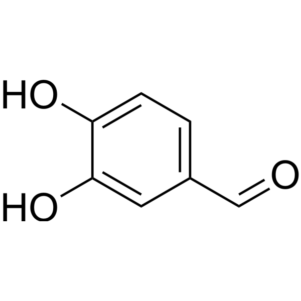
HY-N0295
ProtocatechualdehydePolyphenol Compound -
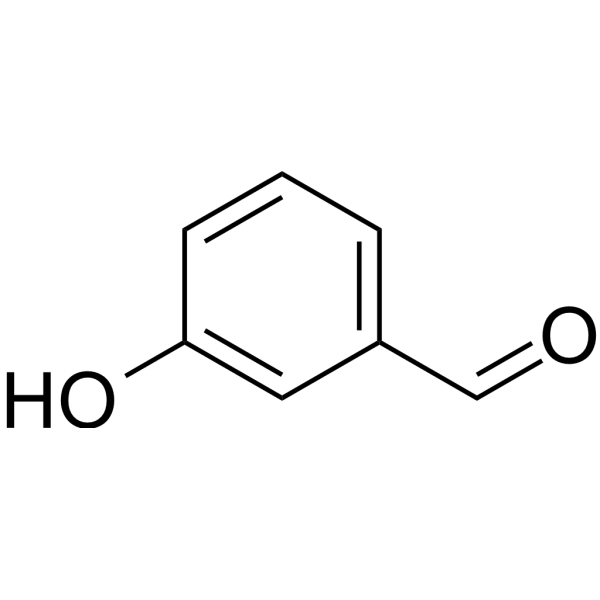
HY-76006
3-HydroxybenzaldehydeSubstrate For ALDH, Precursor Compound for Phenolic Compounds -
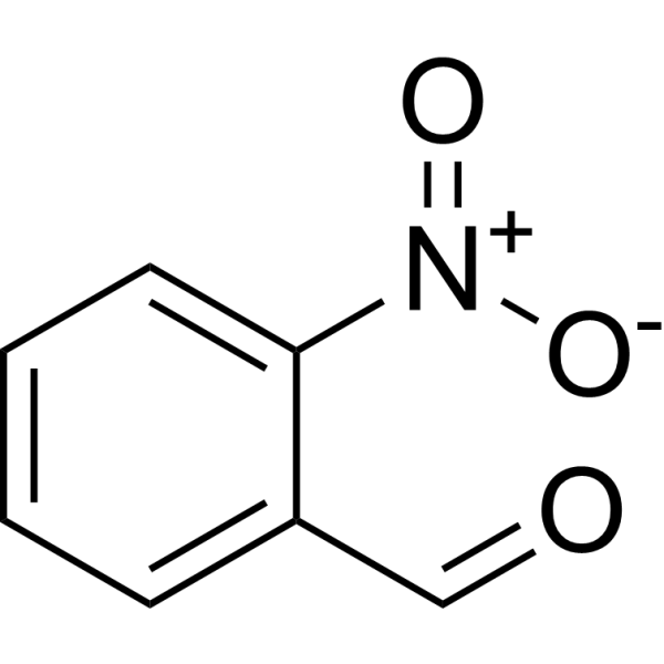
HY-W007454
2-NitrobenzaldehydeBiochemical Reagent -
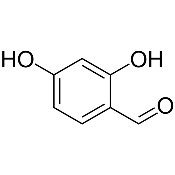
HY-W007539
2,4-DihydroxybenzaldehydeDrug Intermediate -
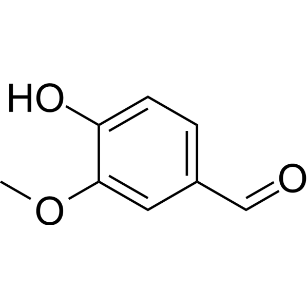
HY-N0098
VanillinPhenolic Aldehyde -

HY-Y1863
3-ChlorobenzaldehydemALDH Substrate -
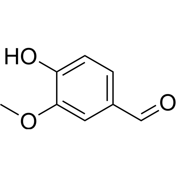
HY-N0098R
Vanillin (Standard) -
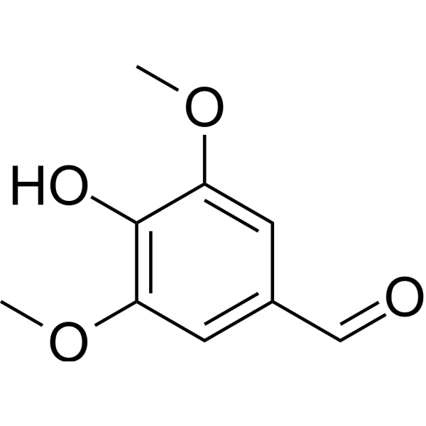
HY-N1390
SyringaldehydeCOX-2 Inhibitor -
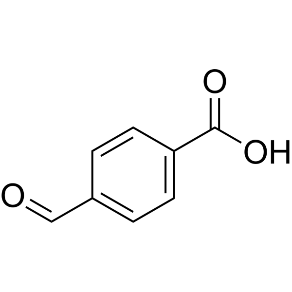
HY-W017446
4-Formylbenzoic acidBiochemical Reagent -
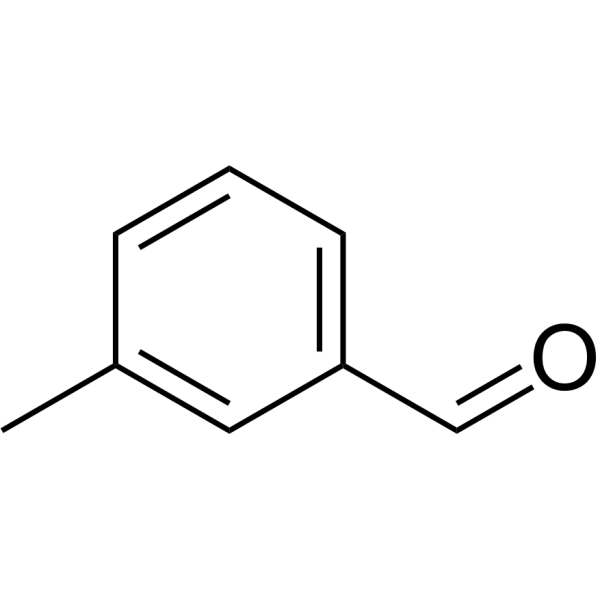
HY-78086
m-TolualdehydeNatural Product -

HY-N0295R
Protocatechualdehyde (Standard)Polyphenol Compound -

HY-76006R
3-Hydroxybenzaldehyde (Standard)Substrate For ALDH, Precursor Compound for Phenolic Compounds


Biological Activity
Description
|
p38 MAPK |
JNK |
ERK |
In Vitro
Glyoxal (1-5 mM; 25-165 min) (40% w/w in water) induces concentration- and time-dependent ROS production in isolated rat hepatocytes[2].
Glyoxal (0.5-10 mM; 25-135 min) (40% w/w in water) induces concentration-dependent GSH depletion and GSSG production in isolated rat hepatocytes[2].
Glyoxal (0.5-5 mM) (40% w/w in water) inhibits cytosolic glutathione reductase activity in rat hepatocyte fractions in a concentration- and time-dependent manner in vitro, with an IC50 of 3 mM[2].
Glyoxal (40% w/w in water)-induced cellular injury, inflammation and oxidative stress can be alleviated by Resveratrol (HY-16561), Luteolin (HY-N0162), morin and Mangiferin (HY-N0290) as well as Clostridium butyricum through regulating immune signaling pathways or cellular detoxification systems, respectively[4].\n
Glyoxal (0.12 mM; 8 days) (40% w/w in water) reduces glutathione levels and mitochondrial membrane potential, and increases the proportion of oxidized Trx1 in human aortic endothelial cells[5].
Glyoxal (1-10 mM; 4-12 h) (40% w/w in water) induces cytotoxicity by reducing mitochondrial activity in bovine pulmonary artery endothelial cells[6].
Glyoxal (40% w/w in water) enhances the fluorescence intensity of STED staining and multiple synaptic/skeletal proteins in rat hippocampal neurons compared with PFA; only the staining signals of LC3B and vimentin are weaker than those with PFA. It is suitable for a variety of cell and tissue samples including cardiac, neural and olfactory epithelial samples. When used for fixation of mouse olfactory epithelium and olfactory bulb, it also yields a higher signal-to-noise ratio for immunostaining[3].
Glyoxal (40% w/w in water) can be sequestered and bound by aspartame to reduce its free content and block in vitro glycation reactions. Additionally, the glyoxal-induced glycation of human serum albumin can be inhibited by Naringin (HY-N0153), Naringenin (HY-N0100), and Quercetin (HY-18085) via competitive occupation of the binding sites on lysine and arginine residues of the protein[4].
Glyoxal (40% w/w in water) can be captured and bound by Gallic acid (HY-N0523), Quercetin, glycine, serine, κ-carrageenan, alginic acid and pectin, thereby reducing the content of free Glyoxal in pH 7.0 phosphate buffer, pH 7.4 phosphate buffer, and lysine-glucose model system, respectively[4].
Glyoxal (GX) (50-200 μM; 8 days) (40% w/w in water) reduces the viability of wild-type and FANC pathway-deficient human aortic endothelial cells, with LD90 values of 0.12 mM and 0.125 mM, respectively[5].
Glyoxal (0.12 mM) (40% w/w in water) activates the mitogen-activated protein kinase pathway in human aortic endothelial cells, increasing the phosphorylation levels of ERK, JNK and p38 to 105%, 314% and 159% of those in the control group, respectively[5].
Glyoxal (1-5 mM; 12 h) (40% w/w in water) induces cytotoxicity in bovine pulmonary artery endothelial cells via membrane damage, with significant LDH release observed[6].
Glyoxal (1 mM; 6-24 h) (40% w/w in water) induces progressive loss of morphology in bovine pulmonary artery endothelial cells[6].
Glyoxal (1-10 mM; 4-12 h) (40% w/w in water) induces actin cytoskeleton rearrangement in bovine pulmonary artery endothelial cells; induces alterations in tight junctions of bovine pulmonary artery endothelial cells; dose-dependently induces the formation of advanced glycation end products (Amadori products) in bovine pulmonary artery endothelial cells; and almost completely inhibits in vitro angiogenesis of bovine pulmonary artery endothelial cells[6].
MedChemExpress (MCE) has not independently confirmed the accuracy of these methods. They are for reference only.
-
Cell Line:wild-type and FANC pathway-deficient human aortic endothelial cells
-
Concentration:0, 50, 100, 150, 200 μM
-
Incubation Time:8 days
-
Result:Reduced the survival rate of wild-type and FANC pathway-deficient human aortic endothelial cells, with LD90 values of 0.12 mM and 0.125 mM, respectively.
-
Cell Line:human aortic endothelial cells (HAECs)
-
Concentration:0.12 mM (LD90)
-
Incubation Time:8 days
-
Result:Caused an upper shift of Trx1 bands on urea-PAGE, indicating altered redox state.
Increased the proportion of oxidized Trx1 from ~35% in untreated control cells to ~90% in treated cells, a statistically significant change.
-
Cell Line:bovine pulmonary artery endothelial cells (BPAECs)
-
Concentration:1, 5, 10 mM
-
Incubation Time:4 h, 6 h, 12 h
-
Result:Caused a significant, dose-dependent decrease in mitochondrial dehydrogenase activity compared to vehicle-treated controls.
-
Cell Line:bovine pulmonary artery endothelial cells (BPAECs)
-
Concentration:1 mM, 5 mM
-
Incubation Time:12 h
-
Result:Caused a significant increase in LDH release compared to vehicle-treated controls.
Induced a slightly lower significant increase in LDH release at 5 mM than at 1 mM.
-
Cell Line:bovine pulmonary artery endothelial cells (BPAECs)
-
Concentration:1, 5, 10 mM
-
Incubation Time:6 h
-
Result:Caused a significant, dose-dependent decline in [3H]-thymidine incorporation compared to controls.
Indicated reduced cellular DNA synthesis and replication in BPAECs.
-
Cell Line:bovine pulmonary artery endothelial cells (BPAECs)
-
Concentration:1 mM
-
Incubation Time:4 h
-
Result:Caused marked disappearance of the regular peripheral localization of ZO-1 tight junction protein, which was maintained in control untreated BPAECs.
-
Cell Line:bovine pulmonary artery endothelial cells (BPAECs)
-
Concentration:1, 5, 10 mM
-
Incubation Time:4 h
-
Result:Induced a dose-dependent increase in intracellular formation of Amadori Products (a type of advanced glycation end product) in BPAECs compared to controls.
- Western Blot
- Mitochondrial membrane-potential fluorescent assay
- Mammalian live/dead viability and cytotoxicity staining
- Annexin V plus membrane-impermeant dye apoptosis staining
- ROS/oxidative-stress fluorescent staining
- Research Protocol for Cardiovascular Diseases
- Research Protocol for Metabolic Diseases
- Cell Viability Determination by MTT Colorimetric Assay
- Kinase activity and phosphorylation assays
- Detection of Apoptosis
- TUNEL staining for apoptotic DNA fragmentation
- Cell Cytotoxicity Assay
- Fluorescent plasma-membrane potential dye assay
- Mitochondrial membrane-potential and mitochondrial mass staining
- Apoptosis Solutions
- Ferroptosis Solutions
- Protocol for Kinase activity and phosphorylation assays
- Genotoxicity/Mutagenicity Study


Chemical Information
-
CAS No. 107-22-2
-
Appearance Liquid (Density: 1.14 g/cm3)
-
Molecular Weight 58.04
-
Formula C2H2O2
-
Color Colorless to light yellow
-
SMILES
O=CC=O
-
Shipping
Room temperature in continental US; may vary elsewhere.
-
Storage
Store at room temperature, keep dry and cool
In solvent -80°C 1 year -20°C 6 months


Solvent & Solubility
In Vitro:
DMSO : 200 mg/mL (3445.90 mM; Need ultrasonic; Hygroscopic DMSO has a significant impact on the solubility of product, please use newly opened DMSO)
Please refer to the solubility information to select the appropriate solvent. Once prepared, please aliquot and store the solution to prevent product inactivation from repeated freeze-thaw cycles.
Storage method and period of stock solution: -80°C, 1 year; -20°C, 6 months. When stored at -80°C, please use it within 1 year. When stored at -20°C, please use it within 6 months.
Please refer to the solubility information to select the appropriate solvent. Once prepared, please aliquot and store the solution to prevent product inactivation from repeated freeze-thaw cycles.
Storage method and period of stock solution: -80°C, 1 year; -20°C, 6 months. When stored at -80°C, please use it within 1 year. When stored at -20°C, please use it within 6 months.
Concentration (start) × Volume (start) = Concentration (final) × Volume (final)
In Vivo:
Select the appropriate dissolution method based on your experimental animal and administration route.
- For the following dissolution methods, please ensure to first prepare a clear stock solution using an In Vitro approach and then sequentially add co-solvents:
- To ensure reliable experimental results, the clarified stock solution can be appropriately stored based on storage conditions. As for the working solution for In Vivo experiments, it is recommended to prepare freshly and use it on the same day.
- The percentages shown for the solvents indicate their volumetric ratio in the final prepared solution. If precipitation or phase separation occurs during preparation, heat and/or sonication can be used to aid dissolution.
Add each solvent one by one: 10% DMSO 40% PEG300 5% Tween-80 45% Saline
Solubility: ≥ 5 mg/mL (86.15 mM); Clear solution
This protocol yields a clear solution of ≥ 5 mg/mL (saturation unknown).
Taking 1 mL working solution as an example, add 100 μL DMSO stock solution (50.0 mg/mL) to 400 μL PEG300, and mix evenly; then add 50 μL Tween-80 and mix evenly; then add 450 μL Saline to adjust the volume to 1 mL.
Preparation of Saline: Dissolve 0.9 g sodium chloride in ddH₂O and dilute to 100 mL to obtain a clear Saline solution.
Add each solvent one by one: 10% DMSO 90% (20% SBE-β-CD in Saline)
Solubility: ≥ 5 mg/mL (86.15 mM); Clear solution
This protocol yields a clear solution of ≥ 5 mg/mL (saturation unknown).
Taking 1 mL working solution as an example, add 100 μL DMSO stock solution (50.0 mg/mL) to 900 μL 20% SBE-β-CD in Saline, and mix evenly.
Preparation of 20% SBE-β-CD in Saline (4°C, storage for one week): 2 g SBE-β-CD powder is dissolved in 10 mL Saline, completely dissolve until clear.
In Vivo Dissolution Calculator
Please enter the basic information of animal experiments:
-
-
-
-
Recommended: Prepare an additional quantity of animals to account for potential losses during experiments.
Please enter your animal formula composition:
-
%DMSO +
Recommended: Keep the proportion of DMSO in working solution below 2% if your animal is weak.
-
%+
-
+%Tween-80 + +
-
%Saline +
The co-solvents required include: DMSO, . All of co-solvents are available by MedChemExpress (MCE). , Tween 80. All of co-solvents are available by MedChemExpress (MCE).
Working solution concentration: 0.22 mg/mL
Method for preparing stock solution: mg drug dissolved in μL DMSO. Stock solution concentration: mg/mL.
1. Take μL DMSO stock solution;
2. Add μL .
μL , mix evenly;
3. Then add μL Tween 80, mix evenly;
4. Then add μL
Please ensure that the stock solution in the first step is dissolved to a clear state, and add co-solvents in sequence. You can use ultrasonic heating (ultrasonic cleaner, recommended frequency 20-40 kHz), vortexing, etc. to assist dissolution.


Purity & Documentation
-
Data Sheet (286 KB)
-
SDS (560 KB)
- English - EN (560 KB)
- Français - FR (560 KB)
- Deutsch - DE (560 KB)
- Norwegian - NO (560 KB)
- Español - ES (560 KB)
- Swedish - SV (560 KB)
- Italian - IT (560 KB)
- Korean - KR (560 KB)
- Portuguese - PT (560 KB)
-
Handling Instructions (2659 KB)
References
[1]. Lange JN, et al. Glyoxal formation and its role in endogenous oxalate synthesis. Advances in urology. 2012;2012:819202. [Content Brief]
[2]. Shangari N, et al. The cytotoxic mechanism of glyoxal involves oxidative stress. Biochemical pharmacology. 2004 Oct 01;68(7):1433-42. [Content Brief]
[3]. Richter KN, et al. Glyoxal as an alternative fixative to formaldehyde in immunostaining and super-resolution microscopy. The EMBO journal. 2018 Jan 04;37(1):139-159. [Content Brief]
[4]. Zhang M, et al. Glyoxal in Foods: Formation, Metabolism, Health Hazards, and Its Control Strategies. Journal of agricultural and food chemistry. 2024 Feb 07;72(5):2434-2450. [Content Brief]
[5]. Xie MZ, et al. Glyoxal damages human aortic endothelial cells by perturbing the glutathione, mitochondrial membrane potential, and mitogen-activated protein kinase pathways. BMC cardiovascular disorders. 2021 Dec 18;21(1):603. [Content Brief]
[6]. Sliman SM, et al. Hyperglycemic oxoaldehyde, glyoxal, causes barrier dysfunction, cytoskeletal alterations, and inhibition of angiogenesis in vascular endothelial cells: aminoguanidine protection. Molecular and cellular biochemistry. 2010 Jan;333(1-2):9-26. [Content Brief]
[7]. Reber F, et al. Alteration of the intracellular pH and apoptosis induction in a retinal cell line by the AGE-inducing agent glyoxal. Graefe's archive for clinical and experimental ophthalmology = Albrecht von Graefes Archiv fur klinische und experimentelle Ophthalmologie. 2002 Dec;240(12):1022-32. [Content Brief]


Complete Stock Solution Preparation Table
Please refer to the solubility information to select the appropriate solvent. Once prepared, please aliquot and store the solution to prevent product inactivation from repeated freeze-thaw cycles.
Storage method and period of stock solution: -80°C, 1 year; -20°C, 6 months. When stored at -80°C, please use it within 1 year. When stored at -20°C, please use it within 6 months.
| Optional Solvent | Concentration Solvent Mass | 1 mg | 5 mg | 10 mg | 25 mg |
|---|---|---|---|---|---|
| DMSO | 1 mM | 17.2295 mL | 86.1475 mL | 172.2950 mL | 430.7374 mL |
| 5 mM | 3.4459 mL | 17.2295 mL | 34.4590 mL | 86.1475 mL | |
| 10 mM | 1.7229 mL | 8.6147 mL | 17.2295 mL | 43.0737 mL | |
| 15 mM | 1.1486 mL | 5.7432 mL | 11.4863 mL | 28.7158 mL | |
| 20 mM | 0.8615 mL | 4.3074 mL | 8.6147 mL | 21.5369 mL | |
| 25 mM | 0.6892 mL | 3.4459 mL | 6.8918 mL | 17.2295 mL | |
| 30 mM | 0.5743 mL | 2.8716 mL | 5.7432 mL | 14.3579 mL | |
| 40 mM | 0.4307 mL | 2.1537 mL | 4.3074 mL | 10.7684 mL | |
| 50 mM | 0.3446 mL | 1.7229 mL | 3.4459 mL | 8.6147 mL | |
| 60 mM | 0.2872 mL | 1.4358 mL | 2.8716 mL | 7.1790 mL | |
| 80 mM | 0.2154 mL | 1.0768 mL | 2.1537 mL | 5.3842 mL | |
| 100 mM | 0.1723 mL | 0.8615 mL | 1.7229 mL | 4.3074 mL |


Glyoxal (40% w/w in water) Related Classifications
HY-Y0785 Related Classifications
 Powered by Bioz
Keywords
- Glyoxal (40% w/w in water)
- 107-22-2
- Aldose Reductase
- Glutathione Reductase (GR)
- p38 MAPK
- ERK
- JNK
- Reactive Oxygen Species (ROS)
- Apoptosis
- Drug Intermediate
- HeLa cells
- aldehyde dehydrogenase
- glutathione reductase
- glyoxalase system
- human aortic endothelial cells
- COS-7 fibroblast cell
- AtT20 cells
- bovine pulmonary artery endothelial cells
- HepG2 hepatoma cells
- aldehyde reductase
- Inhibitor
- inhibitor
- inhibit