



GNE-490
Based on 1 publication(s) in Google Scholar
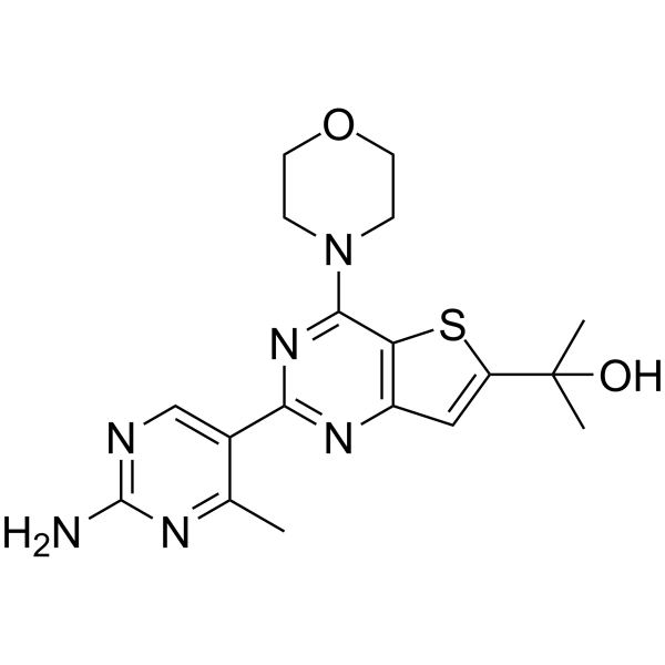
GNE-490, a (thienopyrimidin-2-yl)aminopyrimidine, is a potent pan-PI3K inhibitor with IC50s of 3.5 nM, 25 nM, 5.2 nM, 15 nM for PI3Kα, PI3Kβ, PI3Kδ and PI3Kγ, respectively. GNE-490 has >200 fold selectivity for mTOR (IC50=750 nM). GNE-490 shows potent suppression efficacy profile against MCF7.1 breast cancer xenograft model.
Nur für Forschungszwecke. Wir verkaufen nicht an Patienten.
- Reinheit : 96.79%
- CAS. Nr.: 1033739-92-2
- Formel: C18H22N6O2S
- Molecular Weight:386.47
-
Speicherung:
4°C, protect from light
* In solvent : -80°C, 6 months; -20°C, 1 month (protect from light)

To place orders, for customer services and technical support, please contact: MedChemExpress USA
Tel: 609-228-6898 E-mail: [email protected] [email protected]


-
 Biologische Aktivität
-
Chemical Information
-
Lösungsmittel & Löslichkeit
- Protokoll
- Reinheit & Dokumentation
- Verweise
-
 Help & FAQs


Publications Citing Use of MedChemExpress (MCE) GNE-490
More

Biologische Aktivität
Beschreibung
IC50 & Target
[1]|
PI3Kα 3.5 nM (IC50) |
PI3Kβ 25 nM (IC50) |
PI3Kδ 5.2 nM (IC50) |
PI3Kγ 15 nM (IC50) |
mTOR 750 nM (IC50) |
Cellular Effect
|
Cell Line

|
Type | Value | Description | References |
|---|---|---|---|---|
| PC-3 | IC50 |
0.49 nM
Compound: 21, GNE-490
|
Antiproliferative activity against human PC3 cells deficient in PTEN in after 3 to 4 days
Antiproliferative activity against human PC3 cells deficient in PTEN in after 3 to 4 days
|
[PMID: 20050669] |


Chemical Information
-
CAS. Nr. 1033739-92-2
-
Appearance Solid
-
Molecular Weight 386.47
-
Formel C18H22N6O2S
-
Color White to off-white
-
SMILES
OC(C)(C)C1=CC(N=C(C2=CN=C(N)N=C2C)N=C3N4CCOCC4)=C3S1
-
Versand
Room temperature in continental US; may vary elsewhere.
-
Speicherung
4°C, protect from light
* In solvent : -80°C, 6 months; -20°C, 1 month (protect from light)


Publications (1)
-
Journal Impact Factor
-
Most Recent
-
Molecules
2026 Jan 23;31(3):397. PMID: 41683377


Lösungsmittel & Löslichkeit
In Vitro:
DMSO : ≥ 100 mg/mL (258.75 mM; Hygroscopic DMSO has a significant impact on the solubility of product, please use newly opened DMSO)
* "≥" means soluble, but saturation unknown.
Please refer to the solubility information to select the appropriate solvent. Once prepared, please aliquot and store the solution to prevent product inactivation from repeated freeze-thaw cycles.
Storage method and period of stock solution: -80°C, 6 months; -20°C, 1 month (protect from light). When stored at -80°C, please use it within 6 months. When stored at -20°C, please use it within 1 month.
Please refer to the solubility information to select the appropriate solvent. Once prepared, please aliquot and store the solution to prevent product inactivation from repeated freeze-thaw cycles.
Storage method and period of stock solution: -80°C, 6 months; -20°C, 1 month (protect from light). When stored at -80°C, please use it within 6 months. When stored at -20°C, please use it within 1 month.
Konzentration (Stammlösung) × Volumen (Stammlösung) = Konzentration (Ziellösung) × Volumen (Ziellösung)


Reinheit & Dokumentation
-
Data Sheet (272 KB)
-
SDS (252 KB)
- English - EN (252 KB)
- Français - FR (252 KB)
- Deutsch - DE (252 KB)
- Norwegian - NO (252 KB)
- Español - ES (252 KB)
- Swedish - SV (252 KB)
- Italian - IT (252 KB)
- Korean - KR (252 KB)
- Portuguese - PT (252 KB)
-
Handling Instructions (2659 KB)
Verweise


Complete Stock Solution Preparation Table
Please refer to the solubility information to select the appropriate solvent. Once prepared, please aliquot and store the solution to prevent product inactivation from repeated freeze-thaw cycles.
Storage method and period of stock solution: -80°C, 6 months; -20°C, 1 month (protect from light). When stored at -80°C, please use it within 6 months. When stored at -20°C, please use it within 1 month.
| Optional Solvent | Concentration Solvent Mass | 1 mg | 5 mg | 10 mg | 25 mg |
|---|---|---|---|---|---|
| DMSO | 1 mM | 2.5875 mL | 12.9376 mL | 25.8752 mL | 64.6881 mL |
| 5 mM | 0.5175 mL | 2.5875 mL | 5.1750 mL | 12.9376 mL | |
| 10 mM | 0.2588 mL | 1.2938 mL | 2.5875 mL | 6.4688 mL | |
| 15 mM | 0.1725 mL | 0.8625 mL | 1.7250 mL | 4.3125 mL | |
| 20 mM | 0.1294 mL | 0.6469 mL | 1.2938 mL | 3.2344 mL | |
| 25 mM | 0.1035 mL | 0.5175 mL | 1.0350 mL | 2.5875 mL | |
| 30 mM | 0.0863 mL | 0.4313 mL | 0.8625 mL | 2.1563 mL | |
| 40 mM | 0.0647 mL | 0.3234 mL | 0.6469 mL | 1.6172 mL | |
| 50 mM | 0.0518 mL | 0.2588 mL | 0.5175 mL | 1.2938 mL | |
| 60 mM | 0.0431 mL | 0.2156 mL | 0.4313 mL | 1.0781 mL | |
| 80 mM | 0.0323 mL | 0.1617 mL | 0.3234 mL | 0.8086 mL | |
| 100 mM | 0.0259 mL | 0.1294 mL | 0.2588 mL | 0.6469 mL |


GNE-490 Related Classifications
HY-10812 Related Classifications
 Powered by Bioz