



gp91 ds-tat
Based on 1 publication(s) in Google Scholar
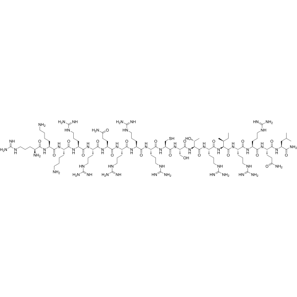
gp91 ds-tat, a biological active peptide, is a NADPH oxidase 2 (Nox2) inhibitor. gp91 ds-tat blocks NADPH oxidase-dependent superoxide production. gp91 ds-tat ameliorates high glucose-induced increase in total ROS, LPOs and iron levels. gp91 ds-tat inhibits homocysteine (Hcy)-induced activation of NLRP3 inflammasomes and restores Hcy-inhibited lysosomal TRPML1 channel activity. gp91 ds-tat improves cerebrovascular and cognitive function in APP/PS1 mice. gp91 ds-tat can be used for the study of Alzheimer’s disease (AD), glomerular inflammation and cardiovascular disease.
For research use only. We do not sell to patients.
- Purity : 99.00%
- CAS No.: 329902-61-6
- Formula: C98H190N50O22S
- Molecular Weight:2452.94
-
Storage:
Sealed storage, away from moisture.
Powder -80°C, 2 years , -20°C, 1 year* In solvent : -80°C, 6 months; -20°C, 1 month (sealed storage, away from moisture)

To place orders, for customer services and technical support, please contact: MedChemExpress USA
Tel: 609-228-6898 E-mail: [email protected] [email protected]


-
 Biological Activity
-
Chemical Information
-
Solvent & Solubility
- Protocols
- Purity & Documentation
- References
-
 Help & FAQs



Publications Citing Use of MedChemExpress (MCE) gp91 ds-tat
More

Biological Activity
Description
IC50 & Target
|
GPX4 |
In Vitro
gp91 ds-tat (5μM, 96 h) ameliorates high glucose-induced increase in total ROS, LPOs and iron levels, prevents inhibition of GPx4 activity, and attenuates cell death in human retinal endothelial cells[1].
gp91 ds-tat (5μM, 96 h) prevents impairments in mitochondrial membrane potential, decrease in oxygen consumption rate (including basal and maximal respiration rates), and reduction in mtDNA transcription and copy numbers in human retinal endothelial cells[1].
gp91 ds-tat (5 μM, 1 h pretreatment before homocysteine (Hcy) treatment) blocks Hcy-induced superoxide production in mouse podocytes[3].
gp91 ds-tat (5 μM, 1 h pretreatment before Hcy treatment) inhibits Hcy-induced activation of NLRP3 inflammasomes and formation of MVBs containing inflammatory cytokines in mouse podocytes[3].
gp91 ds-tat peptide (5 μM, 1 h pretreatment before Hcy treatment) restores Hcy-inhibited TRPML1 channel activity in mouse podocytes, reduces Hcy-enhanced exosome release from mouse podocytes and reverses the impaired lysosome-MVB interaction induced by Homocysteine in mouse podocytes[3].
gp91 ds-tat (1-3 μM, 2 h pretreatment) significantly inhibits chemerin-induced reactive oxygen species (ROS) production and inhibits chemerin-induced proliferation and migration in rat mesenteric arterial smooth muscle cells (SMCs)[4].
MedChemExpress (MCE) has not independently confirmed the accuracy of these methods. They are for reference only. Further protocols information, click here.
-
Cell Line:mouse podocytes
-
Concentration:5 μM
-
Incubation Time:1 h pretreatment before Hcy treatment
-
Result:Increased colocalization of NLRP3 with ASC and caspase-1 via confocal microscopy.
Enhanced colocalization of Rab7a/VPS16 with IL-1β.
Recovered ML-SA1-induced GCaMP3 fluorescence elevation.
Increased colocalization of Rab7a (MVB marker) and Lamp-1 (lysosome marker).
In Vivo
MedChemExpress (MCE) has not independently confirmed the accuracy of these methods. They are for reference only.
-
Animal Model:APP/PS1 transgenic mice (10-11 months old, both sexes) were used as a mouse model of Alzheimer's disease[2]
-
Dosage:10 mg/kg
-
Administration:i.p. every other day for 1 or 2 weeks
-
Result:Reduced ROS accumulation and NOX2 mRNA expression in the neocortex of APP/PS1 mice.
Decreased capillary stalling incidence by 67% in APP/PS1 mice after 1 week.
Increased volumetric blood flow and RBC flow speed in penetrating arterioles of APP/PS1 mice.
Improved performance in object displacement and Y-maze tests (short-term and spatial memory) in APP/PS1 mice, with significant effects after 2 weeks.
Reduced GFAP expression in the hippocampus and IBA1 expression in cortex/hippocampus of APP/PS1 mice (neuroinflammation attenuation), without affecting amyloid-β levels.
Downregulated inflammatory and leukocyte adhesion-related pathways in cerebral microvessels of APP/PS1 mice.
Decreased VCAM-1 and ICAM-1 expression in penetrating arterioles and capillaries of APP/PS1 mice.


Chemical Information
-
CAS No. 329902-61-6
-
Appearance Solid
-
Molecular Weight 2452.94
-
Formula C98H190N50O22S
-
Color White to off-white
-
Sequence
Arg-Lys-Lys-Arg-Arg-Gln-Arg-Arg-Arg-Cys-Ser-Thr-Arg-Ile-Arg-Arg-Gln-Leu-NH2
-
Sequence Shortening
RKKRRQRRRCSTRIRRQL-NH2
-
Shipping
Room temperature in continental US; may vary elsewhere.
-
Storage
Sealed storage, away from moisture
Powder -80°C 2 years -20°C 1 year * In solvent : -80°C, 6 months; -20°C, 1 month (sealed storage, away from moisture)


Publications (1)
-
Journal Impact Factor
-
Most Recent
-
Rheumatology (Oxford)
Autophagy inhibitors block pathogenic NET release in immune-mediated inflammatory disease without impairing host defence. [Abstract]2025 Aug 13:keaf437. PMID: 40802538


Solvent & Solubility
In Vitro:
DMSO : 100 mg/mL (40.77 mM; Need ultrasonic; Hygroscopic DMSO has a significant impact on the solubility of product, please use newly opened DMSO)
H2O : ≥ 50 mg/mL (20.38 mM)
* "≥" means soluble, but saturation unknown.
Please refer to the solubility information to select the appropriate solvent. Once prepared, please aliquot and store the solution to prevent product inactivation from repeated freeze-thaw cycles.
Storage method and period of stock solution: -80°C, 6 months; -20°C, 1 month (sealed storage, away from moisture). When stored at -80°C, please use it within 6 months. When stored at -20°C, please use it within 1 month.
* Note: If you choose water as the stock solution, please dilute it to the working solution, then filter and sterilize it with a 0.22 μm filter before use.
Please refer to the solubility information to select the appropriate solvent. Once prepared, please aliquot and store the solution to prevent product inactivation from repeated freeze-thaw cycles.
Storage method and period of stock solution: -80°C, 6 months; -20°C, 1 month (sealed storage, away from moisture). When stored at -80°C, please use it within 6 months. When stored at -20°C, please use it within 1 month.
* Note: If you choose water as the stock solution, please dilute it to the working solution, then filter and sterilize it with a 0.22 μm filter before use.
Concentration (start) × Volume (start) = Concentration (final) × Volume (final)

Protocols
-
ROS/oxidative-stress fluorescent staining
ROS/oxidative-stress fluorescent staining uses cell-permeant fluorogenic probes that become fluorescent after oxidation inside cells or tissues; commonly used examples include DCFH-DA/DCFDA for broad cellular oxidant detection, DHE for superoxide-related signal detection, MitoSOX for mitochondrial superoxide-related signal detection, and CellROX probes for oxidative-stress-associated fluorescence readouts. The assay detects probe oxidation rather than a single ROS species unless the probe and analysis method have been chemically validated for that species. DCFH-DA enters cells, is deacetylated by intracellular esterases to DCFH, and produces fluorescent DCF after oxidation, so the readout is used as an operational measure of total cellular oxidative stress rather than a species-specific ROS measurement. DHE and MitoSOX can report superoxide-related oxidation, but red fluorescence alone can include non-specific ethidium-like oxidation products; HPLC or optimized spectral approaches are
-
Protocol for Water Maze
The Morris Water Maze is a rodent spatial learning and memory assay in which a mouse or rat swims in opaque water to find an escape platform; in the hidden-platform version, the animal cannot see the platform and must use distal extra-maze cues to learn its fixed spatial location. The assay primarily measures hippocampus-dependent spatial learning during acquisition trials and spatial reference memory during probe trials after platform removal; readouts include escape latency, swim path length, swim speed, quadrant occupancy, platform-site crossings, and proximity to the former platform location.
-
Research Protocol for Cardiovascular Diseases
Cardiovascular disease can be modeled as maladaptive cardiac remodeling, where ischemic injury or pressure overload activates inflammatory signaling, fibroblast activation, extracellular-matrix deposition, cardiomyocyte hypertrophy, vascular remodeling, and progressive ventricular dysfunction. The TGF-β/SMAD axis is a central profibrotic pathway after myocardial injury and pressure overload, while innate immune and cytokine pathways regulate leukocyte recruitment, scar formation, and adverse remodeling. Key unresolved questions include which inflammatory signals are reparative versus harmful, when fibrosis is protective versus maladaptive, and whether pathway inhibition improves function without weakening necessary infarct healing or compensatory remodeling.
-
Lysosome and acidic-vesicle live-cell staining
Lysosome and acidic-vesicle live-cell staining detects acidic intracellular compartments by using membrane-permeant acidotropic probes that accumulate in low-pH vesicles, including lysosomes, late endosomes, autolysosomes, and acidic phagosomes. LysoTracker staining is commonly used as an intensity-based readout of acidic lysosomal compartment abundance or enlargement, while acridine orange produces green fluorescence in less concentrated compartments and red fluorescence after concentration-dependent accumulation in acidic vesicular organelles. Loss or reduction of acridine-orange red signal can be used as a readout of lysosomal membrane permeabilization or reduced acidic-vesicle integrity. This protocol is designed for live cultured cells and can be adapted for fluorescence microscopy, high-content imaging, plate-reader readout, or flow cytometry when the selected literature supports the readout. Because these dyes report acidotropic accumulation rather than lysosome identity alone,
-
Research Protocol for Inflammation-related Diseases
The NLRP3 inflammasome is a cytosolic innate immune signaling platform that integrates priming signals and danger-signal activation to promote caspase-1 activation, maturation of IL-1β and IL-18, and gasdermin D-mediated pyroptotic cell death. The core experimental logic is to determine whether inflammatory disease phenotypes are driven by increased NLRP3 expression, ASC-containing inflammasome assembly, caspase-1 cleavage, GSDMD cleavage, and extracellular release of IL-1β/IL-18 rather than by nonspecific cell injury alone. The pathway is strongly linked to inflammation-related disease phenotypes because monosodium urate crystals activate NALP3/NLRP3 inflammasome signaling in gout-like crystal inflammation, cholesterol crystals activate NLRP3 inflammasomes in atherogenesis models, and DSS-induced intestinal inflammation has been reported to involve NLRP3 inflammasome activity. However, experimental colitis studies also show context-dependent protective effects of NLRP3 inflammasome co


Purity & Documentation
-
Data Sheet (283 KB)
-
SDS (252 KB)
- English - EN (252 KB)
- Français - FR (252 KB)
- Deutsch - DE (252 KB)
- Norwegian - NO (252 KB)
- Español - ES (252 KB)
- Swedish - SV (252 KB)
- Italian - IT (252 KB)
- Korean - KR (252 KB)
- Portuguese - PT (252 KB)
-
Handling Instructions (2659 KB)
References
[1]. Malaviya P, et al. Role of ferroptosis in mitochondrial damage in diabetic retinopathy. Free Radic Biol Med. 2024 Nov 20;225:821-832. [Content Brief]
[2]. Ruiz-Uribe NE, et al. Vascular oxidative stress causes neutrophil arrest in brain capillaries, leading to decreased cerebral blood flow and contributing to memory impairment in a mouse model of Alzheimer’s disease. bioRxiv [Preprint]. 2023 Feb 15:2023.02.15.528710. [Content Brief]
[3]. Li G, et al. Regulation of TRPML1 channel activity and inflammatory exosome release by endogenously produced reactive oxygen species in mouse podocytes. Redox Biol. 2021 Jul;43:102013. [Content Brief]


Complete Stock Solution Preparation Table
Please refer to the solubility information to select the appropriate solvent. Once prepared, please aliquot and store the solution to prevent product inactivation from repeated freeze-thaw cycles.
Storage method and period of stock solution: -80°C, 6 months; -20°C, 1 month (sealed storage, away from moisture). When stored at -80°C, please use it within 6 months. When stored at -20°C, please use it within 1 month.
| Optional Solvent | Concentration Solvent Mass | 1 mg | 5 mg | 10 mg | 25 mg |
|---|---|---|---|---|---|
| H2O / DMSO | 1 mM | 0.4077 mL | 2.0384 mL | 4.0767 mL | 10.1919 mL |
| 5 mM | 0.0815 mL | 0.4077 mL | 0.8153 mL | 2.0384 mL | |
| 10 mM | 0.0408 mL | 0.2038 mL | 0.4077 mL | 1.0192 mL | |
| 15 mM | 0.0272 mL | 0.1359 mL | 0.2718 mL | 0.6795 mL | |
| 20 mM | 0.0204 mL | 0.1019 mL | 0.2038 mL | 0.5096 mL | |
| DMSO | 25 mM | 0.0163 mL | 0.0815 mL | 0.1631 mL | 0.4077 mL |
| 30 mM | 0.0136 mL | 0.0679 mL | 0.1359 mL | 0.3397 mL | |
| 40 mM | 0.0102 mL | 0.0510 mL | 0.1019 mL | 0.2548 mL |
* Note: If you choose water as the stock solution, please dilute it to the working solution, then filter and sterilize it with a 0.22 μm filter before use.


gp91 ds-tat Related Classifications
HY-P5381 Related Classifications
 Powered by Bioz