



Emamectin Benzoate
Based on 3 publication(s) in Google Scholar
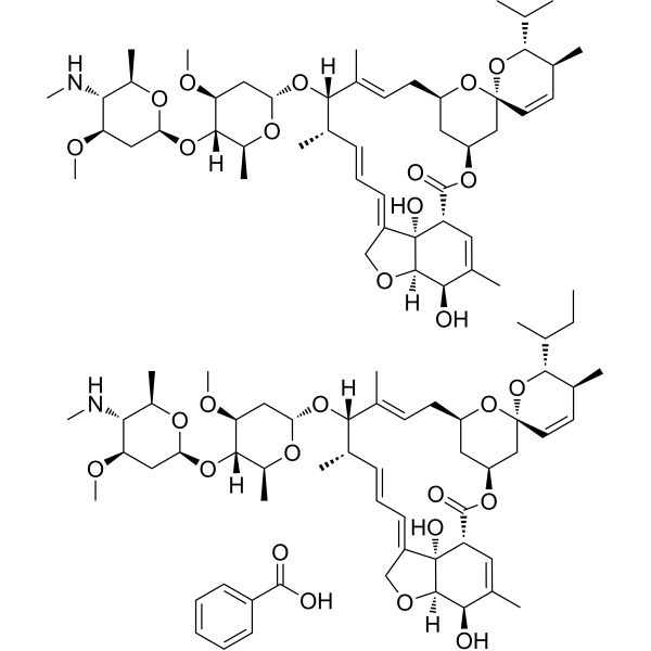
Emamectin Benzoate (MK-244) is an orally active nervoussystem toxicant by binding g-aminobutyric (GABA) receptor in insects. Emamectin Benzoate is one of semi-synthetic derivative of Avermectin (HY-15311) with a broadspectrum of insecticidal and acaricidal activity. Emamectin Benzoate induces ROS-mediated DNA damage and cell apoptosis. Emamectin Benzoate, a mixture of the natural Emamectin B1a benzoate and Emamectin B1b benzoate, has the main component of Emamectin B1a benzoate.
For research use only. We do not sell to patients.
- Purity : 98.83%
- CAS No.: 155569-91-8
- Formula: C49H75NO13.C7H6O2
- Molecular Weight:1008.24(Based on Emamectin B1a Benzoate)
-
Storage:
4°C, sealed storage, away from moisture
* In solvent : -80°C, 6 months; -20°C, 1 month (sealed storage, away from moisture)

To place orders, for customer services and technical support, please contact: MedChemExpress USA
Tel: 609-228-6898 E-mail: [email protected] [email protected]


-
 Biological Activity
-
Chemical Information
-
Solvent & Solubility
- Protocols
- Purity & Documentation
- References
-
 Help & FAQs


-

HY-18739
Phorbol 12-myristate 13-acetatePKC Activator -
HY-134813
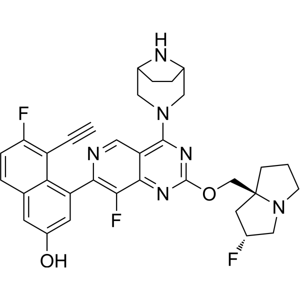
MRTX1133KRAS G12D Inhibitor -
HY-15311
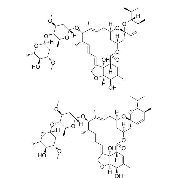
Avermectin B1Anti-infection Agent -
HY-B1422
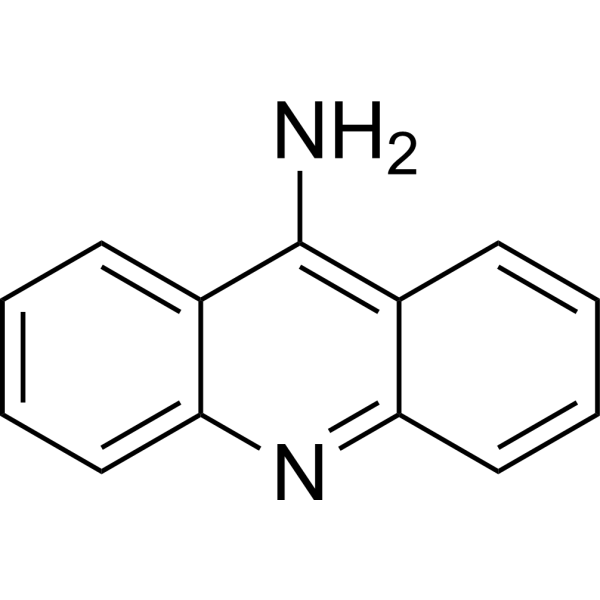
9-AminoacridineAntimicrobial -
HY-14953
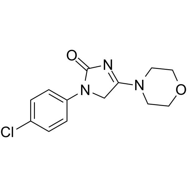
ImepitoinBenzodiazepine Receptor Agonist -
HY-13643
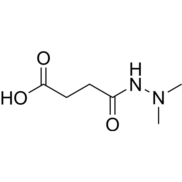
DaminozideKDM2/7 Inhibitor -
HY-B1841
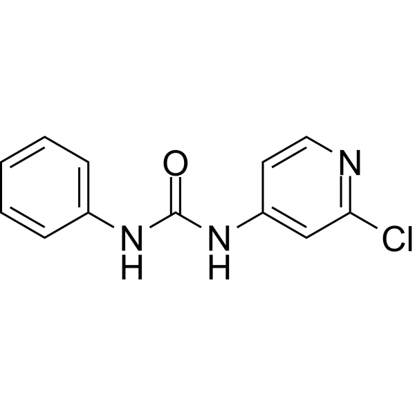
ForchlorfenuronPlant Growth Regulator -
HY-W131845
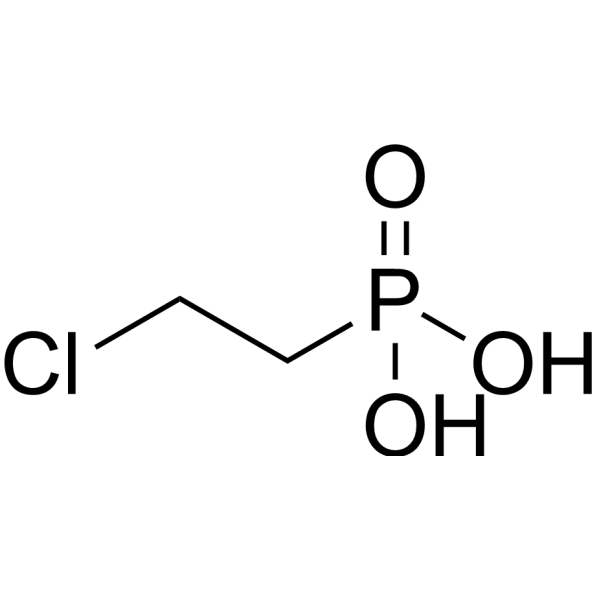
EthephonPlant Growth Regulator -

HY-W131845R
Ethephon (Standard)Plant Growth Regulator -

HY-14953R
Imepitoin (Standard)Benzodiazepine Receptor Agonist -
HY-B0007
BaclofenGABABR Agonist -
HY-B0941
6-BenzylaminopurineCytokinin
Publications Citing Use of MedChemExpress (MCE) Emamectin Benzoate
MoreAll Parasite Isoforms
More

Biological Activity
Description
IC50 & Target
|
Mite |
In Vitro
Emamectin Benzoate (MK-244; 2.5-40 μM; 12 and 24 h) decreases cell viability in a time- and dose-dependent manner[1].
Emamectin Benzoate (2.5-20 μM; 24 hours) induces apoptosis and DNA damage in 16HBE cells. Emamectin Benzoate induces ROS generation in 16HBE cells[1].
Emamectin Benzoate (2.5-20 μM; 12 hours) increases the amounts of cytochrome-c, caspase-3, cas-pase-9, cleaved-PARP, Bax/Bcl-2[1].
Emamectin Benzoate (2.5, 5, 10, 15 μM; 72 h) inhibits cell viability with an IC50 of 3.72 μM in Trichoplusia Tn5B1-4 cell. Emamectin Benzoat induces chromatin condensation in nuclei and cell apoptosis[2].
MedChemExpress (MCE) has not independently confirmed the accuracy of these methods. They are for reference only.
-
Cell Line:human normal bronchial epithelial cell line 16HBE
-
Concentration:2.5, 5, 7.5,10,15, 20, 40 μM
-
Incubation Time:12 and 24 hours
-
Result:Decreased cell viability in a time- and dose-dependent manner wirh IC50s of 11.88 μM and 9.67 μM in 12 and 24 hours, respectively.
-
Cell Line:human normal bronchial epithelial cell line 16HBE
-
Concentration:2.5, 5, 10, 20 μM
-
Incubation Time:24 hours
-
Result:Induced apoptosis and caused chromatin shrinkage and nuclear fragmentation.
-
Cell Line:human normal bronchial epithelial cell line 16HBE
-
Concentration:2.5, 5, 10, 20 μM
-
Incubation Time:12 hours
-
Result:Increased the amounts of cytochrome-c, caspase-3, cas-pase-9, cleaved-PARP, Bax/Bcl-2.
In Vivo
MedChemExpress (MCE) has not independently confirmed the accuracy of these methods. They are for reference only.
-
Animal Model:10 weeks old Swiss albino male mice (25-30 g)[3]
-
Dosage:25, 50, 100 mg/kg
-
Administration:Oral; daily; for 14 days
-
Result:Caused a marked induction of oxidative damage in liver tissue as demonstrated by an increased level of TBARS and reduced GSH level.


Chemical Information
-
CAS No. 155569-91-8
-
Appearance Solid
-
Molecular Weight 1008.24(Based on Emamectin B1a Benzoate)
-
Formula C49H75NO13.C7H6O2
-
Color White to off-white
-
SMILES
C[C@H]1O[C@@H](O[C@@]2([H])[C@H](C)O[C@@H](O[C@]([C@@H](C)/C=C/C=C3CO[C@@]4([H])[C@]\3(O)[C@H]5C=C(C)[C@H]4O)([H])/C(C)=C/C[C@@H]6C[C@H](OC5=O)C[C@]7(C=C[C@H](C)[C@@H](C(C)C)O7)O6)C[C@@H]2OC)C[C@@H](OC)[C@@H]1NC.C[C@H]8O[C@@H](O[C@@]9([H])[C@H](C)O[C@@H](O[C@]([C@@H](C)/C=C/C=C%10CO[C@@]%11([H])[C@]\%10(O)[C@H]%12C=C(C)[C@H]%11O)([H])/C(C)=C/C[C@@H]%13C[C@H](OC%12=O)C[C@]%14(C=C[C@H](C)[C@@H]([C@H](CC)C)O%14)O%13)C[C@@H]9OC)C[C@@H](OC)[C@@H]8NC.O=C(O)C%15=CC=CC=C%15
-
Synonyms
MK-244
-
Shipping
Room temperature in continental US; may vary elsewhere.
-
Storage
4°C, sealed storage, away from moisture
* In solvent : -80°C, 6 months; -20°C, 1 month (sealed storage, away from moisture)


Publications (3)
-
Journal Impact Factor
-
Most Recent
-
Nat Commun
Enhancing tandem mass spectrometry-based metabolite annotation with online chemical labeling. [Abstract]2025 Jul 26;16(1):6911. PMID: 40715063 -
-
Toxicol Appl Pharmacol
Proteome profiling of emamectin-treated primary rat cortical cells following up to twelve days in vitro exposure. [Abstract]2026 Jun:511:117824. PMID: 42009082


Solvent & Solubility
In Vitro:
DMSO : 100 mg/mL (Need ultrasonic; Hygroscopic DMSO has a significant impact on the solubility of product, please use newly opened DMSO)
In Vivo:
Select the appropriate dissolution method based on your experimental animal and administration route.
- For the following dissolution methods, please ensure to first prepare a clear stock solution using an In Vitro approach and then sequentially add co-solvents:
- To ensure reliable experimental results, the clarified stock solution can be appropriately stored based on storage conditions. As for the working solution for In Vivo experiments, it is recommended to prepare freshly and use it on the same day.
- The percentages shown for the solvents indicate their volumetric ratio in the final prepared solution. If precipitation or phase separation occurs during preparation, heat and/or sonication can be used to aid dissolution.
Add each solvent one by one: 10% DMSO 40% PEG300 5% Tween-80 45% Saline
Solubility: ≥ 2.08 mg/mL; Clear solution
This protocol yields a clear solution of ≥ 2.08 mg/mL (saturation unknown).
Taking 1 mL working solution as an example, add 100 μL DMSO stock solution (20.8 mg/mL) to 400 μL PEG300, and mix evenly; then add 50 μL Tween-80 and mix evenly; then add 450 μL Saline to adjust the volume to 1 mL.
Preparation of Saline: Dissolve 0.9 g sodium chloride in ddH₂O and dilute to 100 mL to obtain a clear Saline solution.
Add each solvent one by one: 10% DMSO 90% (20% SBE-β-CD in Saline)
Solubility: ≥ 2.08 mg/mL; Clear solution
This protocol yields a clear solution of ≥ 2.08 mg/mL (saturation unknown).
Taking 1 mL working solution as an example, add 100 μL DMSO stock solution (20.8 mg/mL) to 900 μL 20% SBE-β-CD in Saline, and mix evenly.
Preparation of 20% SBE-β-CD in Saline (4°C, storage for one week): 2 g SBE-β-CD powder is dissolved in 10 mL Saline, completely dissolve until clear.
In Vivo Dissolution Calculator
Please enter the basic information of animal experiments:
-
-
-
-
Recommended: Prepare an additional quantity of animals to account for potential losses during experiments.
Please enter your animal formula composition:
-
%DMSO +
Recommended: Keep the proportion of DMSO in working solution below 2% if your animal is weak.
-
%+
-
+%Tween-80 + +
-
%Saline +
The co-solvents required include: DMSO, . All of co-solvents are available by MedChemExpress (MCE). , Tween 80. All of co-solvents are available by MedChemExpress (MCE).
Working solution concentration: 0.22 mg/mL
Method for preparing stock solution: mg drug dissolved in μL DMSO. Stock solution concentration: mg/mL. * In solvent : -80°C, 6 months; -20°C, 1 month (sealed storage, away from moisture)
1. Take μL DMSO stock solution;
2. Add μL .
μL , mix evenly;
3. Then add μL Tween 80, mix evenly;
4. Then add μL
Please ensure that the stock solution in the first step is dissolved to a clear state, and add co-solvents in sequence. You can use ultrasonic heating (ultrasonic cleaner, recommended frequency 20-40 kHz), vortexing, etc. to assist dissolution.


Purity & Documentation
-
Data Sheet (290 KB)
-
SDS (623 KB)
- English - EN (623 KB)
- Français - FR (623 KB)
- Deutsch - DE (623 KB)
- Norwegian - NO (623 KB)
- Español - ES (623 KB)
- Swedish - SV (623 KB)
- Italian - IT (623 KB)
- Korean - KR (623 KB)
- Portuguese - PT (623 KB)
-
Handling Instructions (2659 KB)
References
[1]. Chenguang Niu, et al. Toxic effects of the Emamectin Benzoate exposure on cultured human bronchial epithelial (16HBE) cells. Environ Pollut. 2020 Feb;257:113618. [Content Brief]
[2]. Shaorong Luan, et al. Emamectin benzoate induces ROS-mediated DNA damage and apoptosis in Trichoplusia Tn5B1-4 cells. Chem Biol Interact. 2017 Aug 1;273:90-98. [Content Brief]
[3]. Özge Temiz, et al. Biopesticide emamectin benzoate in the liver of male mice: evaluation of oxidative toxicity with stress protein, DNA oxidation, and apoptosis biomarkers. Environ Sci Pollut Res Int. 2020 Jun;27(18):23199-23205. [Content Brief]

Calculators
Concentration (start) × Volume (start) = Concentration (final) × Volume (final)


Emamectin Benzoate Related Classifications
HY-B0837 Related Classifications
 Powered by Bioz