



Ciwujianoside B
Based on 1 publication(s) in Google Scholar
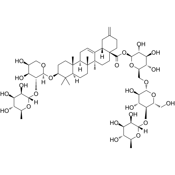
Ciwujianoside B is an orally active, blood-brain barrier penetrable radioprotective agent and memory enhancer. Ciwujianoside B reduces radiation-induced DNA damage, cell cycle arrest and apoptosis, downregulates NF-κB and the Bax/Bcl-2 ratio, and enhances the proliferative capacity of bone marrow cells. Ciwujianoside B enhances object recognition memory in normal mice and induces dendritic extension in primary cultured cortical neurons. Ciwujianoside B can be used in studies related to hematopoietic system radiation injury and memory enhancement.
For research use only. We do not sell to patients.
- Purity : 99.68%
- CAS No.: 114902-16-8
- Formula: C58H92O25
- Molecular Weight:1189.34
-
Storage:
-20°C, sealed storage, away from moisture and light
* In solvent : -80°C, 6 months; -20°C, 1 month (sealed storage, away from moisture and light)

To place orders, for customer services and technical support, please contact: MedChemExpress USA
Tel: 609-228-6898 E-mail: [email protected] [email protected]


-
 Biological Activity
-
Chemical Information
-
Solvent & Solubility
- Protocols
- Purity & Documentation
- References
-
 Help & FAQs


-
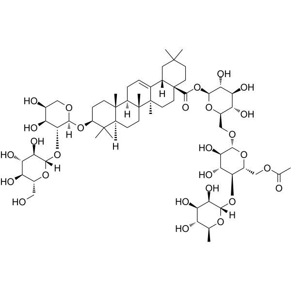
HY-N4135
Acanthopanaxoside B -
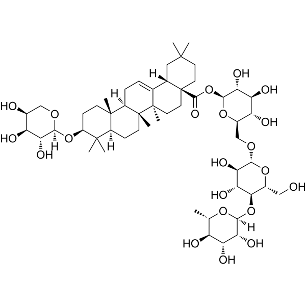
HY-N4134
Ciwujianoside C3Anti-inflammatory Agent -

HY-N0307R
Ciwujianoside B (Standard)Standard of Ciwujianoside B -
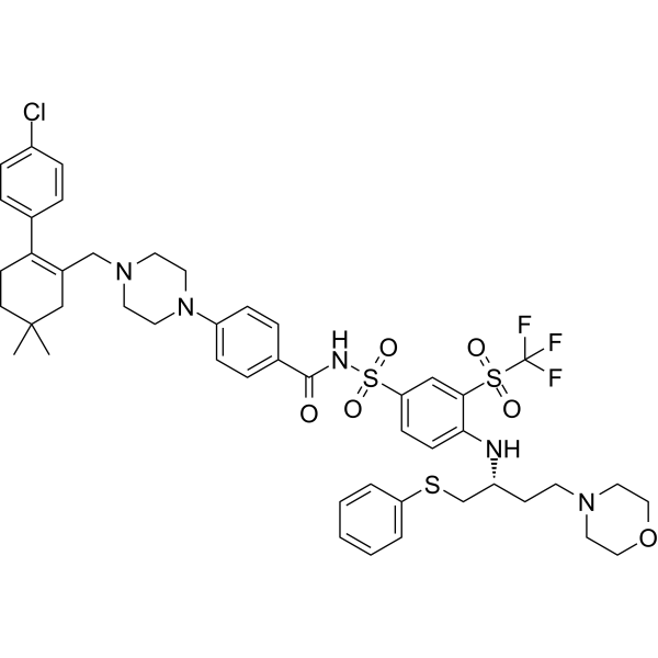
HY-10087
NavitoclaxBcl-2 Inhibitor -
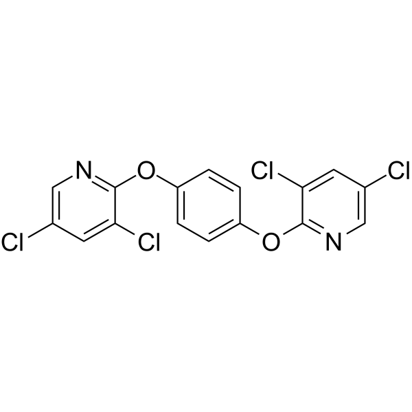
HY-103243
TCPOBOPCAR Agonist -
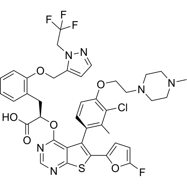
HY-100741
S63845MCL1 Inhibitor -
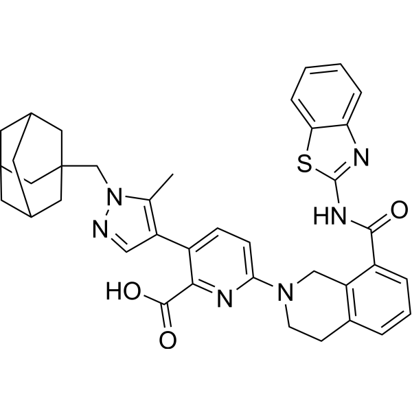
HY-19741
A-1331852Bcl-xL Inhibitor -
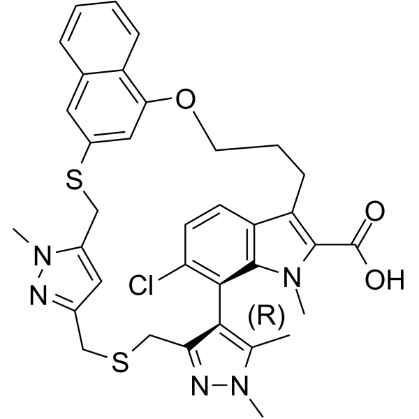
HY-101533
AZD-5991Mcl-1 Inhibitor -
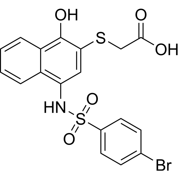
HY-18628
UMI-77Bcl-2 Family Inhibitor -

HY-10087R
Navitoclax (Standard)Reference Standard -
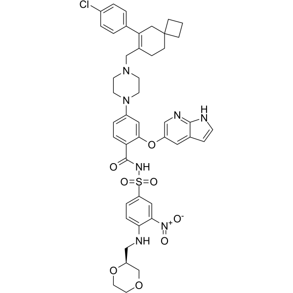
HY-129179
LisaftoclaxBCL-2 Inhibitor -
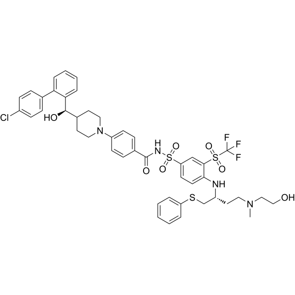
HY-112416
AZD4320BCL2/BCLxL Inhibitor
Publications Citing Use of MedChemExpress (MCE) Ciwujianoside B
MoreAll DNA/RNA Synthesis Isoforms
More

Biological Activity
Description
IC50 & Target
[1]|
Bax |
Bcl-2 |
NF-κB |
In Vitro
Ciwujianoside B (1-10 μM; 4 days) significantly enhances dendrite growth in primary cultured mouse cortical neurons[2].
MedChemExpress (MCE) has not independently confirmed the accuracy of these methods. They are for reference only.
In Vivo
Ciwujianoside B (0.5 mg/kg; p.o.; administered daily for 17 consecutive days) significantly enhances object recognition memory in 8-week-old male ddY mice[2].
MedChemExpress (MCE) has not independently confirmed the accuracy of these methods. They are for reference only.
-
Animal Model:BALB/cAnN (male, 8-10 weeks old)[1]
-
Dosage:10 mg/kg; 20 mg/kg; 40 mg/kg; 80 mg/kg
-
Administration:i.g.; daily; 7 days
-
Result:Enhanced the proliferation ability of bone marrow cells and reduced the proportion of cells in the G0/G1 phase.
Was able to decrease DNA damage and the increment in the Bax/Bcl-2 ratio induced by radiation.
-
Animal Model:ddY mice (male, 8 weeks old)[2]
-
Dosage:0.5 mg/kg
-
Administration:p.o.; daily; 17 days
-
Result:Significantly enhanced object recognition memory.
- Detection of Apoptosis
- TUNEL staining for apoptotic DNA fragmentation
- Flow cytometric DNA-content cell-cycle staining
- Annexin V plus membrane-impermeant dye apoptosis staining
- BrdU Incorporation Assay
- Apoptosis Solutions
- Primary Embryonic Cortical Neuron Culture
- Protocol for Cell Cycle
- Research Protocol for Inflammation-related Diseases
- Genotoxicity/Mutagenicity Study


Chemical Information
-
CAS No. 114902-16-8
-
Appearance Solid
-
Molecular Weight 1189.34
-
Formula C58H92O25
-
Color White to off-white
-
SMILES
O[C@@H]1[C@H](O)[C@@H](O[C@@]2([H])[C@H](O)[C@H](O)[C@@H](O)[C@H](C)O2)[C@]([H])(O[C@@H]3C(C)(C)[C@@](CC[C@]4(C)[C@@]5(CC=C6[C@]4(CC[C@](CC7)(C(O[C@@H]8O[C@H](CO[C@H]9[C@H](O)[C@@H](O)[C@H](O[C@@]%10([H])[C@H](O)[C@H](O)[C@@H](O)[C@H](C)O%10)[C@@H](CO)O9)[C@@H](O)[C@H](O)[C@H]8O)=O)[C@@]6([H])CC7=C)C)[H])([H])[C@]5(C)CC3)OC1
-
Structure Classification
-
Shipping
Room temperature in continental US; may vary elsewhere.
-
Storage
-20°C, sealed storage, away from moisture and light
* In solvent : -80°C, 6 months; -20°C, 1 month (sealed storage, away from moisture and light)


Publications (1)
-
Journal Impact Factor
-
Most Recent
-
Phytomedicine
Ciwujianoside B alleviates cholestatic liver injury by regulating TMAO synthesis via remodeling of the gut microbiota. [Abstract]2026 Jun 11:159:158426. PMID: 42335639


Solvent & Solubility
In Vitro:
DMSO : 100 mg/mL (84.08 mM; Need ultrasonic; Hygroscopic DMSO has a significant impact on the solubility of product, please use newly opened DMSO)
Please refer to the solubility information to select the appropriate solvent. Once prepared, please aliquot and store the solution to prevent product inactivation from repeated freeze-thaw cycles.
Storage method and period of stock solution: -80°C, 6 months; -20°C, 1 month (sealed storage, away from moisture and light). When stored at -80°C, please use it within 6 months. When stored at -20°C, please use it within 1 month.
Please refer to the solubility information to select the appropriate solvent. Once prepared, please aliquot and store the solution to prevent product inactivation from repeated freeze-thaw cycles.
Storage method and period of stock solution: -80°C, 6 months; -20°C, 1 month (sealed storage, away from moisture and light). When stored at -80°C, please use it within 6 months. When stored at -20°C, please use it within 1 month.
Concentration (start) × Volume (start) = Concentration (final) × Volume (final)
In Vivo:
Select the appropriate dissolution method based on your experimental animal and administration route.
- For the following dissolution methods, please ensure to first prepare a clear stock solution using an In Vitro approach and then sequentially add co-solvents:
- To ensure reliable experimental results, the clarified stock solution can be appropriately stored based on storage conditions. As for the working solution for In Vivo experiments, it is recommended to prepare freshly and use it on the same day.
- The percentages shown for the solvents indicate their volumetric ratio in the final prepared solution. If precipitation or phase separation occurs during preparation, heat and/or sonication can be used to aid dissolution.
Add each solvent one by one: 10% DMSO 40% PEG300 5% Tween-80 45% Saline
Solubility: ≥ 2.5 mg/mL (2.10 mM); Clear solution
This protocol yields a clear solution of ≥ 2.5 mg/mL (saturation unknown).
Taking 1 mL working solution as an example, add 100 μL DMSO stock solution (25.0 mg/mL) to 400 μL PEG300, and mix evenly; then add 50 μL Tween-80 and mix evenly; then add 450 μL Saline to adjust the volume to 1 mL.
Preparation of Saline: Dissolve 0.9 g sodium chloride in ddH₂O and dilute to 100 mL to obtain a clear Saline solution.
Add each solvent one by one: 10% DMSO 90% (20% SBE-β-CD in Saline)
Solubility: ≥ 2.5 mg/mL (2.10 mM); Clear solution
This protocol yields a clear solution of ≥ 2.5 mg/mL (saturation unknown).
Taking 1 mL working solution as an example, add 100 μL DMSO stock solution (25.0 mg/mL) to 900 μL 20% SBE-β-CD in Saline, and mix evenly.
Preparation of 20% SBE-β-CD in Saline (4°C, storage for one week): 2 g SBE-β-CD powder is dissolved in 10 mL Saline, completely dissolve until clear.
In Vivo Dissolution Calculator
Please enter the basic information of animal experiments:
-
-
-
-
Recommended: Prepare an additional quantity of animals to account for potential losses during experiments.
Please enter your animal formula composition:
-
%DMSO +
Recommended: Keep the proportion of DMSO in working solution below 2% if your animal is weak.
-
%+
-
+%Tween-80 + +
-
%Saline +
The co-solvents required include: DMSO, . All of co-solvents are available by MedChemExpress (MCE). , Tween 80. All of co-solvents are available by MedChemExpress (MCE).
Working solution concentration: 0.22 mg/mL
Method for preparing stock solution: mg drug dissolved in μL DMSO. Stock solution concentration: mg/mL. * In solvent : -80°C, 6 months; -20°C, 1 month (sealed storage, away from moisture and light)
1. Take μL DMSO stock solution;
2. Add μL .
μL , mix evenly;
3. Then add μL Tween 80, mix evenly;
4. Then add μL
Please ensure that the stock solution in the first step is dissolved to a clear state, and add co-solvents in sequence. You can use ultrasonic heating (ultrasonic cleaner, recommended frequency 20-40 kHz), vortexing, etc. to assist dissolution.


Purity & Documentation
-
Data Sheet (277 KB)
-
SDS (252 KB)
- English - EN (252 KB)
- Français - FR (252 KB)
- Deutsch - DE (252 KB)
- Norwegian - NO (252 KB)
- Español - ES (252 KB)
- Swedish - SV (252 KB)
- Italian - IT (252 KB)
- Korean - KR (252 KB)
- Portuguese - PT (252 KB)
-
Handling Instructions (2659 KB)
References
[1].
Li YR, et al. Comparative investigations on the protective effects of rhodioside, ciwujianoside-B and astragaloside IV on radiation injuries of the hematopoietic system in mice. Phytother Res. 2011 May;25(5):644-53.
[Content Brief]
[2].
Yamauchi Y, et al. Memory Enhancement by Oral Administration of Extract of Eleutherococcus senticosus Leaves and Active Compounds Transferred in the Brain. Nutrients. 2019 May 22;11(5):1142.
[Content Brief]


Complete Stock Solution Preparation Table
Please refer to the solubility information to select the appropriate solvent. Once prepared, please aliquot and store the solution to prevent product inactivation from repeated freeze-thaw cycles.
Storage method and period of stock solution: -80°C, 6 months; -20°C, 1 month (sealed storage, away from moisture and light). When stored at -80°C, please use it within 6 months. When stored at -20°C, please use it within 1 month.
| Optional Solvent | Concentration Solvent Mass | 1 mg | 5 mg | 10 mg | 25 mg |
|---|---|---|---|---|---|
| DMSO | 1 mM | 0.8408 mL | 4.2040 mL | 8.4080 mL | 21.0201 mL |
| 5 mM | 0.1682 mL | 0.8408 mL | 1.6816 mL | 4.2040 mL | |
| 10 mM | 0.0841 mL | 0.4204 mL | 0.8408 mL | 2.1020 mL | |
| 15 mM | 0.0561 mL | 0.2803 mL | 0.5605 mL | 1.4013 mL | |
| 20 mM | 0.0420 mL | 0.2102 mL | 0.4204 mL | 1.0510 mL | |
| 25 mM | 0.0336 mL | 0.1682 mL | 0.3363 mL | 0.8408 mL | |
| 30 mM | 0.0280 mL | 0.1401 mL | 0.2803 mL | 0.7007 mL | |
| 40 mM | 0.0210 mL | 0.1051 mL | 0.2102 mL | 0.5255 mL | |
| 50 mM | 0.0168 mL | 0.0841 mL | 0.1682 mL | 0.4204 mL | |
| 60 mM | 0.0140 mL | 0.0701 mL | 0.1401 mL | 0.3503 mL | |
| 80 mM | 0.0105 mL | 0.0526 mL | 0.1051 mL | 0.2628 mL |


Ciwujianoside B Related Classifications
HY-N0307 Related Classifications
 Powered by Bioz