



PXS-4728A
Based on 3 publication(s) in Google Scholar
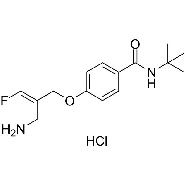
PXS-4728A is an orally active, selective VAP-1/SSAO inhibitor with an IC50 of 5 nM and a Ki of 175 nM. PXS-4728A inhibits the expression of MMP-9. It reduces cell rolling, adhesion and migration, decreases cell infiltration levels, pro-inflammatory cytokines, chemokines and collagen deposition, and improves pulmonary function. PXS-4728A reduces the formation and progression of atherosclerotic plaques, and decreases blood lipid and blood glucose levels as well as body weight gain. PXS-4728A can be used in research related to chronic obstructive pulmonary disease, cystic fibrosis, acute pulmonary inflammation, acute lung injury, bronchiolitis obliterans syndrome, rhinovirus-exacerbated asthma, sepsis, Klebsiella pneumoniae pulmonary infection and atherosclerosis.
For research use only. We do not sell to patients.
- Purity : 98.79%
- CAS No.: 1478364-68-9
- Formula: C15H22ClFN2O2
- Molecular Weight:316.80
-
Storage:
4°C, sealed storage, away from moisture
* In solvent : -80°C, 6 months; -20°C, 1 month (sealed storage, away from moisture)

To place orders, for customer services and technical support, please contact: MedChemExpress USA
Tel: 609-228-6898 E-mail: [email protected] [email protected]


-
 Biological Activity
-
Chemical Information
-
Solvent & Solubility
- Protocols
- Purity & Documentation
- References
-
 Help & FAQs


-
HY-14197A
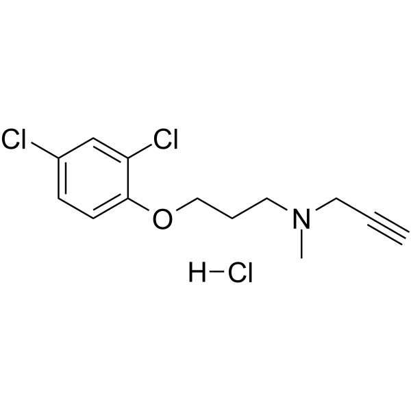
Clorgyline hydrochlorideMAO-A Inhibitor -

HY-Y0882
Hydroxylamine hydrochlorideMAO Inhibitor -
HY-A0091A
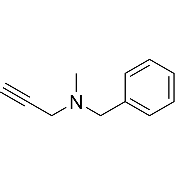
PargylineMonoamine Oxidase Inhibitor -
HY-70057
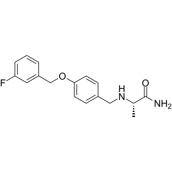
SafinamideMAO-B Inhibitor -
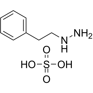
HY-B1018A
Phenelzine sulfateMAO Inhibitor -
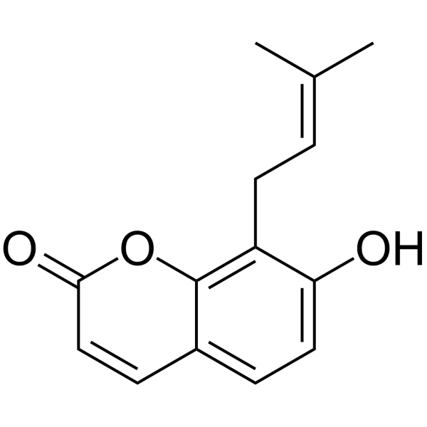
HY-N2554
OsthenolhMAO-A Inhibitor -
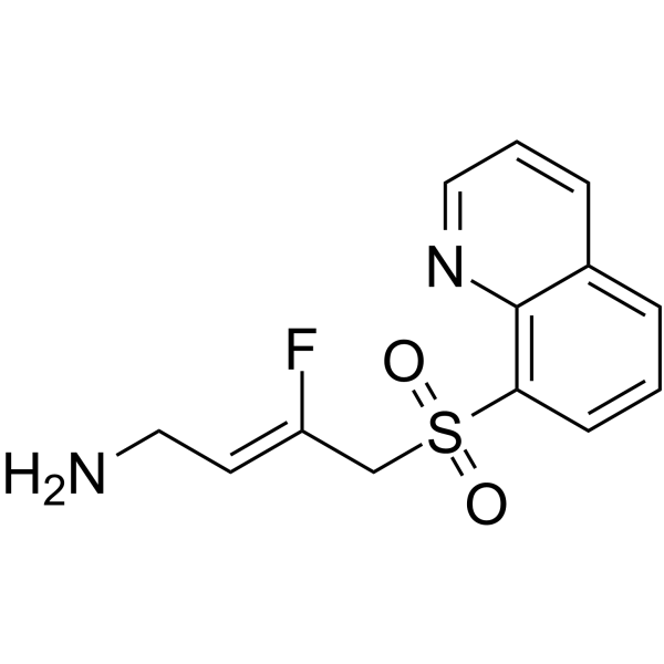
HY-138625
PXS-5505 free baseLysyl Oxidase Inhibitor -
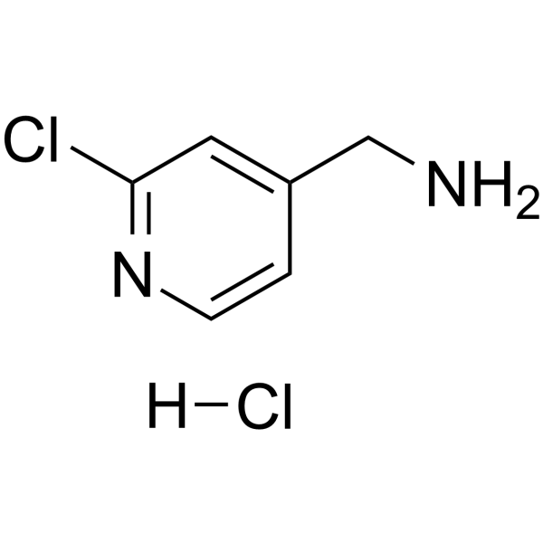
HY-101771A
(2-Chloropyridin-4-yl)methanamine hydrochlorideLOXL2 Inhibitor -
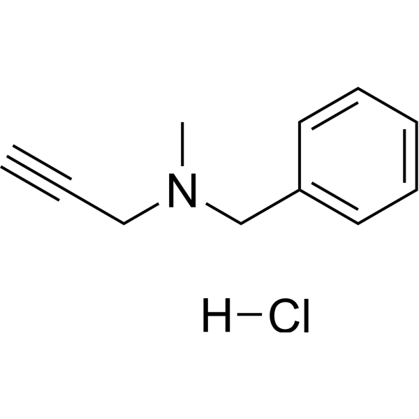
HY-A0091
Pargyline hydrochlorideMonoamine Oxidase Inhibitor -
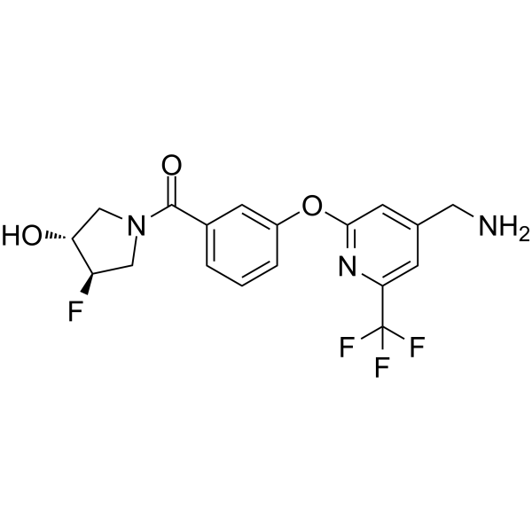
HY-107422
LenumlostatLOXL2 Inhibitor -
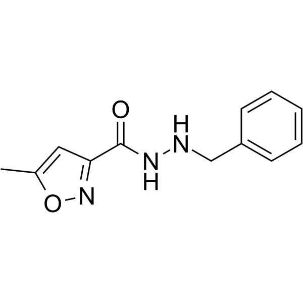
HY-13929
IsocarboxazidMonoamine Oxidase Inhibitor -
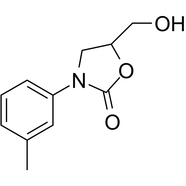
HY-14196
ToloxatoneMAO-A Inhibitor
Publications Citing Use of MedChemExpress (MCE) PXS-4728A
More

Biological Activity
Description
IC50 & Target
|
MMP-9 |
In Vitro
PXS-4728A potently inhibits recombinant human VAP-1/SSAO with an IC50 of 5 nM and acts as a mechanism-based inhibitor with a Ki of 175 nM and Kinact of 0.68 min-1[1].
PXS-4728A shows >500-fold selectivity for VAP-1/SSAO over all tested related human amine oxidases, with IC50 values ranging from 2.7 μM to >100 μM for off-target enzymes[1].
PXS-4728A potently inhibits VAP-1/SSAO activity in gonadal fat tissue homogenates from mice, rats, dogs, and rabbits with an IC50 of <10 nM[1].
PXS-4728A (100 μM) does not induce cell toxicity or phospholipidosis in HepG2 cells at concentrations up to 100 μM[1].
MedChemExpress (MCE) has not independently confirmed the accuracy of these methods. They are for reference only. Further protocols information, click here.
In Vivo
PXS-4728A (0.2-2 mg/kg; p.o.; single dose) dose-dependently reduces LPS-induced neutrophilic acute lung inflammation in BALB/c mice, with 2 mg/kg achieving a near-complete reduction in BALF neutrophils[1].
PXS-4728A (2 mg/kg; i.p.; single dose) reduces neutrophilic inflammation and airway hyperreactivity in BALB/c mice with rhinovirus-exacerbated allergic asthma[1].
PXS-4728A (2-20 mg/kg; p.o.; daily; 4 days) potently inhibits lung and peripheral SSAO activity, reduces airway inflammatory cell influx, and lowers pro-inflammatory mediator levels in mice with acute cigarette smoke-induced COPD-like inflammation[2].
PXS-4728A (1-10 mg/kg/day; p.o.; daily; 4-12 weeks) reduces atherosclerotic plaque area by 37.59% in cholesterol-fed New Zealand White rabbits, alongside inhibiting SSAO activity, lowering plasma lipids and glucose, and suppressing inflammatory, macrophage, and smooth muscle cell-mediated atherogenic pathways[4].
MedChemExpress (MCE) has not independently confirmed the accuracy of these methods. They are for reference only.
-
Animal Model:C57BL/6 (female, 6-8 weeks old, acute cigarette smoke-induced model)[2]
-
Dosage:2 mg/kg; 6 mg/kg; 12 mg/kg; 20 mg/kg
-
Administration:p.o.; daily; 4 days
-
Result:Completely inhibited constitutive lung SSAO activity at 2, 6, and 20 mg/kg.
Completely inhibited SSAO activity in inguinal fat at 20 mg/kg.
Reduced total leukocyte numbers in bronchoalveolar lavage fluid (BALF) at 12 and 20 mg/kg.
Reduced macrophage and lymphocyte counts in BALF at 12 mg/kg.
Suppressed neutrophil counts in BALF at 20 mg/kg.
Reduced BALF CXCL1 levels at 12 and 20 mg/kg.
Reduced BALF TNFα levels back to baseline at 20 mg/kg.
Reduced BALF TNFα levels in a dose-dependent manner at 12 mg/kg.
-
Animal Model:New Zealand White rabbits (male, 2 kg weight, atherosclerosis model via 0.5% cholesterol-enriched diet for 12 weeks)[4]
-
Dosage:1 mg/kg/day (dose-finding, 4 days); 10 mg/kg/day (dose-finding, 4 days); 10 mg/kg/day (12 weeks)
-
Administration:p.o.; daily; 4 consecutive days (dose-finding); p.o.; daily; 12 weeks
-
Result:Inhibited SSAO activity by 80% at 1 mg/kg/day for 4 days in abdominal fat.
Inhibited SSAO activity by >90% at 10 mg/kg/day for 4 days in abdominal fat.
Significantly inhibited SSAO-specific hydrogen peroxide production in thoracic aorta, lung, and epididymal fat at 10 mg/kg/day for 12 weeks relative to cholesterol-fed controls.
Reduced body weight to 3.28 kg vs. 3.58 kg in cholesterol-fed controls.
Reduced plasma total cholesterol to 508.4 mg/dl vs. 695.9 mg/dl in cholesterol-fed controls.
Reduced LDL-cholesterol to 403.2 mg/dl vs. 576.8 mg/dl in cholesterol-fed controls.
Reduced glucose to 195.3 mg/dl vs. 258.7 mg/dl in cholesterol-fed controls.
Reduced AST to 209.2 U/l vs. 502.8 U/l in cholesterol-fed controls.
Reduced atherosclerotic plaque area by 37.59% (30.72% of vessel area vs. 68.31% in cholesterol-fed controls).
Significantly decreased intima/media area ratio to 0.29 vs. 0.91 in cholesterol-fed controls.
Reduced aortic wall thickness to 1.22-fold of control vs. 2.12-fold in cholesterol-fed controls.
Significantly reduced aortic expression of adhesion molecules (ICAM-1, VCAM-1, E-selectin), pro-inflammatory cytokines (COX-2, IL-6, MCP-1), macrophage activation markers (RAGE, LOX-1, CD36, TLR-4), macrophage recruitment, MMP-9 expression, smooth muscle cell accumulation in intima, and PCNA-positive proliferating cells relative to cholesterol-fed controls.
Clinical Trial
| NCT Number | Sponsor | Condition | Start Date |
Phase

|
|---|---|---|---|---|
| NCT01329991 | Plexxikon| | 2011-05 | PHASE1 |


Chemical Information
-
CAS No. 1478364-68-9
-
Appearance Solid
-
Molecular Weight 316.80
-
Formula C15H22ClFN2O2
-
Color White to yellow
-
SMILES
O=C(NC(C)(C)C)C1=CC=C(OC/C(CN)=C/F)C=C1.Cl
-
Synonyms
BI-1467335
-
Shipping
Room temperature in continental US; may vary elsewhere.
-
Storage
4°C, sealed storage, away from moisture
* In solvent : -80°C, 6 months; -20°C, 1 month (sealed storage, away from moisture)


Publications (3)
-
Journal Impact Factor
-
Most Recent
-
Nat Commun
Rational design of potent small-molecule SMARCA2/A4 degraders acting via the recruitment of FBXO22. [Abstract]2025 Nov 3;16(1):9679. PMID: 41184243 -
Exp Mol Med
2026 Apr;58(4):1284-1296. PMID: 42009953 -


Solvent & Solubility
In Vitro:
DMSO : 233.33 mg/mL (736.52 mM; Need ultrasonic; Hygroscopic DMSO has a significant impact on the solubility of product, please use newly opened DMSO)
H2O : 100 mg/mL (315.66 mM; Need ultrasonic)
Please refer to the solubility information to select the appropriate solvent. Once prepared, please aliquot and store the solution to prevent product inactivation from repeated freeze-thaw cycles.
Storage method and period of stock solution: -80°C, 6 months; -20°C, 1 month (sealed storage, away from moisture). When stored at -80°C, please use it within 6 months. When stored at -20°C, please use it within 1 month.
* Note: If you choose water as the stock solution, please dilute it to the working solution, then filter and sterilize it with a 0.22 μm filter before use.
Please refer to the solubility information to select the appropriate solvent. Once prepared, please aliquot and store the solution to prevent product inactivation from repeated freeze-thaw cycles.
Storage method and period of stock solution: -80°C, 6 months; -20°C, 1 month (sealed storage, away from moisture). When stored at -80°C, please use it within 6 months. When stored at -20°C, please use it within 1 month.
* Note: If you choose water as the stock solution, please dilute it to the working solution, then filter and sterilize it with a 0.22 μm filter before use.
Concentration (start) × Volume (start) = Concentration (final) × Volume (final)
In Vivo:
Select the appropriate dissolution method based on your experimental animal and administration route.
- For the following dissolution methods, please ensure to first prepare a clear stock solution using an In Vitro approach and then sequentially add co-solvents:
- To ensure reliable experimental results, the clarified stock solution can be appropriately stored based on storage conditions. As for the working solution for In Vivo experiments, it is recommended to prepare freshly and use it on the same day.
- The percentages shown for the solvents indicate their volumetric ratio in the final prepared solution. If precipitation or phase separation occurs during preparation, heat and/or sonication can be used to aid dissolution.
Add each solvent one by one: 10% DMSO 40% PEG300 5% Tween-80 45% Saline
Solubility: ≥ 2.08 mg/mL (6.57 mM); Clear solution
This protocol yields a clear solution of ≥ 2.08 mg/mL (saturation unknown).
Taking 1 mL working solution as an example, add 100 μL DMSO stock solution (20.8 mg/mL) to 400 μL PEG300, and mix evenly; then add 50 μL Tween-80 and mix evenly; then add 450 μL Saline to adjust the volume to 1 mL.
Preparation of Saline: Dissolve 0.9 g sodium chloride in ddH₂O and dilute to 100 mL to obtain a clear Saline solution.
Add each solvent one by one: 10% DMSO 90% (20% SBE-β-CD in Saline)
Solubility: ≥ 2.08 mg/mL (6.57 mM); Clear solution
This protocol yields a clear solution of ≥ 2.08 mg/mL (saturation unknown).
Taking 1 mL working solution as an example, add 100 μL DMSO stock solution (20.8 mg/mL) to 900 μL 20% SBE-β-CD in Saline, and mix evenly.
Preparation of 20% SBE-β-CD in Saline (4°C, storage for one week): 2 g SBE-β-CD powder is dissolved in 10 mL Saline, completely dissolve until clear.
For the following dissolution methods, please prepare the working solution directly:
It is recommended to prepare fresh solutions and use them promptly within a short period of time.
The percentages shown for the solvents indicate their volumetric ratio in the final prepared solution. If precipitation or phase separation occurs during preparation, heat and/or sonication can be used to aid dissolution.
Add each solvent one by one: PBS
Solubility: 25 mg/mL (78.91 mM); Clear solution; Need ultrasonic and warming
In Vivo Dissolution Calculator
Please enter the basic information of animal experiments:
-
-
-
-
Recommended: Prepare an additional quantity of animals to account for potential losses during experiments.
Working solution concentration: 0.22 mg/mL
This product has good water solubility, please refer to the measured solubility data in water/PBS/Saline for details.

Protocols
-
RNA extraction experimental
By lysing cells, releasing RNA, and removing impurities such as proteins and DNA, high-purity RNA products are finally obtained. The commonly used traditional method is the guanidine isothiocyanate/phenol/chloroform method (Trizol), which is suitable for a variety of animal materials including animal tissues, microorganisms, cultured cells, etc., and most plant materials.
-
Collagen: Sirius Red Staining
Sirius Red or picrosirius red staining is a histochemical method for visualizing collagen-rich extracellular matrix in tissue sections, and collagen fibers are detected as red-stained structures under bright-field microscopy with enhanced birefringence under polarized light. Picrosirius red is useful for assessing total collagen organization, distribution, and fibrosis burden, but polarized color should not be interpreted as a definitive collagen type I versus type III readout because color is affected by fiber orientation, thickness, and packing.
-
Research Protocol for Infectious Diseases
Infectious-disease experiments test how pathogens interact with host barriers, innate immune receptors, inflammatory signaling, pathogen replication, and tissue injury; pattern-recognition receptors such as TLRs, RIG-I-like receptors, NOD-like receptors, and inflammasomes detect microbial molecules and activate NF-κB, interferon, and cytokine responses. The central hypothesis is that infection severity reflects the balance between pathogen burden and host response: protective inflammation restricts pathogen growth, whereas excessive or mislocalized inflammation contributes to tissue damage and disease phenotype. Unresolved questions include which host pathways are protective versus pathogenic, why some infection models fail to translate to human disease, and which combined readouts best predict clinically relevant infection outcomes.
-
Mitochondrial membrane-potential fluorescent assay
Mitochondrial membrane potential fluorescent assays estimate ΔΨm in living cells using lipophilic cationic dyes such as TMRM, TMRE, rhodamine 123, and JC-1, which accumulate in mitochondria according to membrane polarization; loss of signal after FCCP or CCCP treatment is interpreted as mitochondrial depolarization. TMRM/TMRE and rhodamine 123 are commonly used for semi-quantitative live-cell microscopy or flow cytometry, while JC-1 can report a shift from red aggregate fluorescence to green monomer fluorescence during depolarization; interpretation requires controls because dye concentration, quenching mode, cell type, dye efflux, and mitochondrial mass can affect fluorescence independently of ΔΨm.
-
Cell migration
Cell migration is a method that plays an important role in wound healing, cell differentiation, embryonic development, etc.
-
Connective Tissue: Masson's Trichrome/Collagen Trichrome Staining
Masson’s Trichrome (collagen/trichrome staining) is a histological technique that differentially stains tissue compartments using sequential acidic dyes to distinguish collagen from muscle and cytoplasmic components based on dye affinity and tissue permeability differences, enabling visualization of fibrosis and connective tissue architecture in histological sections. The classical formulation typically uses Weigert's iron hematoxylin for nuclear staining, Biebrich scarlet-acid fuchsin for cytoplasm and muscle, and aniline blue (or light green variants) for collagen, producing a characteristic blue/green collagen signal contrasted against red cytoplasm and dark nuclei. The staining principle relies on selective displacement of smaller dye molecules by larger anionic dyes in collagen-rich regions under controlled acidified conditions, which enhances collagen-specific dye retention. This property makes the method widely used for fibrosis assessment in organs such as heart, liver, lung, a
-
Research Protocol for Cardiovascular Diseases
Cardiovascular disease can be modeled as maladaptive cardiac remodeling, where ischemic injury or pressure overload activates inflammatory signaling, fibroblast activation, extracellular-matrix deposition, cardiomyocyte hypertrophy, vascular remodeling, and progressive ventricular dysfunction. The TGF-β/SMAD axis is a central profibrotic pathway after myocardial injury and pressure overload, while innate immune and cytokine pathways regulate leukocyte recruitment, scar formation, and adverse remodeling. Key unresolved questions include which inflammatory signals are reparative versus harmful, when fibrosis is protective versus maladaptive, and whether pathway inhibition improves function without weakening necessary infarct healing or compensatory remodeling.
-
Research Protocol for Inflammation-related Diseases
The NLRP3 inflammasome is a cytosolic innate immune signaling platform that integrates priming signals and danger-signal activation to promote caspase-1 activation, maturation of IL-1β and IL-18, and gasdermin D-mediated pyroptotic cell death. The core experimental logic is to determine whether inflammatory disease phenotypes are driven by increased NLRP3 expression, ASC-containing inflammasome assembly, caspase-1 cleavage, GSDMD cleavage, and extracellular release of IL-1β/IL-18 rather than by nonspecific cell injury alone. The pathway is strongly linked to inflammation-related disease phenotypes because monosodium urate crystals activate NALP3/NLRP3 inflammasome signaling in gout-like crystal inflammation, cholesterol crystals activate NLRP3 inflammasomes in atherogenesis models, and DSS-induced intestinal inflammation has been reported to involve NLRP3 inflammasome activity. However, experimental colitis studies also show context-dependent protective effects of NLRP3 inflammasome co
-
Fibrosis/Collagen Morphometry
Fibrosis and collagen morphometry is based on the quantitative visualization of fibrillar collagen deposition in tissue sections using histochemical stains such as Sirius Red (Picrosirius Red) or Masson's trichrome, followed by image-based or polarization-enhanced analysis to estimate collagen proportional area as a surrogate of extracellular matrix accumulation during fibrotic remodeling. Sirius Red combined with polarized light microscopy enhances detection of collagen fibers due to birefringence properties, enabling more specific visualization of collagen type I and III fibrils compared to conventional bright-field histology, while whole-section or region-restricted digital morphometry reduces field-selection bias in fibrosis assessment. Alternative quantitative approaches include second harmonic generation (SHG) and two-photon excited fluorescence microscopy, which enable label-free detection of fibrillar collagen and have been validated against histological staining and biochemica
-
Cell Cytotoxicity Assay
Cytotoxicity assays are usually based on the assessment of cell membrane damage, which can also be indirectly detected by measuring cell viability. Detection methods include MTT assay, CKK-8 assay, LDH assay and ATP assay, etc.
-
LPS-Induced Endotoxemia/Systemic Inflammation
Lipopolysaccharide (LPS)-induced endotoxemia is a widely used in vivo model of acute systemic inflammation in which LPS, a Gram-negative bacterial endotoxin, activates innate immune signaling primarily through TLR4, leading to rapid and transient induction of pro-inflammatory cytokines such as TNF-α, IL-6, and IL-1β in circulation and tissues. This cytokine surge is commonly used as a measurable readout of systemic inflammatory activation and immune dysregulation, and is typically assessed within hours after intraperitoneal LPS administration in mouse models of endotoxemia. The model captures key features of systemic inflammatory response syndrome, including cytokine release, immune cell activation, and downstream tissue responses, and has been used to evaluate anti-inflammatory interventions such as cytokine modulation, lipid mediators, and immune cell-targeting therapies.
-
Mitochondrial membrane-potential and mitochondrial mass staining
Mitochondrial membrane potential staining measures the electrochemical polarization across the mitochondrial inner membrane in live cells using lipophilic cationic fluorescent probes; early rhodamine-based work showed that selective mitochondrial dye accumulation is lost when the mitochondrial transmembrane potential is dissipated. JC-1 reports mitochondrial polarization by shifting from green monomer fluorescence to red J-aggregate fluorescence as dye concentration increases within energized mitochondria; therefore, the red/green fluorescence ratio is used as a relative readout of mitochondrial membrane potential. TMRE or TMRM staining provides a single-channel relative readout because these cationic rhodamine esters accumulate in polarized mitochondria, and lower fluorescence indicates reduced mitochondrial polarization when acquisition and dye-loading conditions are controlled. Mitochondrial mass staining is commonly performed with MitoTracker Green FM or related MitoTracker dyes as
-
Inhalation Toxicity Study
Inhalation toxicity studies expose rodents to a controlled aerosol, vapor, gas, or smoke atmosphere and assess respiratory and systemic toxicity using exposure-atmosphere characterization, clinical observations, body and organ weights, bronchoalveolar lavage fluid, histopathology, blood chemistry, hematology, and, when included, molecular endpoints such as transcriptomics, proteomics, lipidomics, or tissue burden analysis. The primary biological readouts are airway irritation, pulmonary inflammation, cytotoxicity, altered surfactant or lipid homeostasis, impaired particle clearance, and tissue remodeling, reflected by BALF cell differentials, BALF protein, LDH, phosphatase activities, cytokines, lung weight, microscopic respiratory-tract lesions, and retained lung burden.


Purity & Documentation
-
Data Sheet (288 KB)
-
SDS (557 KB)
- English - EN (557 KB)
- Français - FR (557 KB)
- Deutsch - DE (557 KB)
- Norwegian - NO (557 KB)
- Español - ES (557 KB)
- Swedish - SV (557 KB)
- Italian - IT (557 KB)
- Korean - KR (557 KB)
- Portuguese - PT (557 KB)
-
Handling Instructions (2659 KB)
References
[1]. Schilter HC, et al. Effects of an anti-inflammatory VAP-1/SSAO inhibitor, PXS-4728A, on pulmonary neutrophil migration. Respiratory research. 2015 Mar 20;16(1):42. [Content Brief]
[2]. Jarnicki AG, et al. The inhibitor of semicarbazide-sensitive amine oxidase, PXS-4728A, ameliorates key features of chronic obstructive pulmonary disease in a mouse model. British journal of pharmacology. 2016 Nov;173(22):3161-3175. [Content Brief]
[3]. Li H, et al. Vascular Adhesion Protein-1 (VAP-1)/Semicarbazide-Sensitive Amine Oxidase (SSAO): A Potential Therapeutic Target for Atherosclerotic Cardiovascular Diseases. Frontiers in pharmacology. 2021;12:679707. [Content Brief]
[4]. Wang SH, et al. Inhibition of Semicarbazide-sensitive Amine Oxidase Reduces Atherosclerosis in Cholesterol-fed New Zealand White Rabbits. Scientific reports. 2018 Jun 18;8(1):9249. [Content Brief]


Complete Stock Solution Preparation Table
Please refer to the solubility information to select the appropriate solvent. Once prepared, please aliquot and store the solution to prevent product inactivation from repeated freeze-thaw cycles.
Storage method and period of stock solution: -80°C, 6 months; -20°C, 1 month (sealed storage, away from moisture). When stored at -80°C, please use it within 6 months. When stored at -20°C, please use it within 1 month.
| Optional Solvent | Concentration Solvent Mass | 1 mg | 5 mg | 10 mg | 25 mg |
|---|---|---|---|---|---|
| H2O / DMSO | 1 mM | 3.1566 mL | 15.7828 mL | 31.5657 mL | 78.9141 mL |
| 5 mM | 0.6313 mL | 3.1566 mL | 6.3131 mL | 15.7828 mL | |
| 10 mM | 0.3157 mL | 1.5783 mL | 3.1566 mL | 7.8914 mL | |
| 15 mM | 0.2104 mL | 1.0522 mL | 2.1044 mL | 5.2609 mL | |
| 20 mM | 0.1578 mL | 0.7891 mL | 1.5783 mL | 3.9457 mL | |
| 25 mM | 0.1263 mL | 0.6313 mL | 1.2626 mL | 3.1566 mL | |
| 30 mM | 0.1052 mL | 0.5261 mL | 1.0522 mL | 2.6305 mL | |
| 40 mM | 0.0789 mL | 0.3946 mL | 0.7891 mL | 1.9729 mL | |
| 50 mM | 0.0631 mL | 0.3157 mL | 0.6313 mL | 1.5783 mL | |
| 60 mM | 0.0526 mL | 0.2630 mL | 0.5261 mL | 1.3152 mL | |
| 80 mM | 0.0395 mL | 0.1973 mL | 0.3946 mL | 0.9864 mL | |
| 100 mM | 0.0316 mL | 0.1578 mL | 0.3157 mL | 0.7891 mL |
* Note: If you choose water as the stock solution, please dilute it to the working solution, then filter and sterilize it with a 0.22 μm filter before use.


PXS-4728A Related Classifications
HY-112726 Related Classifications
 Powered by Bioz