



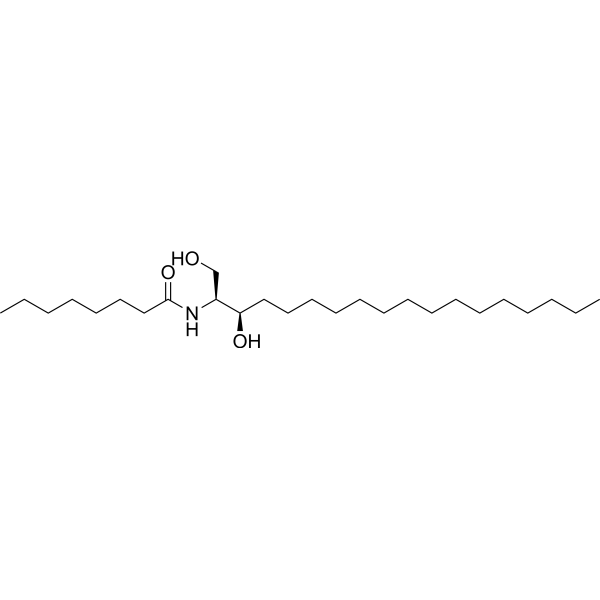
C8 Dihydroceramide

Based on 1 Customer Validation
C8 Dihydroceramide is a negative control of C8 Ceramide. C8-Ceramide (N-Octanoyl-D-erythro-sphingosine) is a cell-permeable analog of naturally occurring ceramides. C8-Ceramide has anti-proliferation properties and acts as a potent chemotherapeutic agent. C8-Ceramide stimulates dendritic cells to promote T cell responses upon virus infections. C8-Ceramide induces slight activation of protein kinase (PKC) in vitro.
商品は「研究用試薬」です。人や動物の医療用・臨床診断用・食品用の製品ではありません。
研究用途以外に使用した場合、当社は一切の責任を負いかねます。
- 純度 : 99.77%
- CAS 番号: 145774-33-0
- 分子式: C26H53NO3
- 分子量:427.70
-
保管条件:Powder -20°C, 3 years ; In solvent -80°C, 6 months , -20°C, 1 month

To place orders, for customer services and technical support, please contact: MedChemExpress USA
Tel: 609-228-6898 E-mail: [email protected] [email protected]


-
 生物活性
生物活性
-
化学情報
- プロトコル
- 純度とドキュメンテーション
- 参考文献
-
 Help & FAQs




生物活性
製品説明
体外実験
C8-dihydro-ceramide had no effect on cell viability in any of the cell lines tested[5].
MedChemExpress (MCE) has not independently confirmed the accuracy of these methods. They are for reference only. Further protocols information, click here.


化学情報
-
CAS 番号 145774-33-0
-
性状 Solid
-
分子量 427.70
-
分子式 C26H53NO3
-
Color White to off-white
-
SMILES
O=C(N[C@H]([C@@H](CCCCCCCCCCCCCCC)O)CO)CCCCCCC
-
輸送条件
Room temperature in continental US; may vary elsewhere.
-
保管条件
Powder -20°C 3 years In solvent -80°C 6 months -20°C 1 month

プロトコル
-
Monocyte-derived dendritic cell differentiation
Human monocyte-derived dendritic cells are generated by isolating PBMC-derived monocytes and culturing them with GM-CSF plus IL-4, which produces cells with dendritic-cell antigen-presenting properties, reduced monocyte phenotype, and increased dendritic-cell functional readouts such as antigen uptake, allogeneic T-cell stimulation, and expression of markers including HLA-DR, CD80, CD86, CD83, CD1a, or CD209 depending on protocol and maturation state. The main readout is phenotypic and functional differentiation: immature MoDCs are commonly evaluated by loss or reduction of CD14 with acquisition of dendritic-cell markers and antigen uptake capacity, whereas mature MoDCs are evaluated by increased CD83, CD80, CD86, HLA-DR, and T-cell stimulatory function after exposure to maturation stimuli such as TNF-α or a cytokine/PGE2 cocktail.
-
Research Protocol for Cancer Immunology
Cancer immunology studies how the immune system recognizes, suppresses, edits, or fails to eliminate malignant cells through tumor antigen release, antigen presentation, T-cell priming, immune trafficking, tumor-cell killing, and feedback inhibition in the tumor microenvironment. The cancer-immunity cycle links tumor antigenicity, dendritic-cell priming, CD8+ T-cell infiltration, cytotoxic function, and immune-checkpoint regulation to tumor rejection or immune escape. Immune-checkpoint pathways such as PD-1/PD-L1 and CTLA-4 suppress antitumor T-cell activity and can be therapeutically blocked, but many tumors remain resistant because of poor antigen presentation, weak T-cell infiltration, suppressive myeloid cells, regulatory T cells, and tumor-intrinsic immune-exclusion programs. Unresolved questions include which immune-cell states predict response, how tumor-intrinsic pathways exclude immune cells, how myeloid suppression limits checkpoint blockade, and which combination strategies
-
Cell Viability Determination by MTT Colorimetric Assay
The following protocol uses the MTT colorimetric assay as a classic literature-established method for assessing cell viability/metabolic activity in cultured mammalian cells. MTT[3-(4,5-dimethylthiazol-2-yl)-2,5-diphenyltetrazolium bromide] is reduced by metabolically active cells to a colored formazan product; the amount of formazan is quantified spectrophotometrically and provides an indirect measure of metabolically active viable cells. Importantly, MTT reduction reflects cellular oxidoreductase/metabolic activity rather than an absolute direct count of living cells, so changes in cellular metabolism can alter the signal independently of cell number.


純度とドキュメンテーション
-
データシート (262 KB)
-
SDS (252 KB)
- English - EN (252 KB)
- Français - FR (252 KB)
- Deutsch - DE (252 KB)
- Norwegian - NO (252 KB)
- Español - ES (252 KB)
- Swedish - SV (252 KB)
- Italian - IT (252 KB)
- Korean - KR (252 KB)
- Portuguese - PT (252 KB)
-
取扱説明書 (2659 KB)
参考文献
[1]. Rebeca López-Marure , et al. Ceramide promotes the death of human cervical tumor cells in the absence of biochemical and morphological markers of apoptosis. Biochem Biophys Res Commun. 2002 May 10;293(3):1028-36. [Content Brief]
[2]. Yuli C. Chang, et al. Exogenous C8-Ceramide Induces Apoptosis by Overproduction of ROS and the Switch of Superoxide Dismutases SOD1 to SOD2 in Human Lung Cancer Cells. Int J Mol Sci. 2018 Oct; 19(10): 3010. [Content Brief]
[3]. H W Huang, et al. Ceramides modulate protein kinase C activity and perturb the structure of Phosphatidylcholine/Phosphatidylserine bilayers. Biophys J. 1999 Sep; 77(3): 1489-1497. [Content Brief]
[4]. Lan Weiss, et al. Ceramide contributes to pathogenesis and may be targeted for therapy in VCP inclusion body myopathy. Hum Mol Genet. 2021 Jan 7;ddaa248. [Content Brief]
[5]. Rebeca López-Marure, et al. Ceramide promotes the death of human cervical tumor cells in the absence of biochemical and morphological markers of apoptosis. Biochem Biophys Res Commun. 2002 May 10;293(3):1028-36. [Content Brief]

Calculators
濃度 (開始) × 体積 (開始) = 濃度 (終了) × 体積 (終了)


C8 Dihydroceramide 関連分類
HY-119312 関連分類
 Powered by Bioz