



DC-U4106
Based on 1 publication(s) in Google Scholar
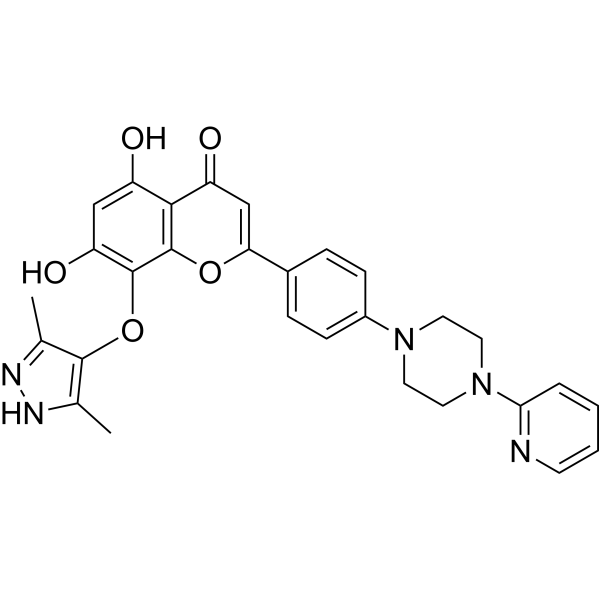
DC-U4106 is a USP8 targeting inhibitor with the Kdvalue of 4.7 μM and the IC50 value of 1.2 μM. DC-U4106 can target the ubiquitin pathway and facilitate the degradation of Erα. DC-U4106 inhibits tumor growth with minimal toxicity and has the potential for the research of breast cancer.
商品は「研究用試薬」です。人や動物の医療用・臨床診断用・食品用の製品ではありません。
研究用途以外に使用した場合、当社は一切の責任を負いかねます。
- 純度 : 98.09%
- CAS 番号: 2410534-62-0
- 分子式: C29H27N5O5
- 分子量:525.56
-
保管条件:
4°C, sealed storage, away from moisture and light
* In solvent : -80°C, 6 months; -20°C, 1 month (sealed storage, away from moisture and light)

To place orders, for customer services and technical support, please contact: MedChemExpress USA
Tel: 609-228-6898 E-mail: [email protected] [email protected]


-
 生物活性
生物活性
-
化学情報
-
溶剤 & 溶解度
- プロトコル
- 純度とドキュメンテーション
- 参考文献
-
 Help & FAQs


-
HY-13989
b-AP15UCHL5/Usp14 Inhibitor -
HY-151563
OTUB1/USP8-IN-1OTUB1/USP8 Inhibitor -
HY-162312
LLK203USP2/USP8 Inhibitor -
HY-145967
FT709USP9X Inhibitor -
HY-101666
HBX 41108USP7 Inhibitor -
HY-18637
LDN-57444Deubiquitinase Inhibitor -
HY-112937
GNE-6640USP7 Inhibitor -
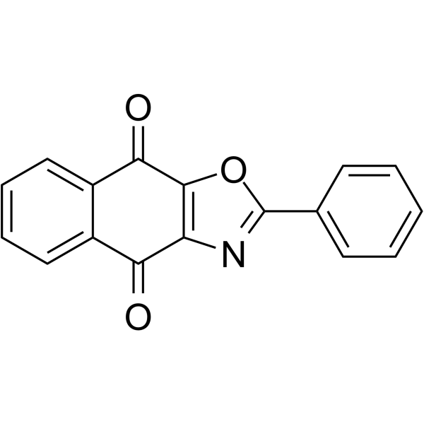
HY-15757
SJB2-043Deubiquitinase Inhibitor -
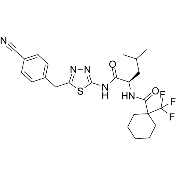
HY-153045
BAY-805USP21 Inhibitor -
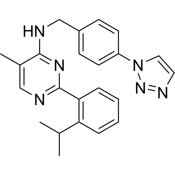
HY-17543
ML-323USP1-UAF1 Inhibitor -

HY-18638
TCIDUCH-L3 Inhibitor -
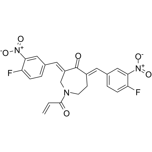
HY-12471
VLX1570Proteasome DUB Inhibitor
MedChemExpress(MCE)の使用を引用している文献 DC-U4106
More

生物活性
製品説明
IC50 & Target
Kd: 4.7 μM (USP8), IC50: 1.2 μM (USP8)[1].
体外実験
DC-U4106 (1.2-45.2 μM) inhibits USP8 and USP2 with the IC50 values of 1.2 μM and 58.4 μM, respectively, and no activity in USP7[1]. DC-U4106 (0-7 μM, 24 hours) reduces mRNA levels of ERα and PR[1]. DC-U4106 (0-5 μM, 24 hours) can regulate the RTK pathway related proteins and the expression of ERα and PR proteins[1]. DC-U4106 (0-5 μM, 12 hours) can induce apoptosis and inhibit cell proliferation[1].
MedChemExpress (MCE) has not independently confirmed the accuracy of these methods. They are for reference only. Further protocols information, click here.
-
Cell Line:USP8-positive cell line MCF-7
-
Concentration:0-5 μM
-
Incubation Time:24 hours
-
Result:Reduced the expression of EGFR, ErbB2, and ErbB3 proteins with increasing concentrations and caused degradation of ERα and PR proteins.
-
Cell Line:USP8-positive cell line MCF-7
-
Concentration:0-7 μM
-
Incubation Time:24 hours
-
Result:Reduced mRNA levels of ERα and PR.
-
Cell Line:USP8-positive cell line MCF-7
-
Concentration:0-5 μM
-
Incubation Time:
-
Result:Inhibited cell growth in a dose-dependent manner.
-
Cell Line:USP8-positive cell line MCF-7
-
Concentration:0-5 μM
-
Incubation Time:12 hours
-
Result:Resulted in increasing in the proportion of apoptotic cells with increasing concentrations.
体内実験
MedChemExpress (MCE) has not independently confirmed the accuracy of these methods. They are for reference only.
-
Animal Model:BALB/c nude mice[1]
-
Dosage:5 mg/kg , 20 mg/kg
-
Administration:Intraperitoneal injection, Every 2 days, 14 days
-
Result:Inhibited tumor growth significantly at a concentration of 20 mg/kg.


化学情報
-
CAS 番号 2410534-62-0
-
性状 Solid
-
分子量 525.56
-
分子式 C29H27N5O5
-
Color Yellow to brown
-
SMILES
O=C1C=C(C2=CC=C(N3CCN(C4=NC=CC=C4)CC3)C=C2)OC5=C1C(O)=CC(O)=C5OC6=C(C)NN=C6C
-
輸送条件
Room temperature in continental US; may vary elsewhere.
-
保管条件
4°C, sealed storage, away from moisture and light
* In solvent : -80°C, 6 months; -20°C, 1 month (sealed storage, away from moisture and light)


Publications (1)
-
Journal Impact Factor
-
Most Recent


溶剤 & 溶解度
体外:
DMSO : 25 mg/mL (47.57 mM; ultrasonic and warming and heat to 60°C; Hygroscopic DMSO has a significant impact on the solubility of product, please use newly opened DMSO)
THF : 10 mg/mL (19.03 mM; Need ultrasonic)
Please refer to the solubility information to select the appropriate solvent. Once prepared, please aliquot and store the solution to prevent product inactivation from repeated freeze-thaw cycles.
Storage method and period of stock solution: -80°C, 6 months; -20°C, 1 month (sealed storage, away from moisture and light). When stored at -80°C, please use it within 6 months. When stored at -20°C, please use it within 1 month.
Please refer to the solubility information to select the appropriate solvent. Once prepared, please aliquot and store the solution to prevent product inactivation from repeated freeze-thaw cycles.
Storage method and period of stock solution: -80°C, 6 months; -20°C, 1 month (sealed storage, away from moisture and light). When stored at -80°C, please use it within 6 months. When stored at -20°C, please use it within 1 month.
濃度 (開始) × 体積 (開始) = 濃度 (終了) × 体積 (終了)

プロトコル
-
Cell Cytotoxicity Assay
Cytotoxicity assays are usually based on the assessment of cell membrane damage, which can also be indirectly detected by measuring cell viability. Detection methods include MTT assay, CKK-8 assay, LDH assay and ATP assay, etc.
-
Patient-Derived Xenograft (PDX)
Patient-derived xenograft (PDX) models are generated by engrafting primary human tumor tissue directly into immunodeficient mice, allowing in vivo propagation of patient tumor biology without initial in vitro adaptation. These models are used to preserve key histopathological and molecular characteristics of the original tumor and enable assessment of tumor growth dynamics and therapeutic response in a living organism. The biological readout is tumor engraftment and subsequent growth in the murine host, which reflects the ability of human tumor cells to survive, vascularize, and expand in an immunocompromised microenvironment.
-
Patient-Derived Orthotopic Xenograft (PDOX)
Patient-derived orthotopic xenograft (PDOX) modeling implants fresh patient tumor tissue or patient-derived tumor cells into the anatomically corresponding organ or tissue site of immunodeficient mice, usually by surgical orthotopic implantation, to preserve patient tumor histology, local microenvironmental context, invasion, metastatic behavior, and treatment-response features better than subcutaneous implantation. PDOX readouts include tumor engraftment, orthotopic tumor growth, local invasion, metastasis, recurrence after resection, histologic similarity to the donor tumor, biomarker retention, molecular concordance, survival, and response or resistance to therapy. PDOX models are used for preclinical drug testing and individualized therapy evaluation, but engraftment success varies by tumor type and specimen quality.
-
Subcutaneous Cell-Line-Derived Xenograft
Subcutaneous cell-line-derived xenograft (CDX) models are established by implanting cultured human cancer cell lines into immunodeficient mice, where the injected cells form localized tumors that can be monitored in vivo as a measure of tumorigenic potential, growth kinetics, and treatment response. These models are widely used in oncology research because they allow reproducible tumor formation and enable comparative assessment of tumor growth between different cell lines or genetic manipulations in a controlled in vivo microenvironment. Subcutaneous implantation of cancer cells in immunodeficient mice is a standard approach for evaluating tumor growth behavior and therapeutic response across multiple cancer types, including prostate, esophageal, pancreatic, and colon cancer models.
-
Breast Cancer Modeling
Breast cancer is a heterogeneous cancer, and it has been distinguished into four subtypes: luminal A, luminal B, HER2-positive and basal-like. Molecular mutations, epigenetic alterations, hormone exposure and immune microenvironment are related to the progression of breast cancer.
-
Orthotopic Cell-Line Xenograft
Orthotopic cell-line xenograft models involve implantation of human cancer cell lines into the anatomically corresponding organ of immunodeficient mice to reproduce tumor growth within a native microenvironment, enabling more clinically relevant tumor behavior compared with subcutaneous models. These models are widely used because orthotopic placement better recapitulates tumor progression, including invasion and metastatic spread, which are often underrepresented in heterotopic implantation systems. Compared with conventional xenografts, orthotopic implantation is described as more technically complex but provides improved simulation of tumor-microenvironment interactions and metastatic behavior, making it particularly valuable for translational oncology research. Surgical orthotopic implantation approaches have been emphasized as enabling faithful reproduction of clinical cancer features, including metastasis and disease progression patterns that align with the tumor’s organ of origi


純度とドキュメンテーション
-
データシート (277 KB)
-
SDS (252 KB)
- English - EN (252 KB)
- Français - FR (252 KB)
- Deutsch - DE (252 KB)
- Norwegian - NO (252 KB)
- Español - ES (252 KB)
- Swedish - SV (252 KB)
- Italian - IT (252 KB)
- Korean - KR (252 KB)
- Portuguese - PT (252 KB)
-
取扱説明書 (2659 KB)
参考文献


Complete Stock Solution Preparation Table
Please refer to the solubility information to select the appropriate solvent. Once prepared, please aliquot and store the solution to prevent product inactivation from repeated freeze-thaw cycles.
Storage method and period of stock solution: -80°C, 6 months; -20°C, 1 month (sealed storage, away from moisture and light). When stored at -80°C, please use it within 6 months. When stored at -20°C, please use it within 1 month.
| Optional Solvent | Concentration Solvent Mass | 1 mg | 5 mg | 10 mg | 25 mg |
|---|---|---|---|---|---|
| THF / DMSO | 1 mM | 1.9027 mL | 9.5137 mL | 19.0273 mL | 47.5683 mL |
| 5 mM | 0.3805 mL | 1.9027 mL | 3.8055 mL | 9.5137 mL | |
| 10 mM | 0.1903 mL | 0.9514 mL | 1.9027 mL | 4.7568 mL | |
| 15 mM | 0.1268 mL | 0.6342 mL | 1.2685 mL | 3.1712 mL | |
| DMSO | 20 mM | 0.0951 mL | 0.4757 mL | 0.9514 mL | 2.3784 mL |
| 25 mM | 0.0761 mL | 0.3805 mL | 0.7611 mL | 1.9027 mL | |
| 30 mM | 0.0634 mL | 0.3171 mL | 0.6342 mL | 1.5856 mL | |
| 40 mM | 0.0476 mL | 0.2378 mL | 0.4757 mL | 1.1892 mL |


DC-U4106 関連分類
HY-150505 関連分類
 Powered by Bioz