



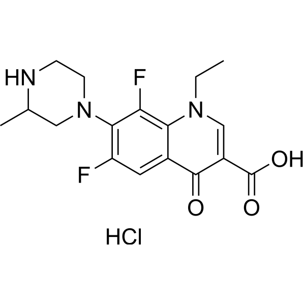
Lomefloxacin hydrochloride

Based on 1 Customer Validation
Lomefloxacin hydrochloride (NY-198 hydrochloride) is an orally active difluoroquinolone antibiotic. Lomefloxacin hydrochloride prevents DNA supercoiling and replication by inhibiting bacterial topoisomerase II. Lomefloxacin hydrochloride induces ROS production and Apoptosis. Lomefloxacin hydrochloride has broad-spectrum bactericidal activity against Gram-positive and Gram-negative bacteria. Lomefloxacin hydrochloride has anticancer effects against melanoma. Lomefloxacin hydrochloride can be used in the study of systemic bacterial infections (such as Salmonella typhimurium infections), skin and melanoma .
For research use only. We do not sell to patients.
- Purity : 99.97%
- CAS No.: 98079-52-8
- Formula: C17H20ClF2N3O3
- Molecular Weight:387.81
-
Storage:
4°C, sealed storage, away from moisture and light
* In solvent : -80°C, 6 months; -20°C, 1 month (sealed storage, away from moisture and light)

To place orders, for customer services and technical support, please contact: MedChemExpress USA
Tel: 609-228-6898 E-mail: [email protected] [email protected]


-
 Biological Activity
-
Chemical Information
-
Solvent & Solubility
- Protocols
- Purity & Documentation
- References
-
 Help & FAQs


-
HY-B0330
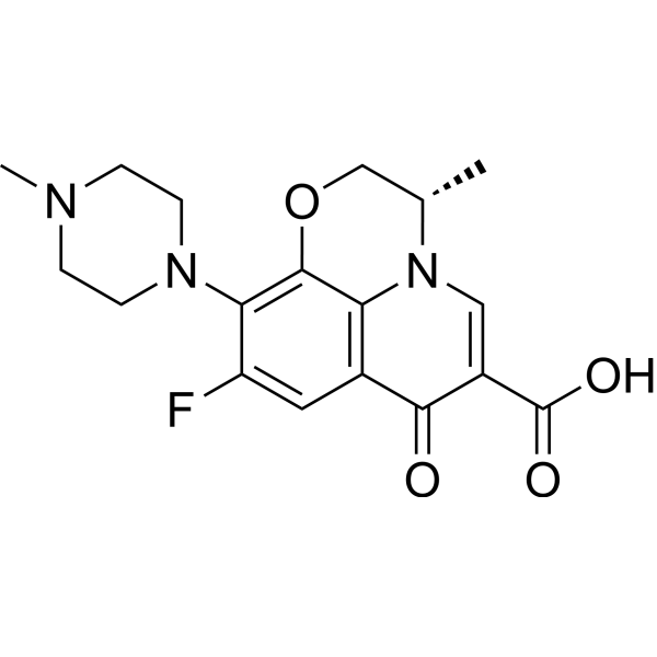
LevofloxacinDNA Gyrase/Topoisomerase Inhibitor -
HY-B0132
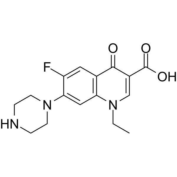
Norfloxacin -
HY-B0125
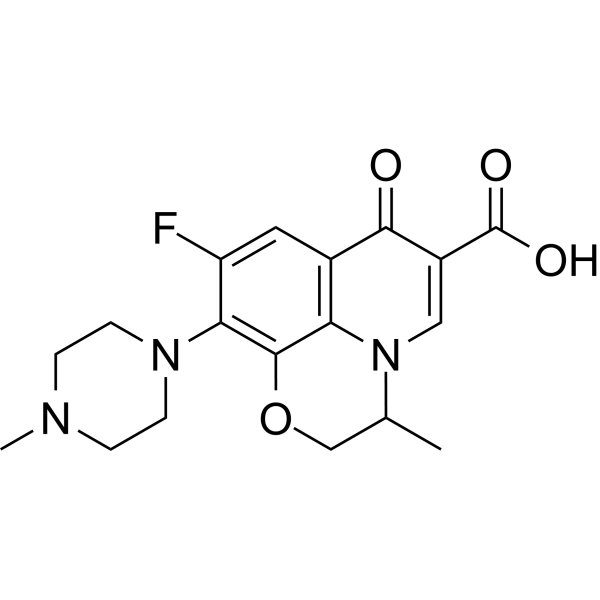
OfloxacinBacterial Inhibitor -
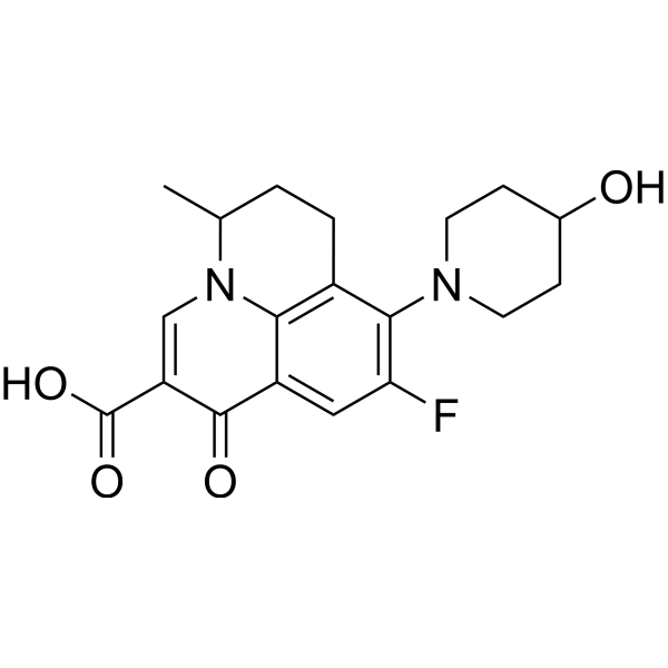
HY-B0506
NadifloxacinAntibiotic -
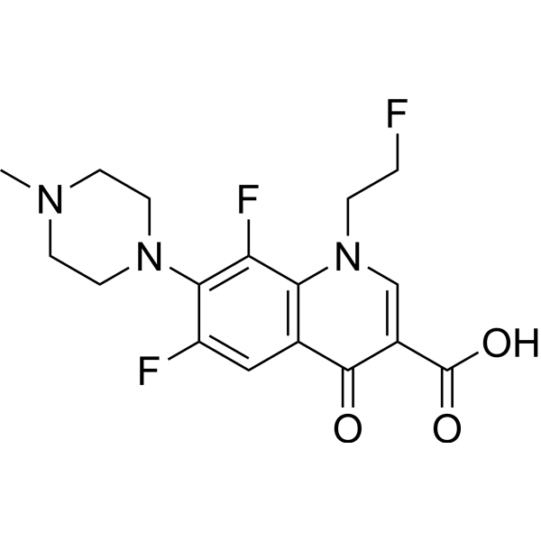
HY-B0414
FleroxacinBacterial Inhibitor -
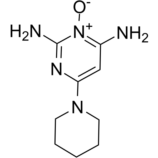
HY-B0112
MinoxidilEndogenous Metabolite -
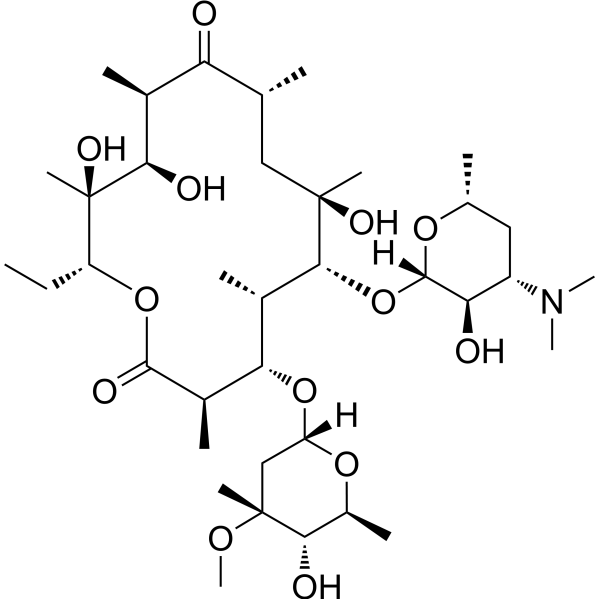
HY-B0220
ErythromycinAntibiotic -
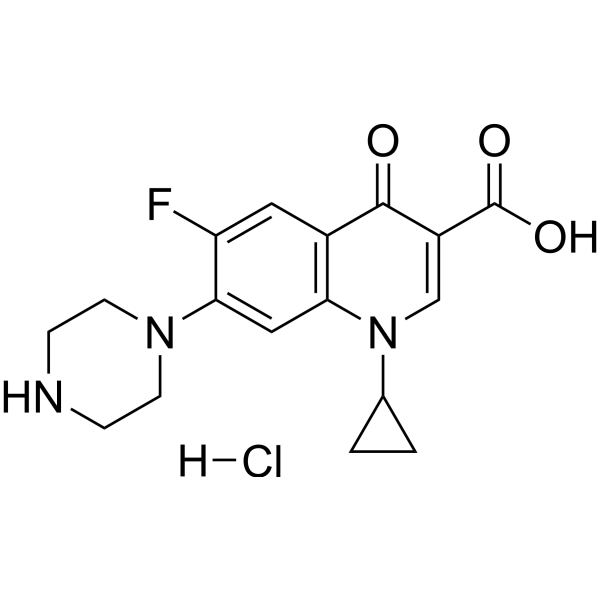
HY-B0356A
Ciprofloxacin monohydrochlorideTopoisomerase IV Inhibitor -
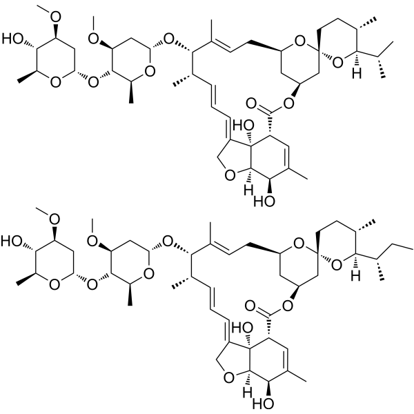
HY-15310
IvermectinAnti-parasite Agent -
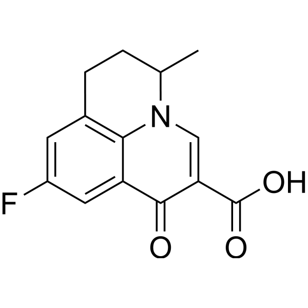
HY-B0526
FlumequineBacterial Inhibitor -
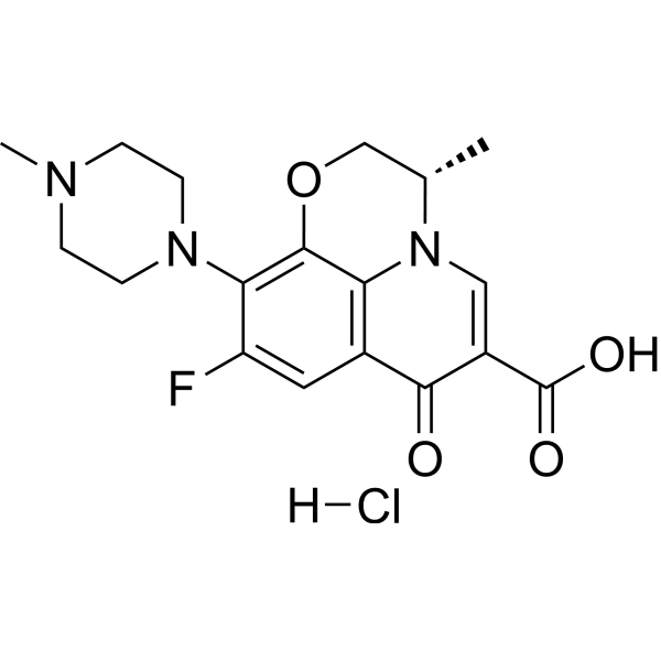
HY-B0330B
Levofloxacin hydrochlorideAntibiotic -
HY-B0147A
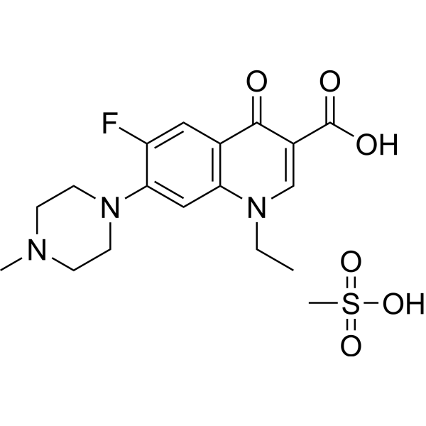
Pefloxacin mesylateAntibiotic


Biological Activity
Description
IC50 & Target
|
Quinolone |
In Vitro
Lomefloxacin (1/4 to 2×MIC concentrations) hydrochloride rapidly reduces viable cell counts of Staphylococcus aureus, Escherichia coli, and Pseudomonas aeruginosa, with no regrowth observed in all strains except P. aeruginosa[1].
Lomefloxacin hydrochloride exhibits MIC values ranging from 0.06-4 μg/mL in susceptibility determination experiments with strains such as Escherichia coli and Staphylococcus aureus[2].
Lomefloxacin (0.1-1.0 mM; 24-72 h) hydrochloride decreases viability of COLO829 cells and induces ROS production in COLO829 cells[3].
Lomefloxacin (50-100 μM) hydrochloride induces apoptosis in keratinocyte[4].
MedChemExpress (MCE) has not independently confirmed the accuracy of these methods. They are for reference only. Further protocols information, click here.
-
Cell Line:COLO829 melanoma cells
-
Concentration:0.0001, 0.001, 0.01, 0.05, 0.1, 0.5, 1.0 mM
-
Incubation Time:24, 48, 72 h
-
Result:Decreased cell viability by 12-68% at 0.1-1.0 mM for 24 h, with IC50 values of 0.51, 0.33, and 0.25 mM at 24, 48, 72 h, respectively.
In Vivo
Lomefloxacin (-80 mg/kg; orogastric administration) hydrochloride reduces mortality, significantly decreases splenic bacterial counts, and inhibits inflammatory response in mice infected with Salmonella typhimurium[5].
Lomefloxacin (16-32 mg/kg; i.p.; 5 days) hydrochloride only at the highest dose slightly reduces the number of implants and live fetuses in the third week of mating in mice, without significant dominant lethal mutation induction[6].
MedChemExpress (MCE) has not independently confirmed the accuracy of these methods. They are for reference only.


Chemical Information
-
CAS No. 98079-52-8
-
Appearance Solid
-
Molecular Weight 387.81
-
Formula C17H20ClF2N3O3
-
Color White to off-white
-
SMILES
CCN(C=C(C(O)=O)C1=O)C(C1=CC(F)=C2N(CCN3)CC3C)=C2F.Cl
-
Synonyms
SC47111A hydrochloride; NY-198 hydrochloride
-
Shipping
Room temperature in continental US; may vary elsewhere.
-
Storage
4°C, sealed storage, away from moisture and light
* In solvent : -80°C, 6 months; -20°C, 1 month (sealed storage, away from moisture and light)


Solvent & Solubility
In Vitro:
H2O : 7.14 mg/mL (18.41 mM; ultrasonic and warming and heat to 60°C)
Please refer to the solubility information to select the appropriate solvent. Once prepared, please aliquot and store the solution to prevent product inactivation from repeated freeze-thaw cycles.
Storage method and period of stock solution: -80°C, 6 months; -20°C, 1 month (sealed storage, away from moisture and light). When stored at -80°C, please use it within 6 months. When stored at -20°C, please use it within 1 month.
* Note: If you choose water as the stock solution, please dilute it to the working solution, then filter and sterilize it with a 0.22 μm filter before use.
Please refer to the solubility information to select the appropriate solvent. Once prepared, please aliquot and store the solution to prevent product inactivation from repeated freeze-thaw cycles.
Storage method and period of stock solution: -80°C, 6 months; -20°C, 1 month (sealed storage, away from moisture and light). When stored at -80°C, please use it within 6 months. When stored at -20°C, please use it within 1 month.
* Note: If you choose water as the stock solution, please dilute it to the working solution, then filter and sterilize it with a 0.22 μm filter before use.
Concentration (start) × Volume (start) = Concentration (final) × Volume (final)

Protocols
-
Apoptosis
Apoptosis, also called programmed cell death, is generally characterized by distinct morphological characteristics.
-
TUNEL staining for apoptotic DNA fragmentation
TUNEL staining detects DNA strand breaks by using terminal deoxynucleotidyl transferase to add labeled nucleotides to exposed 3′-OH DNA termini, generating either microscopic staining in fixed cells or tissue sections, or fluorescence/cytometric signal in cell suspensions. TUNEL positivity reflects DNA fragmentation but should not be interpreted alone as definitive apoptosis, because TUNEL can also label necrotic, autolytic, mechanically damaged, or DNA-repair-associated DNA breaks.
-
Annexin V plus membrane-impermeant dye apoptosis staining
Annexin V-based apoptosis assays rely on the detection of phosphatidylserine (PS) externalization from the inner leaflet of the plasma membrane to the outer leaflet, an early biochemical hallmark of apoptosis. Fluorescently labeled Annexin V binds PS in a calcium-dependent manner, enabling identification of early apoptotic cells by flow cytometry or fluorescence microscopy. When combined with a membrane-impermeant DNA-binding dye (e. g. , propidium iodide), this approach allows discrimination between viable (Annexin V−/dye−), early apoptotic (Annexin V+/dye−), and late apoptotic or necrotic (Annexin V+/dye+) cell populations by assessing membrane integrity and PS exposure.
-
ROS/oxidative-stress fluorescent staining
ROS/oxidative-stress fluorescent staining uses cell-permeant fluorogenic probes that become fluorescent after oxidation inside cells or tissues; commonly used examples include DCFH-DA/DCFDA for broad cellular oxidant detection, DHE for superoxide-related signal detection, MitoSOX for mitochondrial superoxide-related signal detection, and CellROX probes for oxidative-stress-associated fluorescence readouts. The assay detects probe oxidation rather than a single ROS species unless the probe and analysis method have been chemically validated for that species. DCFH-DA enters cells, is deacetylated by intracellular esterases to DCFH, and produces fluorescent DCF after oxidation, so the readout is used as an operational measure of total cellular oxidative stress rather than a species-specific ROS measurement. DHE and MitoSOX can report superoxide-related oxidation, but red fluorescence alone can include non-specific ethidium-like oxidation products; HPLC or optimized spectral approaches are
-
Apoptosis Solutions
Apoptosis is a regulated, generally non-lytic cell-death pathway that removes unwanted, damaged, infected, or abnormal cells through coordinated morphological changes, caspase activation, DNA fragmentation, and membrane remodeling. The intrinsic apoptosis pathway is controlled mainly by mitochondrial outer membrane permeabilization, BCL-2 family proteins, cytochrome c release, apoptosome formation, caspase-9 activation, and downstream executioner caspase-3/7 activation. The extrinsic apoptosis pathway is initiated by death receptors such as Fas, TNFR, and TRAIL receptors, which recruit adaptor proteins and activate caspase-8 before engaging executioner caspases or mitochondrial amplification through BID cleavage. Apoptosis is linked to many phenotypes, including cancer cell killing, tissue homeostasis, immune regulation, neurodegeneration, infection response, and treatment-induced cytotoxicity; unresolved questions include how apoptosis interacts with necroptosis, pyroptosis, ferroptos
-
Gram Staining of Tissue Sections
Gram staining of tissue sections is a histochemical technique used to differentiate Gram-positive and Gram-negative bacteria within histological specimens based on differences in bacterial cell wall structure and dye retention, adapted from classical bacteriological Gram staining into tissue-compatible “histological Gram stain” variants. In tissue applications, modifications of the Brown-Hopps and Brown-Brenn methods are commonly used to improve differentiation of microorganisms embedded within host connective tissue and to reduce overstaining or loss of Gram-negative signal, which are known limitations of earlier approaches. The principle relies on crystal violet-iodine complex retention in Gram-positive organisms and subsequent decolorization and counterstaining steps that allow contrast visualization of Gram-negative organisms against tissue background.
-
Bacterial live/dead nucleic-acid viability staining
The LIVE/DEAD bacterial viability staining method is based on differential permeability of nucleic-acid-binding fluorescent dyes, most commonly SYTO 9 and propidium iodide (PI), which enables discrimination of bacterial populations with intact versus compromised cytoplasmic membranes. SYTO 9 penetrates both intact and damaged bacterial membranes and binds nucleic acids to produce green fluorescence, whereas propidium iodide penetrates only cells with compromised membranes and fluoresces red while also reducing SYTO 9 signal through competitive binding and fluorescence interactions. The resulting fluorescence pattern is interpreted as a proxy for membrane integrity, which is widely used as an indicator of bacterial viability in microscopy, flow cytometry, and spectroscopic platforms. However, mechanistic studies show that SYTO 9 and PI interactions involve displacement and fluorescence resonance energy transfer effects, which can influence signal interpretation depending on dye ratios a


Purity & Documentation
-
Data Sheet (276 KB)
-
SDS (393 KB)
- English - EN (393 KB)
- Français - FR (393 KB)
- Deutsch - DE (393 KB)
- Norwegian - NO (393 KB)
- Español - ES (393 KB)
- Swedish - SV (393 KB)
- Italian - IT (393 KB)
- Korean - KR (393 KB)
- Portuguese - PT (393 KB)
-
Handling Instructions (2659 KB)
References
[1]. Hirose T, et al. In vitro and in vivo activity of NY-198, a new difluorinated quinolone. Antimicrob Agents Chemother. 1987 Jun;31(6):854-9. [Content Brief]
[2]. Piddock LJ, et al. Mechanism of action of lomefloxacin. Antimicrob Agents Chemother. 1990 Jun;34(6):1088-93. [Content Brief]
[3]. Beberok A, et al. Lomefloxacin Induces Oxidative Stress and Apoptosis in COLO829 Melanoma Cells. Int J Mol Sci. 2017 Oct 20;18(10):2194. [Content Brief]
[4]. Marrot L, et al. Molecular responses to photogenotoxic stress induced by the antibiotic lomefloxacin in human skin cells: from DNA damage to apoptosis. J Invest Dermatol. 2003 Sep;121(3):596-606. [Content Brief]
[5]. Butler T, et al. Treatment of experimental Salmonella typhimurium infection in mice with lomefloxacin. J Antimicrob Chemother. 1990 Apr;25(4):629-34. [Content Brief]
[6]. Singh AC, et al. Genotoxicity of lomefloxacin--an antibacterial drug in somatic and germ cells of Swiss albino mice in vivo. Mutat Res. 2003 Feb 5;535(1):35-42. [Content Brief]


Complete Stock Solution Preparation Table
Please refer to the solubility information to select the appropriate solvent. Once prepared, please aliquot and store the solution to prevent product inactivation from repeated freeze-thaw cycles.
Storage method and period of stock solution: -80°C, 6 months; -20°C, 1 month (sealed storage, away from moisture and light). When stored at -80°C, please use it within 6 months. When stored at -20°C, please use it within 1 month.
| Optional Solvent | Concentration Solvent Mass | 1 mg | 5 mg | 10 mg | 25 mg |
|---|---|---|---|---|---|
| H2O | 1 mM | 2.5786 mL | 12.8929 mL | 25.7858 mL | 64.4646 mL |
| 5 mM | 0.5157 mL | 2.5786 mL | 5.1572 mL | 12.8929 mL | |
| 10 mM | 0.2579 mL | 1.2893 mL | 2.5786 mL | 6.4465 mL | |
| 15 mM | 0.1719 mL | 0.8595 mL | 1.7191 mL | 4.2976 mL |
* Note: If you choose water as the stock solution, please dilute it to the working solution, then filter and sterilize it with a 0.22 μm filter before use.


Lomefloxacin hydrochloride Related Classifications
HY-B0455 Related Classifications
 Powered by Bioz