



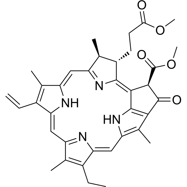
Methyl pheophorbide a

Based on 1 Customer Validation
Methyl pheophorbide a is a natural product, one of the chlorophyll-a derivatives. Methyl pheophorbide a is a photosensitizer that produces singlet oxygen and reactive oxygen species (ROS) under light of specific wavelength, thereby leading to cell death.
Nos produits utilisent uniquement pour la recherche. Nous ne vendons pas aux patients.
- Pureté : 99.57%
- CAS No.: 5594-30-9
- Formule: C36H38N4O5
- Masse moléculaire:606.71
-
Stockage:Powder -20°C, 3 years ; In solvent -80°C, 6 months , -20°C, 1 month

To place orders, for customer services and technical support, please contact: MedChemExpress USA
Tel: 609-228-6898 E-mail: [email protected] [email protected]


-
 Activité biologique
-
Chemical Information
- Protocole
- Pureté et documentation
- Références
-
 Help & FAQs


Voir tous les produits spécifiques à Isoform Caspase
More

Activité biologique
Description
Cellular Effect
|
Cell Line

|
Type | Value | Description | References |
|---|---|---|---|---|
| 1A9 | ED50 |
0.26 μg/mL
Compound: 5
|
Cytotoxicity against human 1A9 cells after 72 hrs by SRB assay
Cytotoxicity against human 1A9 cells after 72 hrs by SRB assay
|
[PMID: 11473423] |
| A549 | ED50 |
0.83 μg/mL
Compound: 5
|
Cytotoxicity against human A549 cells after 72 hrs by SRB assay
Cytotoxicity against human A549 cells after 72 hrs by SRB assay
|
[PMID: 11473423] |
| Daudi | IC50 |
25 μM
Compound: 6, mPheo
|
Cytotoxicity against human Daudi cells under deem light after 96 hrs by MTT assay
Cytotoxicity against human Daudi cells under deem light after 96 hrs by MTT assay
|
[PMID: 17993275] |
| HCT-8 | ED50 |
1 μg/mL
Compound: 5
|
Cytotoxicity against human HCT8 cells after 72 hrs by SRB assay
Cytotoxicity against human HCT8 cells after 72 hrs by SRB assay
|
[PMID: 11473423] |
| HeLa | IC50 |
0.182 μM
Compound: 7, MPa
|
Photosensitizing activity against human HeLa cells assessed as reduction in cell viability pre-incubated for 24 hrs followed by 15 mins exposure to 670 to 710 nm light at dose of 2.0 J/cm2 and measured 12 hrs after radiation by WST-8 assay
Photosensitizing activity against human HeLa cells assessed as reduction in cell viability pre-incubated for 24 hrs followed by 15 mins exposure to 670 to 710 nm light at dose of 2.0 J/cm2 and measured 12 hrs after radiation by WST-8 assay
|
[PMID: 25797162] |
| HeLa | IC50 |
25 μM
Compound: 6, mPheo
|
Cytotoxicity against human HeLa cells under deem light after 96 hrs by MTT assay
Cytotoxicity against human HeLa cells under deem light after 96 hrs by MTT assay
|
[PMID: 17993275] |
| K562 | IC50 |
>100 μM
Compound: 6, mPheo
|
Cytotoxicity against human K562 cells under deem light after 96 hrs by MTT assay
Cytotoxicity against human K562 cells under deem light after 96 hrs by MTT assay
|
[PMID: 17993275] |
| KB | ED50 |
0.23 μg/mL
Compound: 5
|
Cytotoxicity against human KB cells after 72 hrs by SRB assay
Cytotoxicity against human KB cells after 72 hrs by SRB assay
|
[PMID: 11473423] |
| MCF7 | ED50 |
0.95 μg/mL
Compound: 5
|
Cytotoxicity against human MCF7 cells after 72 hrs by SRB assay
Cytotoxicity against human MCF7 cells after 72 hrs by SRB assay
|
[PMID: 11473423] |
| NCI-H69 | IC50 |
>100 μM
Compound: 6, mPheo
|
Cytotoxicity against human H69 cells under deem light after 96 hrs by MTT assay
Cytotoxicity against human H69 cells under deem light after 96 hrs by MTT assay
|
[PMID: 17993275] |
| Raji | IC50 |
>100 μM
Compound: 6, mPheo
|
Cytotoxicity against human Raji cells under deem light after 96 hrs by MTT assay
Cytotoxicity against human Raji cells under deem light after 96 hrs by MTT assay
|
[PMID: 17993275] |
| SiHa | IC50 |
>100 μM
Compound: 6, mPheo
|
Cytotoxicity against human SIHA cells under deem light after 96 hrs by MTT assay
Cytotoxicity against human SIHA cells under deem light after 96 hrs by MTT assay
|
[PMID: 17993275] |
| SK-MEL | ED50 |
1.2 μg/mL
Compound: 5
|
Cytotoxicity against human SK-MEL cells after 72 hrs by SRB assay
Cytotoxicity against human SK-MEL cells after 72 hrs by SRB assay
|
[PMID: 11473423] |
In Vitro
Methyl pheophorbide a (0.25-2 μg/mL, 4 h) reduces the cell viability of U937 and SK-HEP-1, activates caspase 3/7, causes the formation of apoptotic bodies and vesicle, and induces apoptosis[3].
MedChemExpress (MCE) has not independently confirmed the accuracy of these methods. They are for reference only.
-
Cell Line:U937 and SK-HEP-1
-
Concentration:0.25-2 μg/mL
-
Incubation Time:4 h
-
Result:Inhibited cell viability under light.


Chemical Information
-
CAS No. 5594-30-9
-
Appearance Solid
-
Masse moléculaire 606.71
-
Formule C36H38N4O5
-
Color Brown to black
-
SMILES
COC([C@@H]1/C2=C([C@H]([C@@H]3C)CCC(OC)=O)/N=C3/C=C(C(C)=C/4C=C)\NC4=C/C5=N/C(C(CC)=C5C)=C\C6=C(C)C(C1=O)=C2N6)=O
-
Structure Classification
-
Initial Source
-
Livraison
Room temperature in continental US; may vary elsewhere.
-
Stockage
Powder -20°C 3 years In solvent -80°C 6 months -20°C 1 month


Pureté et documentation
-
Fiche technique (279 KB)
-
SDS (251 KB)
- English - EN (251 KB)
- Français - FR (251 KB)
- Deutsch - DE (251 KB)
- Norwegian - NO (251 KB)
- Español - ES (251 KB)
- Swedish - SV (251 KB)
- Italian - IT (251 KB)
- Korean - KR (251 KB)
- Portuguese - PT (251 KB)
-
Instruction de manipulation (2659 KB)
Références

Calculators
Concentration (start) × Volume (start) = Concentration (final) × Volume (final)


Methyl pheophorbide a Related Classifications
HY-N10902 Related Classifications
 Powered by Bioz