



Obtusin

Based on 1 Customer Validation
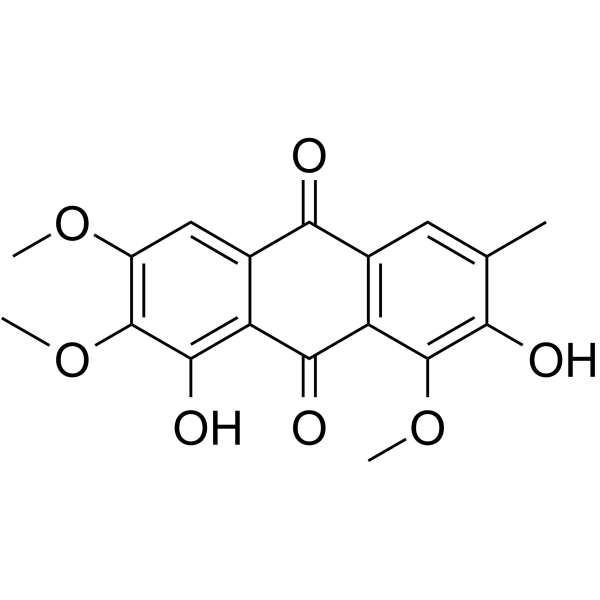
Obtusin is an orally active human monoamine oxidase A (hMAO-A) inhibitor discovered from the seeds of Cassia obtusifolia, with a Ki value of 6.15 μM. Obtusin inhibits high glucose-induced activation of the MAPKs/NF-κB/VEGF pathway and downregulates the expression of Poldip2, Nox4, VCAM-1, and HIF-1α, thereby alleviating oxidative stress, endothelial activation, and angiogenesis-related functions. Obtusin ameliorates diabetic retinopathy in vivo. Obtusin is used in research related to diabetes and neurodegenerative diseases (anxiety and depression).
For research use only. We do not sell to patients.
- Purity : 99.10%
- CAS No.: 70588-05-5
- Formula: C18H16O7
- Molecular Weight:344.32
-
Storage:
4°C, protect from light
* In solvent : -80°C, 6 months; -20°C, 1 month (protect from light)

To place orders, for customer services and technical support, please contact: MedChemExpress USA
Tel: 609-228-6898 E-mail: [email protected] [email protected]


-
 Biological Activity
-
Chemical Information
-
Solvent & Solubility
- Protocols
- Purity & Documentation
- References
-
 Help & FAQs


-

HY-Y0882
Hydroxylamine hydrochlorideMAO Inhibitor -
HY-N9329
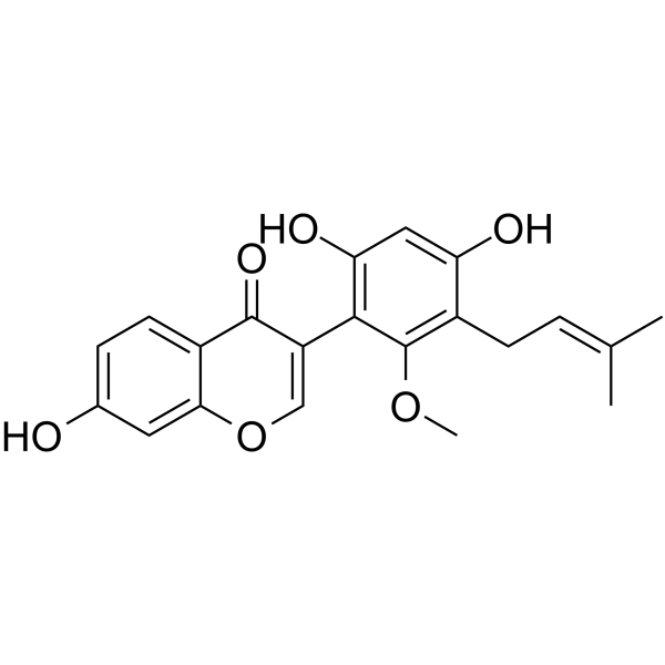
Glicoricone -
HY-A0091A
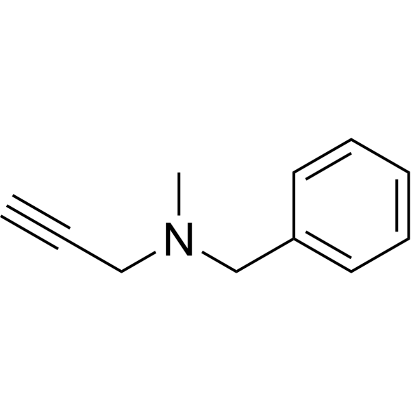
PargylineMonoamine Oxidase Inhibitor -
HY-70057
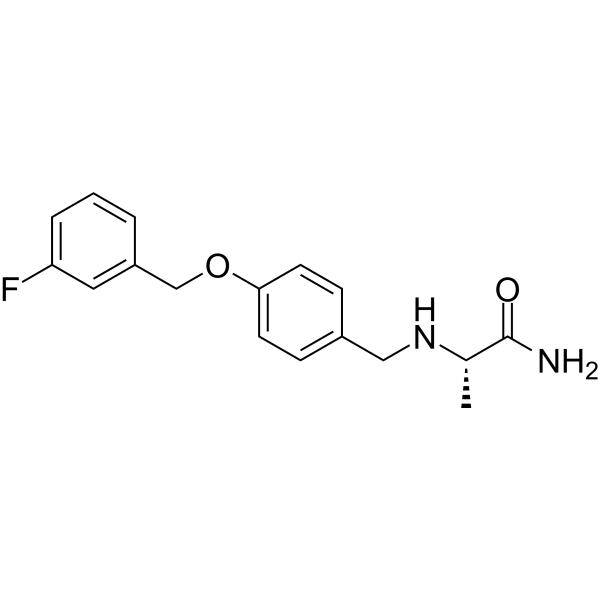
SafinamideMAO-B Inhibitor -
HY-14197A
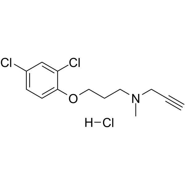
Clorgyline hydrochlorideMAO-A Inhibitor -
HY-A0091
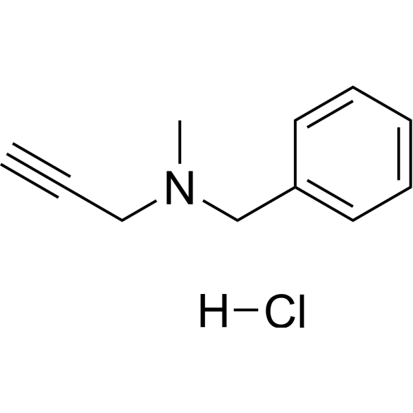
Pargyline hydrochlorideMonoamine Oxidase Inhibitor -
HY-14196
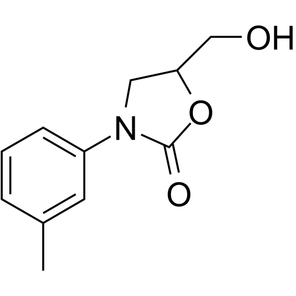
ToloxatoneMAO-A Inhibitor -
HY-B1199
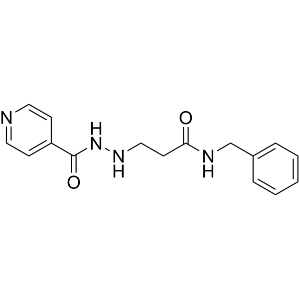
NialamideMonoamine Oxidase Inhibitor -
HY-D0004
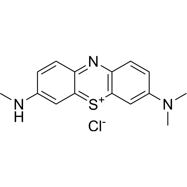
Azure BMAO-A Inhibitor -
HY-B1018A
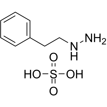
Phenelzine sulfateMAO Inhibitor -
HY-B1413
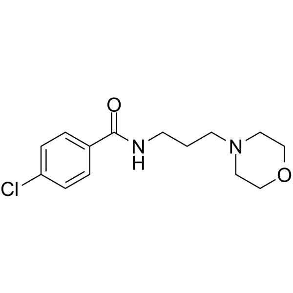
EprobemideMonoamine Oxidase Inhibitor -
HY-N2554
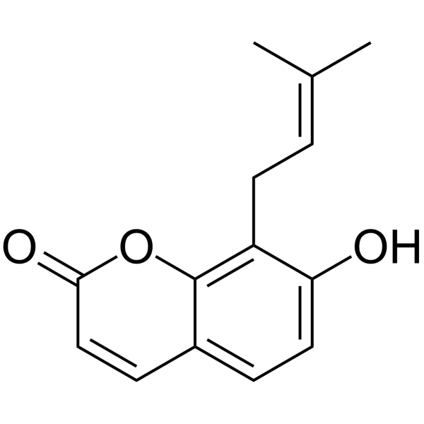
OsthenolhMAO-A Inhibitor
All VEGFR Isoforms
More

Biological Activity
Description
|
hMAO-A 11.12 μM (IC50) |
hMAO-A 6.15 μM (Ki) |
HIF-1α |
In Vitro
Obtusin (10-20 μM; 48 h) alleviates high glucose-induced HRMEC dysfunction, oxidative stress, and endothelial activation by inhibiting proliferation, migration, and tube formation, and downregulating the expression of Poldip2, Nox4, VCAM-1, and HIF-1α[1].
Obtusin (10-20 μM; 2 h) inhibits the activation of high glucose-induced MAPKs/NF-κB/VEGF pathway in HRMECs and decreases the expression of phosphorylated p38 MAPK, phosphorylated NF-κB p65, and VEGFA[1].
Obtusin selectively inhibits hMAO-A activity with an IC50 of 11.12 μM[2].
Obtusin competitively inhibits hMAO-A with a Ki value of 6.15 μM[2].
Obtusin binds to the catalytic site of hMAO-A with a binding energy of -8.51 kcal/mol, forming key hydrogen bond interactions with Cys323 and Thr336[2].
MedChemExpress (MCE) has not independently confirmed the accuracy of these methods. They are for reference only. Further protocols information, click here.
-
Cell Line:HRMECs
-
Concentration:10 and 20 μM
-
Incubation Time:48 h
-
Result:Determined 10 μM and 20 μM obtusin for 48 h as optimal conditions.
Dose-dependently suppressed proliferation and migration.
Inhibited tube-forming ability dose-dependently.
-
Cell Line:HRMECs
-
Concentration:10 and 20 μM
-
Incubation Time:2 h
-
Result:Significantly inhibited VEGFA mRNA expression.
Markedly reduced intracellular ROS levels.
Effectively reversed changes in Poldip2, Nox4, VCAM-1, and HIF-1α protein expression.
Reversed changes in MAPKs/NF-κB/VEGF pathway proteins (p-p38, p-p65, VEGFA).
In Vivo
MedChemExpress (MCE) has not independently confirmed the accuracy of these methods. They are for reference only.
-
Animal Model:C57BLKS/J Lepr db mutant mice (seven-week-old, male)[1]
-
Dosage:5 and 10 mg/kg/day
-
Administration:p.o.; daily; 12 weeks
-
Result:Reduced blood glucose levels from weeks 12 to 20 and improved body weight by week 14.
Corrected abnormal lipid profiles by reducing serum TC, TG, and LDL levels at 20 weeks.
Reduced GCL edema, increased cell numbers in the INL and ONL, and improved retinal thickness and morphology.
Reduced vascular leaks, improved retinal vascular permeability, and diminished overall fluorescence intensity.
Significantly reduced pericyte loss and the formation of acellular capillaries.
Reduced ROS levels across all retinal layers.
Alleviated structural abnormalities in retinal ganglion cells and vascular endothelial cells in a dose-dependent manner.
Reduced glial cell activation (IBA1 and GFAP) and preserved ganglion cell numbers (BRN3A) in a dose-dependent manner.
Significantly reduced the elevated levels of iNOS, IL-1β, IL-6, and VEGFA in the retinas.


Chemical Information
-
CAS No. 70588-05-5
-
Appearance Solid
-
Molecular Weight 344.32
-
Formula C18H16O7
-
Color Yellow to orange
-
SMILES
O=C1C2=C(C=C(C)C(O)=C2OC)C(C3=CC(OC)=C(OC)C(O)=C13)=O
-
Structure Classification
-
Shipping
Room temperature in continental US; may vary elsewhere.
-
Storage
4°C, protect from light
* In solvent : -80°C, 6 months; -20°C, 1 month (protect from light)


Solvent & Solubility
In Vitro:
DMSO : 5 mg/mL (14.52 mM; ultrasonic and warming and heat to 60°C; Hygroscopic DMSO has a significant impact on the solubility of product, please use newly opened DMSO)
Please refer to the solubility information to select the appropriate solvent. Once prepared, please aliquot and store the solution to prevent product inactivation from repeated freeze-thaw cycles.
Storage method and period of stock solution: -80°C, 6 months; -20°C, 1 month (protect from light). When stored at -80°C, please use it within 6 months. When stored at -20°C, please use it within 1 month.
Please refer to the solubility information to select the appropriate solvent. Once prepared, please aliquot and store the solution to prevent product inactivation from repeated freeze-thaw cycles.
Storage method and period of stock solution: -80°C, 6 months; -20°C, 1 month (protect from light). When stored at -80°C, please use it within 6 months. When stored at -20°C, please use it within 1 month.
Concentration (start) × Volume (start) = Concentration (final) × Volume (final)

Protocols
-
RNA extraction experimental
By lysing cells, releasing RNA, and removing impurities such as proteins and DNA, high-purity RNA products are finally obtained. The commonly used traditional method is the guanidine isothiocyanate/phenol/chloroform method (Trizol), which is suitable for a variety of animal materials including animal tissues, microorganisms, cultured cells, etc., and most plant materials.
-
Human pluripotent stem cell endothelial-cell differentiation
Human pluripotent stem cell endothelial differentiation is based on stepwise developmental patterning: early activation of WNT/GSK3β inhibition promotes mesodermal or vascular progenitor entry, followed by endothelial specification using VEGF-related signaling, BMP4, FGF2, Notch modulation, or cAMP depending on the published protocol. Endothelial differentiation is read out by acquisition of CD31, CD34, VE-cadherin/CD144, KDR/VEGFR2, vWF, Tie2, NOS3, acetylated LDL uptake, tube/network formation, barrier function, and in vivo vessel-forming capacity where tested.
-
Vascular/Branching Fractal Analysis
Vascular/branching fractal analysis quantifies the geometric complexity of vessel trees or vascular networks from segmented 2D images, commonly by converting vessels into binary and/or skeletonized maps and estimating fractal dimension using box-counting or related approaches. Fractal dimension is interpreted as an image-derived readout of vascular branching complexity, space filling, or density, and has been applied to retinal photographs, fluorescein angiography, OCT angiography, capillary perfusion maps, and in vitro Matrigel angiogenesis networks. The assay readout is generated from vessel-positive pixels after image preprocessing, vessel segmentation, binarization, and optional skeletonization; reported outputs include fractal dimension, vessel density, branchpoint density, endpoint density, vessel length density, tortuosity, and generation-based branching metrics when VESGEN-style analysis is used. The biological interpretation is limited to quantitative vascular patterning and s
-
ROS/oxidative-stress fluorescent staining
ROS/oxidative-stress fluorescent staining uses cell-permeant fluorogenic probes that become fluorescent after oxidation inside cells or tissues; commonly used examples include DCFH-DA/DCFDA for broad cellular oxidant detection, DHE for superoxide-related signal detection, MitoSOX for mitochondrial superoxide-related signal detection, and CellROX probes for oxidative-stress-associated fluorescence readouts. The assay detects probe oxidation rather than a single ROS species unless the probe and analysis method have been chemically validated for that species. DCFH-DA enters cells, is deacetylated by intracellular esterases to DCFH, and produces fluorescent DCF after oxidation, so the readout is used as an operational measure of total cellular oxidative stress rather than a species-specific ROS measurement. DHE and MitoSOX can report superoxide-related oxidation, but red fluorescence alone can include non-specific ethidium-like oxidation products; HPLC or optimized spectral approaches are
-
Endothelial Tube Formation Assay
Endothelial tube formation assay evaluates the ability of endothelial cells to attach, migrate, align, and organize into capillary-like networks when cultured on gelled basement membrane extract or Matrigel; the readout is the morphology and quantity of tube-like networks, which reflects an in vitro endothelial morphogenesis step related to angiogenesis. Basement membrane extract/Matrigel provides laminin-rich extracellular matrix cues that support endothelial differentiation into capillary-like structures, but it can contain biologically active growth factors, so growth-factor-reduced matrix is preferred when testing defined angiogenic stimulators or inhibitors.
-
Research Protocol for Inflammation-related Diseases
The NLRP3 inflammasome is a cytosolic innate immune signaling platform that integrates priming signals and danger-signal activation to promote caspase-1 activation, maturation of IL-1β and IL-18, and gasdermin D-mediated pyroptotic cell death. The core experimental logic is to determine whether inflammatory disease phenotypes are driven by increased NLRP3 expression, ASC-containing inflammasome assembly, caspase-1 cleavage, GSDMD cleavage, and extracellular release of IL-1β/IL-18 rather than by nonspecific cell injury alone. The pathway is strongly linked to inflammation-related disease phenotypes because monosodium urate crystals activate NALP3/NLRP3 inflammasome signaling in gout-like crystal inflammation, cholesterol crystals activate NLRP3 inflammasomes in atherogenesis models, and DSS-induced intestinal inflammation has been reported to involve NLRP3 inflammasome activity. However, experimental colitis studies also show context-dependent protective effects of NLRP3 inflammasome co
-
Research Protocol for Metabolic Diseases
AMP-activated protein kinase, AMPK, is a conserved cellular energy sensor that responds to reduced cellular energy status and coordinates metabolism by increasing ATP-generating catabolic pathways while suppressing ATP-consuming anabolic processes. In metabolic disease research, the AMPK pathway is experimentally relevant because it regulates hepatic lipid synthesis, fatty acid oxidation, glucose production, skeletal-muscle glucose disposal, mTORC1-linked biosynthesis, autophagy, mitochondrial homeostasis, and whole-body energy balance. The central pathway logic is that energy stress, metformin, exercise-like stimulation, or direct AMPK activators increase AMPKα Thr172 phosphorylation and downstream substrate phosphorylation, including ACC and RAPTOR. Phosphorylation of ACC suppresses lipogenesis and supports fatty acid oxidation, whereas phosphorylation of RAPTOR suppresses mTORC1 signaling and links cellular energy status to growth and protein synthesis control. The pathway is linked
-
Endothelial Cell Migration/Angiogenic Sprouting Assay
Endothelial cell migration and angiogenic sprouting assays are in vitro (and partially ex vivo-adapted) functional models that quantify the ability of endothelial cells to undergo coordinated migration, extracellular matrix invasion, and multicellular organization into capillary-like sprouts in response to pro-angiogenic stimuli such as VEGF, bFGF, or conditioned microenvironments. These assays are used to model early angiogenic events including tip-cell formation, directional migration, and lumen-like sprout extension, which collectively reflect angiogenic activation and vascular morphogenesis processes observed in vivo.


Purity & Documentation
-
Data Sheet (291 KB)
-
SDS (252 KB)
- English - EN (252 KB)
- Français - FR (252 KB)
- Deutsch - DE (252 KB)
- Norwegian - NO (252 KB)
- Español - ES (252 KB)
- Swedish - SV (252 KB)
- Italian - IT (252 KB)
- Korean - KR (252 KB)
- Portuguese - PT (252 KB)
-
Handling Instructions (2659 KB)
References


Complete Stock Solution Preparation Table
Please refer to the solubility information to select the appropriate solvent. Once prepared, please aliquot and store the solution to prevent product inactivation from repeated freeze-thaw cycles.
Storage method and period of stock solution: -80°C, 6 months; -20°C, 1 month (protect from light). When stored at -80°C, please use it within 6 months. When stored at -20°C, please use it within 1 month.
| Optional Solvent | Concentration Solvent Mass | 1 mg | 5 mg | 10 mg | 25 mg |
|---|---|---|---|---|---|
| DMSO | 1 mM | 2.9043 mL | 14.5214 mL | 29.0428 mL | 72.6069 mL |
| 5 mM | 0.5809 mL | 2.9043 mL | 5.8086 mL | 14.5214 mL | |
| 10 mM | 0.2904 mL | 1.4521 mL | 2.9043 mL | 7.2607 mL |


Obtusin Related Classifications
HY-N6057 Related Classifications
 Powered by Bioz