



PEAQX
Based on 8 publication(s) in Google Scholar
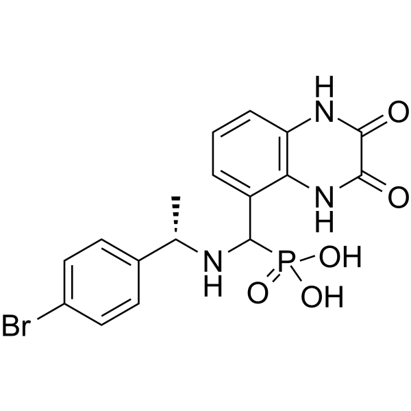
PEAQX (NVP-AAM077) is an orally active and selective NMDA antagonist, with IC50 values of 270 nM and 29.6 μM for hNMDAR 1A/2A and hNMDAR 1A/2B, respectively. PEAQX can promote the activation of caspase-3 and induce cell apoptosis in cortical striatal slice cultures.
For research use only. We do not sell to patients.
- CAS No.: 459836-30-7
- Formula: C17H17BrN3O5P
- Molecular Weight:454.21
-
Storage:
Please store the product under the recommended conditions in the Certificate of Analysis.

To place orders, for customer services and technical support, please contact: MedChemExpress USA
Tel: 609-228-6898 E-mail: [email protected] [email protected]


-
 Biological Activity
-
Chemical Information
-
Solvent & Solubility
- Protocols
- Purity & Documentation
- References
-
 Help & FAQs


-
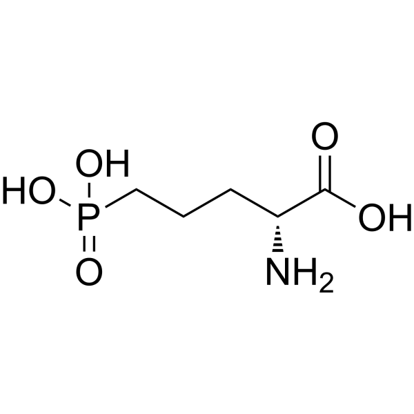
HY-100714A
D-AP5NMDA Antagonist -
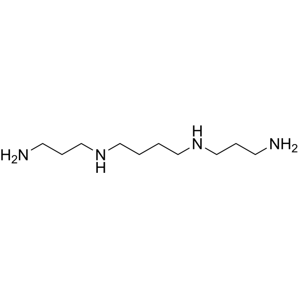
HY-B1777
SpermineEndogenous Metabolite -

HY-B1777R
Spermine (Standard)Endogenous Metabolite -
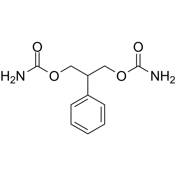
HY-B0184
FelbamateiGluR Inhibitor -
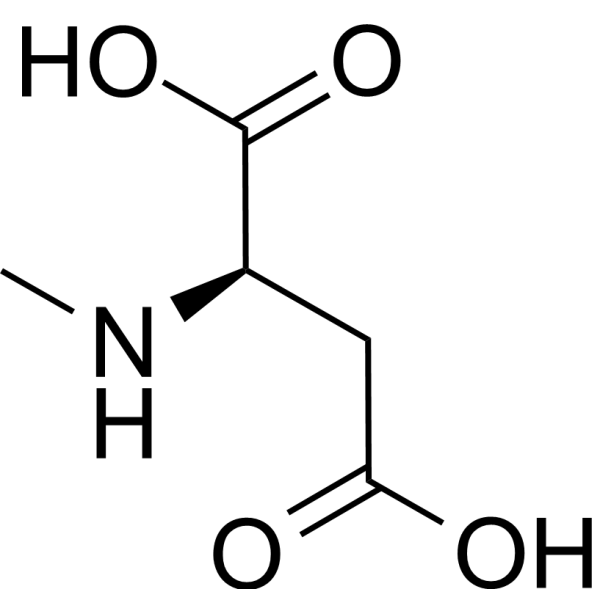
HY-17551
NMDAEndogenous Metabolite -
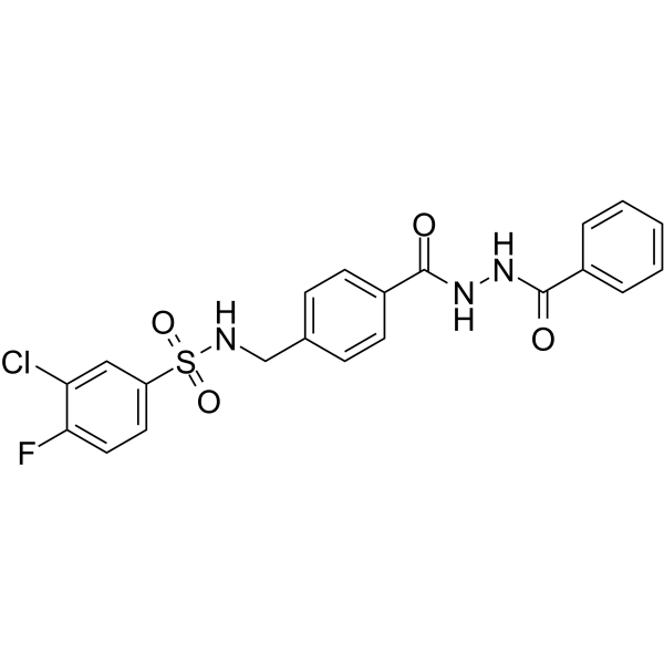
HY-13457
TCN 201NMDA Receptor Antagonist -
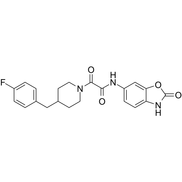
HY-14777
RadiprodilNMDA NR2B Antagonist -
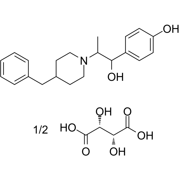
HY-12882A
Ifenprodil tartrateNMDA Receptor Antagonist -
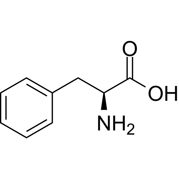
HY-N0215
L-PhenylalanineCalcium Channels Antagonist -
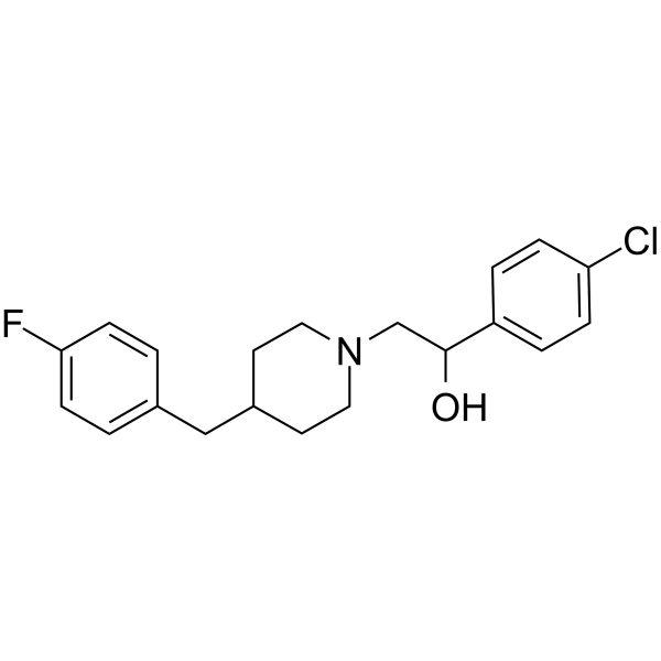
HY-12881
EliprodilNR2B-NMDA Inhibitor -
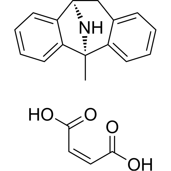
HY-15084
Dizocilpine maleateNMDA Antagonist
Publications Citing Use of MedChemExpress (MCE) PEAQX
More- J Clin Invest. 2019 Sep 3;129(9):3864-3876. [Abstract]
- Brain Behav Immun. 2026 Jul:135:106529. [Abstract]
- Neurosci Bull. 2025 Apr 28. [Abstract]
- Cells. 2023 Apr 22;12(9):1212. [Abstract]
- Front Neurosci. 2021 Sep 30:15:703044. [Abstract]
- Neuroscience. 2018 Nov 1:391:1-12. [Abstract]
- Am J Hypertens. 2021 Aug 9;34(8):840-850. [Abstract]
- bioRxiv. 2025 Sep 27.
 Customer Validation & Images

-
WB
All Caspase Isoforms
MoreAll iGluR Isoforms
More

Biological Activity
Description
IC50 & Target
|
NMDA Receptor |
In Vitro
In Vivo
PEAQX (10 mg/kg; once daily; i.p.) can block the CaMKIV-TORC1-CREB pathway signal induced by sigma-1 receptor (σ 1r) agonists in mice with cerebral ischemia and improve learning and memory impairments[3].
MedChemExpress (MCE) has not independently confirmed the accuracy of these methods. They are for reference only.
-
Animal Model:Pups obtained from deliveries of pregnant female Sprague-Dawley rats[2].
-
Dosage:10, 20, 40 mg/kg
-
Administration:Subcutaneous injection (s.c.)
-
Result:Resulted in an 8-fold increase in caspase-3 activity in the striatum when administered at a dose of 20 mg/kg.
-
Animal Model:11-week old C57BL/6 mice with cerebral ischemia[3].
-
Dosage:10 mg/kg
-
Administration:Intraperitoneal injection (i.p.); once daily; 4 days
-
Result:Reduced protein expression levels in the CaMKIV-TORC1-p-CREB-BDNF pathway.


Chemical Information
-
CAS No. 459836-30-7
-
Molecular Weight 454.21
-
Formula C17H17BrN3O5P
-
SMILES
O=C1C(NC2=C(C(C(P(O)(O)=O)N[C@H](C3=CC=C(Br)C=C3)C)=CC=C2)N1)=O
-
Synonyms
NVP-AAM077
-
Shipping
Room temperature in continental US; may vary elsewhere.
-
Storage
Please store the product under the recommended conditions in the Certificate of Analysis.


Publications (8)
-
Journal Impact Factor
-
Most Recent
-
J Clin Invest
2019 Sep 3;129(9):3864-3876. PMID: 31424425 -
Brain Behav Immun
NR2B-CaMKII signaling in the dentate gyrus driven by astrocytic P2Y1Rs mediates the antidepressant effect of low-dose LPS. [Abstract]2026 Jul:135:106529. PMID: 41796645 -
Neurosci Bull
Upregulation of NR2A in Glutamatergic VTA Neurons Contributes to Chronic Visceral Pain in Male Mice. [Abstract]2025 Apr 28. PMID: 40293685 -
Cells
2023 Apr 22;12(9):1212. PMID: 37174612 -
Front Neurosci
GluN2A/ERK/CREB Signaling Pathway Involved in Electroacupuncture Regulating Hypothalamic-Pituitary-Adrenal Axis Hyperactivity. [Abstract]2021 Sep 30:15:703044. PMID: 34658758 -
Neuroscience
Src is Implicated in Hepatic Ischemia Reperfusion-Induced Hippocampus Injury and Long-Term Cognitive Impairment in Young Mice via NMDA Receptor Subunit 2A Activation. [Abstract]2018 Nov 1:391:1-12. PMID: 30213765
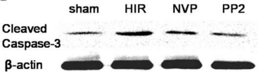
PEAQX purchased from MedChemExpress. Usage Cited in: Neuroscience. 2018 Nov 1:391:1-12. [Abstract]
PP2 and NVP-AAM077 pretreatment decreases the expression level of Cleaved Caspase-3 significantly.
-
Am J Hypertens
Chronic Blockade of NMDAR Subunit 2A in the Hypothalamic Paraventricular Nucleus Alleviates Hypertension Through Suppression of MEK/ERK/CREB Pathway. [Abstract]2021 Aug 9;34(8):840-850. PMID: 33856436 -


Solvent & Solubility
In Vivo:
The following protocol is derived from the literature and is for reference only. It is recommended to first try a small sample.
In Vivo Dissolution Calculator
Please enter the basic information of animal experiments:
-
-
-
-
Recommended: Prepare an additional quantity of animals to account for potential losses during experiments.
Working solution concentration: 0.22 mg/mL

Protocols
-
Apoptosis
Apoptosis, also called programmed cell death, is generally characterized by distinct morphological characteristics.
-
TUNEL staining for apoptotic DNA fragmentation
TUNEL staining detects DNA strand breaks by using terminal deoxynucleotidyl transferase to add labeled nucleotides to exposed 3′-OH DNA termini, generating either microscopic staining in fixed cells or tissue sections, or fluorescence/cytometric signal in cell suspensions. TUNEL positivity reflects DNA fragmentation but should not be interpreted alone as definitive apoptosis, because TUNEL can also label necrotic, autolytic, mechanically damaged, or DNA-repair-associated DNA breaks.
-
Cell Cytotoxicity Assay
Cytotoxicity assays are usually based on the assessment of cell membrane damage, which can also be indirectly detected by measuring cell viability. Detection methods include MTT assay, CKK-8 assay, LDH assay and ATP assay, etc.
-
Annexin V plus membrane-impermeant dye apoptosis staining
Annexin V-based apoptosis assays rely on the detection of phosphatidylserine (PS) externalization from the inner leaflet of the plasma membrane to the outer leaflet, an early biochemical hallmark of apoptosis. Fluorescently labeled Annexin V binds PS in a calcium-dependent manner, enabling identification of early apoptotic cells by flow cytometry or fluorescence microscopy. When combined with a membrane-impermeant DNA-binding dye (e. g. , propidium iodide), this approach allows discrimination between viable (Annexin V−/dye−), early apoptotic (Annexin V+/dye−), and late apoptotic or necrotic (Annexin V+/dye+) cell populations by assessing membrane integrity and PS exposure.
-
Apoptosis Solutions
Apoptosis is a regulated, generally non-lytic cell-death pathway that removes unwanted, damaged, infected, or abnormal cells through coordinated morphological changes, caspase activation, DNA fragmentation, and membrane remodeling. The intrinsic apoptosis pathway is controlled mainly by mitochondrial outer membrane permeabilization, BCL-2 family proteins, cytochrome c release, apoptosome formation, caspase-9 activation, and downstream executioner caspase-3/7 activation. The extrinsic apoptosis pathway is initiated by death receptors such as Fas, TNFR, and TRAIL receptors, which recruit adaptor proteins and activate caspase-8 before engaging executioner caspases or mitochondrial amplification through BID cleavage. Apoptosis is linked to many phenotypes, including cancer cell killing, tissue homeostasis, immune regulation, neurodegeneration, infection response, and treatment-induced cytotoxicity; unresolved questions include how apoptosis interacts with necroptosis, pyroptosis, ferroptos


Purity & Documentation
References
[1]. Auberson YP, et al. 5-Phosphonomethylquinoxalinediones as competitive NMDA receptor antagonists with a preference for the human 1A/2A, rather than 1A/2B receptor composition. Bioorg Med Chem Lett. 2002 Apr 8;12(7):1099-102. [Content Brief]
[2]. Anastasio NC, et al. Differential role of N-methyl-D-aspartate receptor subunits 2A and 2B in mediating phencyclidine-induced perinatal neuronal apoptosis and behavioral deficits. Neuroscience. 2009 Nov 10;163(4):1181-91. [Content Brief]
[3]. Qian Xu, et al. Sigma-1 receptor in brain ischemia/reperfusion: Possible role in the NR2A-induced pathway to regulate brain-derived neurotrophic factor. J Neurol Sci. 2017 May 15;376:166-175. [Content Brief]

Calculators
Concentration (start) × Volume (start) = Concentration (final) × Volume (final)


PEAQX Related Classifications
HY-12294 Related Classifications
 Powered by Bioz