



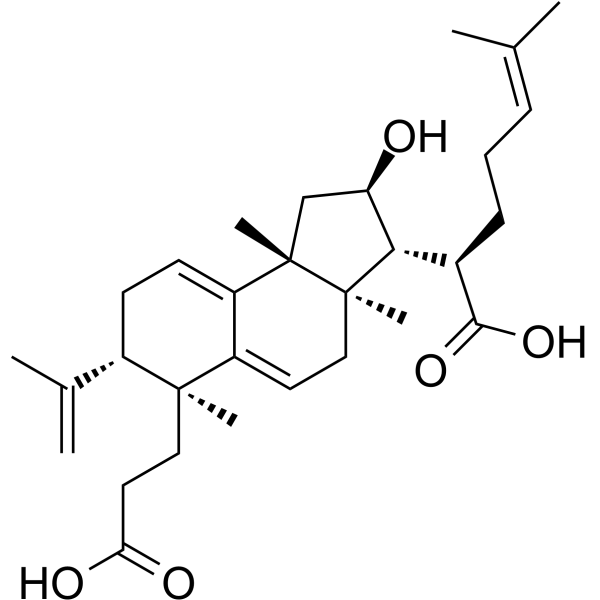
Poricoic acid A
Based on 1 publication(s) in Google Scholar
Poricoic acid A can be isolated from Poria cocos. Poricoic acid A is an orally active anti-tumor agent. Poricoic acid A enhances melatonin inhibition of AKI-to-CKD transition by regulating Gas6/AxlNFκB/Nrf2 axis. Poricoic acid A also attenuatea fibroblast activation and abnormal extracellular matrix remodeling in renal fibrosis by activating AMPK and inhibiting Smad3. Poricoic acid A significantly reduces the magnitude of rise in serum creatinine and urea levels in rat model when combined with Melatonin. Poricoic acid A ameliorates renal fibrosis and podocyte injury by attenuating oxidative stress and inflammation through regulating NF-κB and Nrf2 in IRI rodent model in combination with Melatonin.
For research use only. We do not sell to patients.
- Purity : 99.32%
- CAS No.: 137551-38-3
- Formula: C31H46O5
- Molecular Weight:498.69
-
Storage:
4°C, protect from light
* In solvent : -80°C, 6 months; -20°C, 1 month (protect from light)

To place orders, for customer services and technical support, please contact: MedChemExpress USA
Tel: 609-228-6898 E-mail: [email protected] [email protected]


-
 Biological Activity
-
Chemical Information
-
Solvent & Solubility
- Protocols
- Purity & Documentation
- References
-
 Help & FAQs


-
HY-N0371
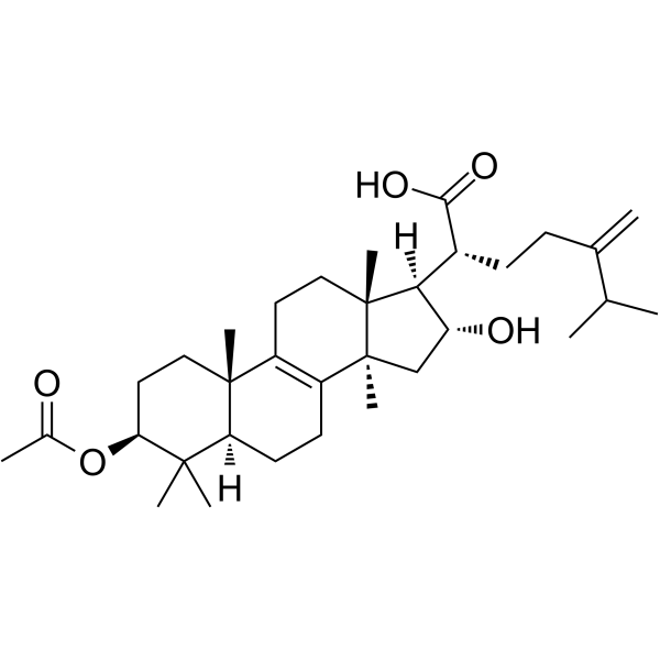
Pachymic acidTriterpenoid Compound -
HY-N0037
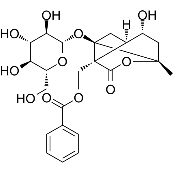
AlbiflorinNeuroprotective Agent -
HY-N0377
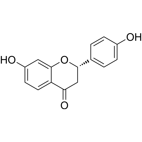
LiquiritigeninEstrogen Receptor/ERR Agonist -
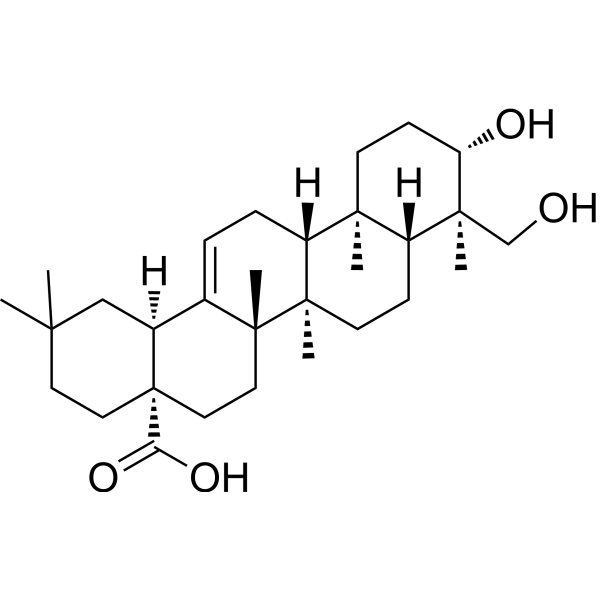
HY-N0256
HederageninCOX Inhibitor -
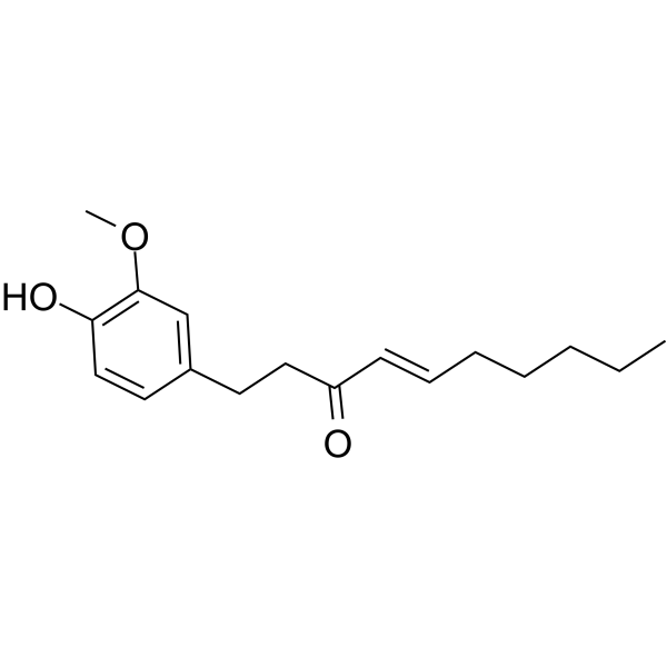
HY-14616
ShogaolAutophagy Inducer -
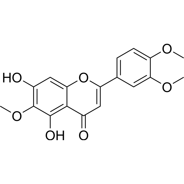
HY-N0783
EupatilinAutophagy Inducer -
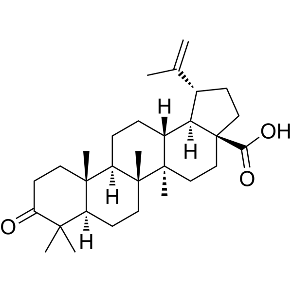
HY-N1451
Betulonic acidParasite Inhibitor -
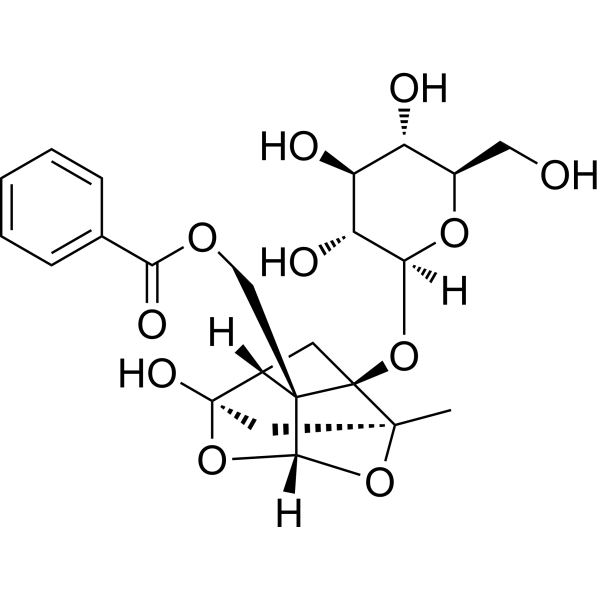
HY-N0293
PaeoniflorinHSP Activator -
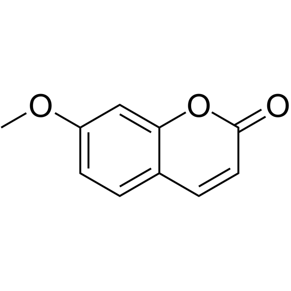
HY-N1366
HerniarinAnticancer Agent -
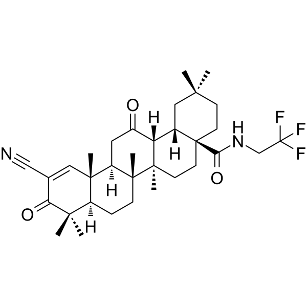
HY-112671
CDDO-dhTFEANrf2 Activator -
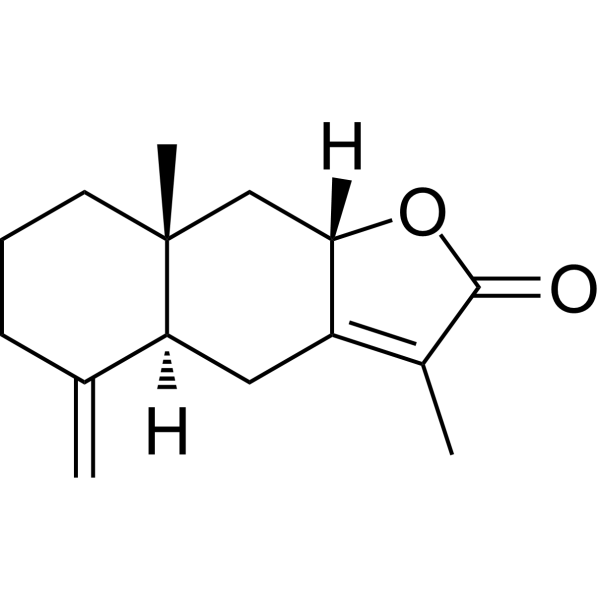
HY-N0202
Atractylenolide IIApoptosis Inducer -
HY-N4124
Poricoic acid BTriterpenoid Compound
Publications Citing Use of MedChemExpress (MCE) Poricoic acid A
MoreAll AMPK Isoforms
More

Biological Activity
Description
Cellular Effect
|
Cell Line

|
Type | Value | Description | References |
|---|---|---|---|---|
| A549 | IC50 |
>100 μM
Compound: 1a
|
Cytotoxicity in human A549 cells assessed as cell viability after 48 hrs by MTT assay
Cytotoxicity in human A549 cells assessed as cell viability after 48 hrs by MTT assay
|
[PMID: 21250700] |
| AZ-521 cell line | IC50 |
>100 μM
Compound: 1a
|
Cytotoxicity in human AZ-521 cells assessed as cell viability after 48 hrs by MTT assay
Cytotoxicity in human AZ-521 cells assessed as cell viability after 48 hrs by MTT assay
|
[PMID: 21250700] |
| B16 | IC50 |
48.01 μg/mL
Compound: 8
|
Cytotoxicity against mouse B16 cells assessed as inhibition of cell proliferation incubated for 48 hrs by MTS assay
Cytotoxicity against mouse B16 cells assessed as inhibition of cell proliferation incubated for 48 hrs by MTS assay
|
[PMID: 39026645] |
| CCRF-CEM | IC50 |
>40 μM
Compound: 22
|
Antiproliferative activity against human CCRF-CEM cells after 72 hrs by MTT assay
Antiproliferative activity against human CCRF-CEM cells after 72 hrs by MTT assay
|
[PMID: 27808511] |
| DU-145 | IC50 |
>100 μM
Compound: 1a
|
Cytotoxicity in human DU145 cells assessed as cell viability after 48 hrs by MTT assay
Cytotoxicity in human DU145 cells assessed as cell viability after 48 hrs by MTT assay
|
[PMID: 21250700] |
| HL-60 | IC50 |
>40 μM
Compound: 22
|
Antiproliferative activity against human HL60 cells after 72 hrs by MTT assay
Antiproliferative activity against human HL60 cells after 72 hrs by MTT assay
|
[PMID: 27808511] |
| HL-60 | IC50 |
38 μM
Compound: 1a
|
Cytotoxicity in human HL60 cells assessed as cell viability after 48 hrs by MTT assay
Cytotoxicity in human HL60 cells assessed as cell viability after 48 hrs by MTT assay
|
[PMID: 21250700] |
| HL-60 | IC50 |
38 μM
Compound: 7
|
Cytotoxicity against human HL60 cells after 48 hrs by MTT assay
Cytotoxicity against human HL60 cells after 48 hrs by MTT assay
|
[PMID: 19746919] |
| K562 | IC50 |
>40 μM
Compound: 22
|
Antiproliferative activity against human K562 cells after 72 hrs by MTT assay
Antiproliferative activity against human K562 cells after 72 hrs by MTT assay
|
[PMID: 27808511] |
| MOLT-4 | IC50 |
>40 μM
Compound: 22
|
Antiproliferative activity against human MOLT4 cells after 72 hrs by MTT assay
Antiproliferative activity against human MOLT4 cells after 72 hrs by MTT assay
|
[PMID: 27808511] |
| OVCAR-3 | IC50 |
>100 μM
Compound: 1a
|
Cytotoxicity in human OVCAR3 cells assessed as cell viability after 48 hrs by MTT assay
Cytotoxicity in human OVCAR3 cells assessed as cell viability after 48 hrs by MTT assay
|
[PMID: 21250700] |
| PANC-1 | IC50 |
>100 μM
Compound: 1a
|
Cytotoxicity in human PANC1 cells assessed as cell viability after 48 hrs by MTT assay
Cytotoxicity in human PANC1 cells assessed as cell viability after 48 hrs by MTT assay
|
[PMID: 21250700] |
| SK-BR-3 | IC50 |
>100 μM
Compound: 1a
|
Cytotoxicity in human SK-BR-3 cells assessed as cell viability after 48 hrs by MTT assay
Cytotoxicity in human SK-BR-3 cells assessed as cell viability after 48 hrs by MTT assay
|
[PMID: 21250700] |
In Vitro
Poricoic acid A (10 μM) protects against hypoxia/reoxygenation (H/R)-induced injury in cultured renal NRK-52E cells[2].
Poricoic acid A (5-10 μM, 24 h) has inhibitory effects on NRK-49F viability dose-dependently[3].
MedChemExpress (MCE) has not independently confirmed the accuracy of these methods. They are for reference only. Further protocols information, click here.
-
Cell Line:Renal NRK-52E cells
-
Concentration:10 μM
-
Incubation Time:24 h
-
Result:Reduced the level of NF-κB that transferred into the nucleus after H/R.
Resulted in stronger inhibitory effect on inflammatory pathway when combined with Melatonin.
Upregulated the HO-1 level that was reduced due to H/R.
In Vivo
Poricoic acid A (5-20 mg/kg, p.o., 1-2 w) ameliorates hyoertension and anaemia as well as improves kidney function in Nx-induced CKD rats[3].
Poricoic acid A (5-20 mg/kg, p.o., 1-2 w) significantly reduced renal fibrosis in UUO rats[3].
MedChemExpress (MCE) has not independently confirmed the accuracy of these methods. They are for reference only.
-
Animal Model:Male IRI SD rats[2]
-
Dosage:10 mg/kg/day
-
Administration:Intragastric (i.g.), day 2 to 13 after reperfusion
-
Result:Significantly downregulated pro-fibrotic Gas6 protein level, and combined therapy with Melatonin performed better.
Significantly ameliorated the activation of NF-κB pathway.
Significantly attenuated the expression of ED-1 and CD3 in renal interstitium compared with IRI group.
Mitigated the downregulation of Nrf2 and its downstream targets in IRI rats.
Markedly reduced the deposition of collagen I and collagen III in renal interstitium.
-
Animal Model:Nx-induced CKD rats[3]
-
Dosage:5, 10, 20 mg/kg
-
Administration:Oral gavage (p.o.)
-
Result:Ameliorated polyuria, elevated blood pressure, increased serum urea and creatinine levesl, reduced creatinine clearance and lowered body weight.
Suppressed aberrant synthesis and enhanced degradation of ECM in Nx rats.
Activated AMPK and suppressed TGF-β1/Smad3 pathway.


Chemical Information
-
CAS No. 137551-38-3
-
Appearance Solid
-
Molecular Weight 498.69
-
Formula C31H46O5
-
Color White to off-white
-
SMILES
C[C@]12[C@@]([C@]([C@H](C(O)=O)CCC(C(C)C)=C)([H])[C@H](O)C1)(CC=C3C2=CC[C@@H](C(C)=C)[C@]3(C)CCC(O)=O)C
-
Synonyms
Poricoic acid A(F)
-
Structure Classification
-
Initial Source
-
Shipping
Room temperature in continental US; may vary elsewhere.
-
Storage
4°C, protect from light
* In solvent : -80°C, 6 months; -20°C, 1 month (protect from light)


Publications (1)
-
Journal Impact Factor
-
Most Recent
-
Int J Mol Sci
Poricoic Acid A Attenuates Osteoarthritis Progression by Stabilizing PTEN and Suppressing PI3K/AKT Signaling. [Abstract]2026 Feb 14;27(4):1835. PMID: 41751971


Solvent & Solubility
In Vitro:
DMSO : 100 mg/mL (200.53 mM; Need ultrasonic; Hygroscopic DMSO has a significant impact on the solubility of product, please use newly opened DMSO)
Please refer to the solubility information to select the appropriate solvent. Once prepared, please aliquot and store the solution to prevent product inactivation from repeated freeze-thaw cycles.
Storage method and period of stock solution: -80°C, 6 months; -20°C, 1 month (protect from light). When stored at -80°C, please use it within 6 months. When stored at -20°C, please use it within 1 month.
Please refer to the solubility information to select the appropriate solvent. Once prepared, please aliquot and store the solution to prevent product inactivation from repeated freeze-thaw cycles.
Storage method and period of stock solution: -80°C, 6 months; -20°C, 1 month (protect from light). When stored at -80°C, please use it within 6 months. When stored at -20°C, please use it within 1 month.
Concentration (start) × Volume (start) = Concentration (final) × Volume (final)
In Vivo:
Select the appropriate dissolution method based on your experimental animal and administration route.
- For the following dissolution methods, please ensure to first prepare a clear stock solution using an In Vitro approach and then sequentially add co-solvents:
- To ensure reliable experimental results, the clarified stock solution can be appropriately stored based on storage conditions. As for the working solution for In Vivo experiments, it is recommended to prepare freshly and use it on the same day.
- The percentages shown for the solvents indicate their volumetric ratio in the final prepared solution. If precipitation or phase separation occurs during preparation, heat and/or sonication can be used to aid dissolution.
In Vivo Dissolution Calculator
Please enter the basic information of animal experiments:
-
-
-
-
Recommended: Prepare an additional quantity of animals to account for potential losses during experiments.
Please enter your animal formula composition:
-
%DMSO +
Recommended: Keep the proportion of DMSO in working solution below 2% if your animal is weak.
-
%+
-
+%Tween-80 + +
-
%Saline +
The co-solvents required include: DMSO, . All of co-solvents are available by MedChemExpress (MCE). , Tween 80. All of co-solvents are available by MedChemExpress (MCE).
Working solution concentration: 0.22 mg/mL
Method for preparing stock solution: mg drug dissolved in μL DMSO. Stock solution concentration: mg/mL. * In solvent : -80°C, 6 months; -20°C, 1 month (protect from light)
1. Take μL DMSO stock solution;
2. Add μL .
μL , mix evenly;
3. Then add μL Tween 80, mix evenly;
4. Then add μL
Please ensure that the stock solution in the first step is dissolved to a clear state, and add co-solvents in sequence. You can use ultrasonic heating (ultrasonic cleaner, recommended frequency 20-40 kHz), vortexing, etc. to assist dissolution.

Protocols
-
Fibrosis/Collagen Morphometry
Fibrosis and collagen morphometry is based on the quantitative visualization of fibrillar collagen deposition in tissue sections using histochemical stains such as Sirius Red (Picrosirius Red) or Masson's trichrome, followed by image-based or polarization-enhanced analysis to estimate collagen proportional area as a surrogate of extracellular matrix accumulation during fibrotic remodeling. Sirius Red combined with polarized light microscopy enhances detection of collagen fibers due to birefringence properties, enabling more specific visualization of collagen type I and III fibrils compared to conventional bright-field histology, while whole-section or region-restricted digital morphometry reduces field-selection bias in fibrosis assessment. Alternative quantitative approaches include second harmonic generation (SHG) and two-photon excited fluorescence microscopy, which enable label-free detection of fibrillar collagen and have been validated against histological staining and biochemica
-
Collagen: Sirius Red Staining
Sirius Red or picrosirius red staining is a histochemical method for visualizing collagen-rich extracellular matrix in tissue sections, and collagen fibers are detected as red-stained structures under bright-field microscopy with enhanced birefringence under polarized light. Picrosirius red is useful for assessing total collagen organization, distribution, and fibrosis burden, but polarized color should not be interpreted as a definitive collagen type I versus type III readout because color is affected by fiber orientation, thickness, and packing.
-
Nephrotoxicity Study
This protocol assesses nephrotoxicity by combining functional kidney injury readouts, urinary/tissue injury biomarkers, and renal histopathology. Serum creatinine and BUN reflect impaired kidney function, while KIM-1, NGAL, clusterin, osteopontin, IL-18, cystatin C, nephrin, Oat5, urinary protein, glucose, and alkaline phosphatase have been used to detect tubular injury in cisplatin-, gentamicin-, and acetaminophen-induced nephrotoxicity models.
-
ROS/oxidative-stress fluorescent staining
ROS/oxidative-stress fluorescent staining uses cell-permeant fluorogenic probes that become fluorescent after oxidation inside cells or tissues; commonly used examples include DCFH-DA/DCFDA for broad cellular oxidant detection, DHE for superoxide-related signal detection, MitoSOX for mitochondrial superoxide-related signal detection, and CellROX probes for oxidative-stress-associated fluorescence readouts. The assay detects probe oxidation rather than a single ROS species unless the probe and analysis method have been chemically validated for that species. DCFH-DA enters cells, is deacetylated by intracellular esterases to DCFH, and produces fluorescent DCF after oxidation, so the readout is used as an operational measure of total cellular oxidative stress rather than a species-specific ROS measurement. DHE and MitoSOX can report superoxide-related oxidation, but red fluorescence alone can include non-specific ethidium-like oxidation products; HPLC or optimized spectral approaches are
-
ECM-Embedded Organoid (Matrigel/Dome) Culture
ECM-embedded organoid dome culture embeds epithelial stem cells, crypts, organoid fragments, or tumor-derived epithelial cells in a basement-membrane-like hydrogel such as Matrigel, allowing 3D growth, self-organization, lumen formation, budding or cystic morphogenesis, and lineage maintenance under defined niche-factor-containing medium. The primary readouts are organoid establishment efficiency, growth, morphology, passaging capacity, lineage-marker expression, and, when fluorescently labeled lines are used, microscopy- or flow-cytometry-based quantification of population behavior in 3D culture.
-
Connective Tissue: Masson's Trichrome/Collagen Trichrome Staining
Masson’s Trichrome (collagen/trichrome staining) is a histological technique that differentially stains tissue compartments using sequential acidic dyes to distinguish collagen from muscle and cytoplasmic components based on dye affinity and tissue permeability differences, enabling visualization of fibrosis and connective tissue architecture in histological sections. The classical formulation typically uses Weigert's iron hematoxylin for nuclear staining, Biebrich scarlet-acid fuchsin for cytoplasm and muscle, and aniline blue (or light green variants) for collagen, producing a characteristic blue/green collagen signal contrasted against red cytoplasm and dark nuclei. The staining principle relies on selective displacement of smaller dye molecules by larger anionic dyes in collagen-rich regions under controlled acidified conditions, which enhances collagen-specific dye retention. This property makes the method widely used for fibrosis assessment in organs such as heart, liver, lung, a
-
How to Select a Suitable Non-Mouse Animal Model
Selecting a suitable non-mouse animal model is a structured decision based on the research question, required anatomy or physiology, disease mechanism, endpoint feasibility, translational relevance, and ethical justification. Non-mouse models are preferred when mice cannot reproduce key human-relevant features, such as organ size, surgical anatomy, cardiovascular physiology, neuroanatomy, immune features, pharmacology, toxicology, or long-term clinical procedures. Candidate species may include rats, rabbits, guinea pigs, ferrets, zebrafish, pigs, sheep, goats, dogs, cats, horses, and non-human primates, but each species must be justified by its specific scientific advantage rather than convenience or tradition. Unresolved questions include how to quantify translational superiority across species, how to balance increased biological relevance against higher ethical burden, and when human-derived systems or new approach methodologies should replace animal use.
-
Matrigel/ECM Transwell Invasion Assay
The Matrigel/ECM Transwell invasion assay measures the ability of cells to move toward a chemoattractant while crossing an extracellular-matrix barrier placed on a porous membrane; therefore, the readout reflects both chemotactic motility and matrix invasion rather than migration alone. Matrigel is a basement-membrane-rich matrix derived from Engelbreth-Holm-Swarm mouse sarcoma and has been used as a reconstituted basement membrane barrier in chemoinvasion assays. The assay readout is generated by quantifying cells that reach the underside of the insert membrane or lower compartment after incubation, commonly by staining and counting invaded cells or by fluorescence-based quantification.
-
Research Protocol for Inflammation-related Diseases
The NLRP3 inflammasome is a cytosolic innate immune signaling platform that integrates priming signals and danger-signal activation to promote caspase-1 activation, maturation of IL-1β and IL-18, and gasdermin D-mediated pyroptotic cell death. The core experimental logic is to determine whether inflammatory disease phenotypes are driven by increased NLRP3 expression, ASC-containing inflammasome assembly, caspase-1 cleavage, GSDMD cleavage, and extracellular release of IL-1β/IL-18 rather than by nonspecific cell injury alone. The pathway is strongly linked to inflammation-related disease phenotypes because monosodium urate crystals activate NALP3/NLRP3 inflammasome signaling in gout-like crystal inflammation, cholesterol crystals activate NLRP3 inflammasomes in atherogenesis models, and DSS-induced intestinal inflammation has been reported to involve NLRP3 inflammasome activity. However, experimental colitis studies also show context-dependent protective effects of NLRP3 inflammasome co
-
Matrigel Transwell/Boyden Chamber Invasion Assay
Matrigel Transwell/Boyden chamber invasion assay measures the ability of cells to degrade or traverse an extracellular matrix-coated porous membrane and move from an upper chamber toward a chemoattractant in a lower chamber. Invasion is distinguished from migration by coating the membrane with Matrigel or basement membrane matrix; uncoated inserts measure migration, while coated inserts require cells to cross an ECM barrier before reaching the underside of the membrane.
-
How to Choose the Right Model Animal
Choosing the right model animal is a validity-driven decision in which the species, strain, sex, age, genetic background, disease-induction method, outcome measures, and welfare burden must match the scientific question rather than laboratory tradition or convenience. A model should be selected by judging face validity, construct validity, and predictive validity: whether it resembles the human phenotype, whether it reproduces relevant mechanisms, and whether results are likely to predict human biology or treatment response. Animal studies often fail to translate because of species differences, weak disease resemblance, poor experimental design, inadequate reporting, publication bias, and underuse of randomization, blinding, and sample-size justification. Unresolved questions include how to rank competing models objectively, how much human-disease complexity must be reproduced for a given objective, and when non-animal systems such as organoids, ex vivo tissue, or computational models


Purity & Documentation
-
Data Sheet (278 KB)
-
SDS (252 KB)
- English - EN (252 KB)
- Français - FR (252 KB)
- Deutsch - DE (252 KB)
- Norwegian - NO (252 KB)
- Español - ES (252 KB)
- Swedish - SV (252 KB)
- Italian - IT (252 KB)
- Korean - KR (252 KB)
- Portuguese - PT (252 KB)
-
Handling Instructions (2659 KB)
References
[1]. Dong H, et al. Enrichment and separation of antitumor triterpene acids from the epidermis of Poria cocos by pH-zone-refining counter-current chromatography and conventional high-speed counter-current chromatography. J Sep Sci. 2015 Jun;38(11):1977-82. [Content Brief]
[2]. Chen DQ, et al. Poricoic acid A enhances melatonin inhibition of AKI-to-CKD transition by regulating Gas6/AxlNFκB/Nrf2 axis. Free Radic Biol Med. 2019 Apr;134:484-497. [Content Brief]


Complete Stock Solution Preparation Table
Please refer to the solubility information to select the appropriate solvent. Once prepared, please aliquot and store the solution to prevent product inactivation from repeated freeze-thaw cycles.
Storage method and period of stock solution: -80°C, 6 months; -20°C, 1 month (protect from light). When stored at -80°C, please use it within 6 months. When stored at -20°C, please use it within 1 month.
| Optional Solvent | Concentration Solvent Mass | 1 mg | 5 mg | 10 mg | 25 mg |
|---|---|---|---|---|---|
| DMSO | 1 mM | 2.0053 mL | 10.0263 mL | 20.0525 mL | 50.1313 mL |
| 5 mM | 0.4011 mL | 2.0053 mL | 4.0105 mL | 10.0263 mL | |
| 10 mM | 0.2005 mL | 1.0026 mL | 2.0053 mL | 5.0131 mL | |
| 15 mM | 0.1337 mL | 0.6684 mL | 1.3368 mL | 3.3421 mL | |
| 20 mM | 0.1003 mL | 0.5013 mL | 1.0026 mL | 2.5066 mL | |
| 25 mM | 0.0802 mL | 0.4011 mL | 0.8021 mL | 2.0053 mL | |
| 30 mM | 0.0668 mL | 0.3342 mL | 0.6684 mL | 1.6710 mL | |
| 40 mM | 0.0501 mL | 0.2507 mL | 0.5013 mL | 1.2533 mL | |
| 50 mM | 0.0401 mL | 0.2005 mL | 0.4011 mL | 1.0026 mL | |
| 60 mM | 0.0334 mL | 0.1671 mL | 0.3342 mL | 0.8355 mL | |
| 80 mM | 0.0251 mL | 0.1253 mL | 0.2507 mL | 0.6266 mL | |
| 100 mM | 0.0201 mL | 0.1003 mL | 0.2005 mL | 0.5013 mL |


Poricoic acid A Related Classifications
HY-N2995 Related Classifications
 Powered by Bioz