



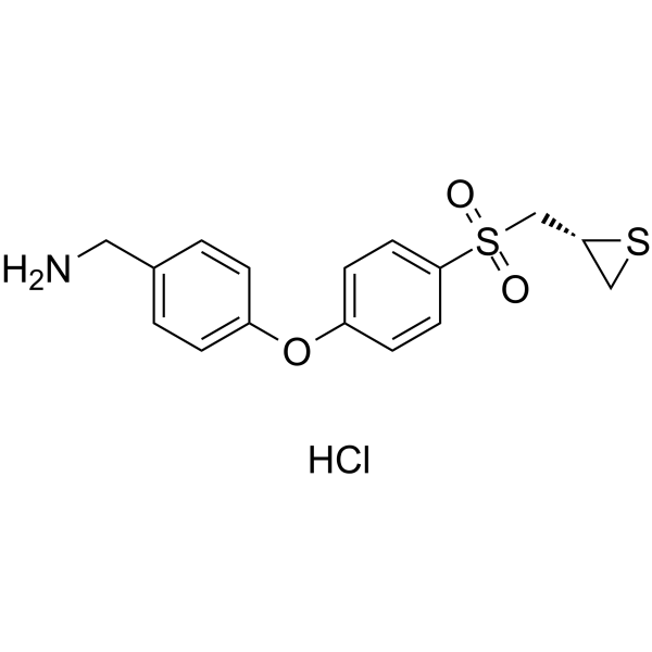
(R)-ND-336
(R)-ND-336 is a potent and selective MMP-9 inhibitor with a Ki of 19 nM. (R)-ND-336 inhibits MMP-2 (Ki=127 nM) and MMP-14 (Ki=119 nM). (R)-ND-336 has the potential for diabetic foot ulcers (DFUs) research.
For research use only. We do not sell to patients.
- CAS No.: 2252493-33-5
- Formula: C16H18ClNO3S2
- Molecular Weight:371.90
-
Storage:
Please store the product under the recommended conditions in the Certificate of Analysis.

To place orders, for customer services and technical support, please contact: MedChemExpress USA
Tel: 609-228-6898 E-mail: [email protected] [email protected]


-
 Biological Activity
-
Chemical Information
- Protocols
- Purity & Documentation
- References
-
 Help & FAQs




Biological Activity
Description
IC50 & Target
[1]|
MMP-9 19 nM (Ki) |
MMP-2 127 nM (Ki) |
MMP-14 119 nM (Ki) |
In Vitro
(R)-ND-336 poorly inhibits other MMPs (Ki>100 μM). (R)-ND-336 has a long residence time (the time the inhibitor is bound to MMP-9, calculated as 1/koff) of 300 min[1].
A favorable IC50value of 143 μM afforded a therapeutic index (IC50/Ki for MMP-9) ratio of 7530, indicating that (R)-ND-336 is not cytotoxic[1].
MedChemExpress (MCE) has not independently confirmed the accuracy of these methods. They are for reference only.
In Vivo
(R)-ND-336 has an AUC of 1.3 μM min after topical administration and 35 μM min following intravenous dosing, for an AUCtop/AUCiv ratio of 3.7%, indicating minimal absorption after topical administration in db/db mice[1].
MedChemExpress (MCE) has not independently confirmed the accuracy of these methods. They are for reference only.
-
Animal Model:Female db/db mice (BKS.Cg-Dock7m+/+Leprdb/J, 8-weeks old, ∼40 g body weight)[1]
-
Dosage:50 μg
-
Administration:Wound; once a day for 14 days
-
Result:Accelerated wound healing faster and showed complete inhibition of MMP-9.


Chemical Information
-
CAS No. 2252493-33-5
-
Molecular Weight 371.90
-
Formula C16H18ClNO3S2
-
SMILES
NCC1=CC=C(OC2=CC=C(C=C2)S(C[C@@H]3SC3)(=O)=O)C=C1.Cl
-
Shipping
Room temperature in continental US; may vary elsewhere.
-
Storage
Please store the product under the recommended conditions in the Certificate of Analysis.


Purity & Documentation
References

Calculators
Concentration (start) × Volume (start) = Concentration (final) × Volume (final)


(R)-ND-336 Related Classifications
HY-124373A Related Classifications
 Powered by Bioz