



173 Results for "

cell staining

" in MedChemExpress (MCE) Product Catalog:
Products (173)

173 Results for "cell staining" in MCE Product Catalog:

188
188 Publications Verification
Cat. No.: HY-D0079
CAS No.: 104821-25-2
Synonyms: Hydroethidine; PD-MY 003
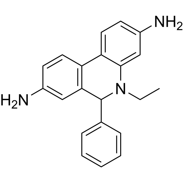
Dihydroethidium, also known as DHE, is a peroxide indicator. Dihydroethidium penetrates cell membranes to form a fluorescent protein complex with blue fluoresces. After entering the cells, Dihydroethidium is mainly localized in the cell membrane, cytoplasm and nucleus, and the staining effect is the strongest in the nucleus. Dihydroethidium produces inherent blue fluorescence with a maximum excitation wavelength of 370 nm and a maximum emission wavelength of 420 nm; after dehydrogenation, Dihydroethidium combines with RNA or DNA to produce red fluorescence with a maximum excitation wavelength of 300 nm and a maximum emission wavelength of 610 nm. 535 nm can also be used as the excitation wavelength for actual observation .
loading...
    loading...
175
175 Cited Publications
Cat. No.: HY-D0814
CAS No.: 28718-90-3
Synonyms: 4',6-Diamidino-2-phenylindole dihydrochloride
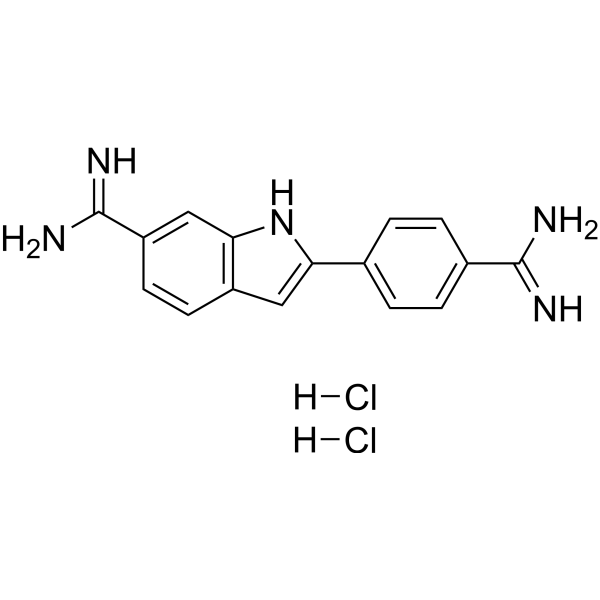
DAPI (4',6-Diamidino-2-phenylindole) dihydrochloride is a DAPI dye. DAPI is a fluorescent dye that binds strongly to DNA. It binds to the AT base pair of the double-stranded DNA minor groove, and one DAPI molecule can occupy three base pair positions. The fluorescence intensity of DAPI molecules bound to double-stranded DNA is increased by about 20 times, and it is commonly observed with fluorescence microscopy, and the amount of DNA can be determined based on the intensity of fluorescence. DAPI cannot penetrate intact cell membranes and is commonly used for staining damaged and fixed cells . DAPI (Compound 3) is an acid-sensing ion channel 3 (ASIC3) inhibitor. DAPI binds to ASIC3 and blocks the channel function. DAPI can be used in the study of chronic pain treatment (Ex/Em = 356/451 nm).
loading...
    loading...
160
160 Cited Publications
Cat. No.: HY-D0815
CAS No.: 25535-16-4
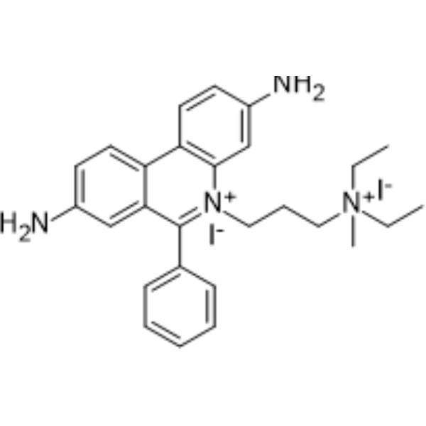
Propidium Iodide (PI) is a nuclear staining agent that stains DNA. Propidium Iodide is an analogue of ethidine bromide that emits red fluorescence upon embedding in double-stranded DNA. Propidium Iodide cannot pass through living cell membranes, but it can pass through damaged cell membranes to stain the nucleus. Propidium Iodide has a fluorescence wavelength of 493/617 nm and a wavelength of 536/635 nm after Mosaic with DNA. Propidium Iodide is commonly used in the detection of apoptosis (apoptosis) or necrosis (necrosis), and is often used in flow cytometry analysis.
loading...
    loading...
159
159 Cited Publications
Cat. No.: HY-D2868
CAS No.: 47165-04-8
Synonyms: 4',6-Diamidino-2-phenylindole
DAPI (4',6-Diamidino-2-phenylindole) is a DAPI dye. DAPI is a fluorescent dye that binds strongly to DNA. It binds to the AT base pair of the double-stranded DNA minor groove, and one DAPI molecule can occupy three base pair positions. The fluorescence intensity of DAPI molecules bound to double-stranded DNA is increased by about 20 times, and it is commonly observed with fluorescence microscopy, and the amount of DNA can be determined based on the intensity of fluorescence. DAPI cannot penetrate intact cell membranes and is commonly used for staining damaged and fixed cells . DAPI (Compound 3) is an acid-sensing ion channel 3 (ASIC3) inhibitor. DAPI binds to ASIC3 and blocks the channel function. DAPI can be used in the study of chronic pain treatment (Ex/Em = 356/451 nm).
loading...
    loading...
129
129 Cited Publications
Cat. No.: HY-D1738
CAS No.: 28718-91-4
Synonyms: 4',6-Diamidino-2-phenylindole dilactate
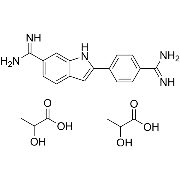
DAPI (4',6-Diamidino-2-phenylindole) dilactate is a DAPI dye. DAPI is a fluorescent dye that binds strongly to DNA. It binds to the AT base pair of the double-stranded DNA minor groove, and one DAPI molecule can occupy three base pair positions. The fluorescence intensity of DAPI molecules bound to double-stranded DNA is increased by about 20 times, and it is commonly observed with fluorescence microscopy, and the amount of DNA can be determined based on the intensity of fluorescence. DAPI cannot penetrate intact cell membranes and is commonly used for staining damaged and fixed cells . DAPI (Compound 3) is an acid-sensing ion channel 3 (ASIC3) inhibitor. DAPI binds to ASIC3 and blocks the channel function. DAPI can be used in the study of chronic pain treatment (Ex/Em = 356/451 nm) .
loading...
    loading...
126
126 Cited Publications
Cat. No.: HY-W090090
CAS No.: 121207-31-6
Synonyms: Pyrromethene 546
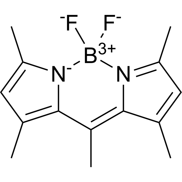
BODIPY493/503 is a BODIPY dye. BODIPY dye is a small molecule dye with strong ultraviolet absorption ability, its fluorescence peak is relatively sharp, and the quantum yield is high. They are relatively insensitive to the polarity and pH of the environment and are relatively stable under different physiological conditions. Due to its structural asymmetry, BODIPY derives a variety of structural products. BODIPY lipid droplet dyes can well pass through the cell membrane into the cell, and localize the neutral lipids in the cell to specifically stain the lipid droplets, which can be used for labeling of live cells and fixed cells . Maximum excitation/emission wavelength: 493/503 nm .
loading...
    loading...
119
119 Cited Publications
Cat. No.: HY-D0938
CAS No.: 150347-59-4
Synonyms: CFSE; 5(6)-Carboxyfluorescein diacetate succinimidyl ester; 5(6)-CFDA N-succinmidyl ester
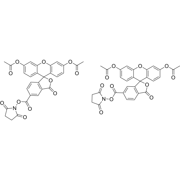
CFDA-SE is a fluorescent dye that can penetrate the cell membrane. It can react with the free amine group in the cytoskeleton protein inside the cell, and finally form a protein complex with fluorescence. After entering the cell, CFDA-SE locates in the cell membrane, cytoplasm and nucleus, and the fluorescence staining is strongest in the nucleus . CFDA-SE dye can be uniformly inherited by the cells with cell division and proliferation, and its attenuation is proportional to the number of cell divisions. This phenomenon can be detected and analyzed by flow cytometry under the excitation light of 488 nm, and can be used to detect the proliferation of cells .
loading...
    loading...
83
83 Cited Publications
Cat. No.: HY-K1073

MCE Annexin V-FITC/PI Apoptosis Detection Kit provides a rapid and convenient method to detect cell apoptosis and necrosis. After staining, live cells show little or no fluorescence (Annexin V-/PI-), early apoptosis cells show green fluorescence(Annexin V+/PI-), late apoptosis cells and necrosis cells show red and green fluorescence (Annexin V+/PI+).


loading...
    loading...
69
69 Cited Publications
Cat. No.: HY-B0324A
CAS No.: 548-62-9
Synonyms: Basic Violet 3; Gentian Violet; Methyl Violet 10B
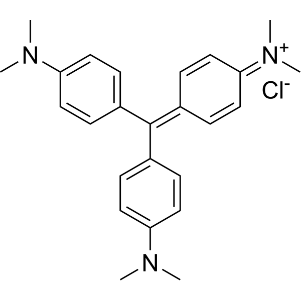
Crystal Violet, also known as Gentian violet, methyl violet 10B, is a triphenyl-methane, an alkaline dye that binds to DNA in the nucleus of a cell, staining it a deep purple. It is often used for Gram staining to classify bacteria, or for cell or histological staining[1].
loading...
    loading...
43
43 Cited Publications
Cat. No.: HY-D0952
CAS No.: 494-38-2
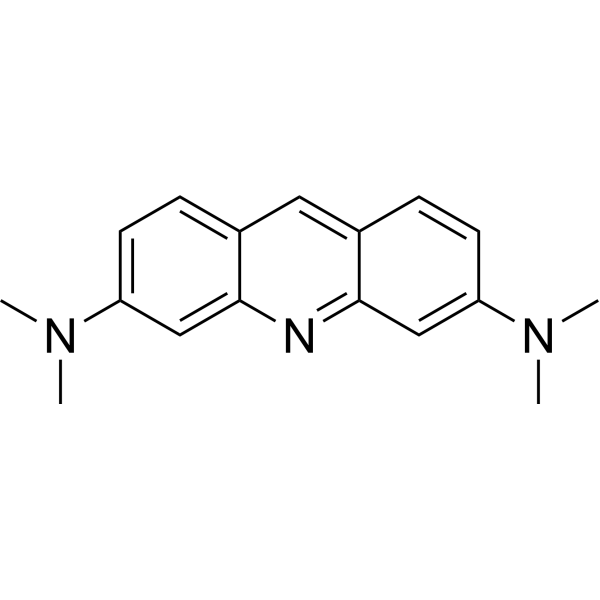
Acridine Orange base is a cell-permeable fluorescent dye that stains organisms (bacteria, parasites, viruses, etc.) bright orange and, when used under appropriate conditions (pH=3.5, Ex=460 nm), distinguishes human cells in green for detection by fluorescence microscopy. Acridine Orange base fluoresces green when bound to dsDNA (Ex=488, Em=520-524) and red when bound to ssDNA (Ex=457, Em=630-644) or ssRNA (Ex=457, Em=630-644), also can be used in cell cycle assays .
loading...
    loading...
42
42 Cited Publications
Cat. No.: HY-K1078

MCE One Step TUNEL Apoptosis Detection Kit (FITC) provides a rapid and convenient method to detect cell apoptosis. After staining cells with this kit, live cells have no fluorescence, apoptosis cells show green fluorescence.

loading...
    loading...
37
37 Cited Publications
Cat. No.: HY-118411
CAS No.: 61135-33-9
Purity:  99.78%
Synonyms: EdU
Target:  

PROTAC Linkers

Research Areas:  

Cancer

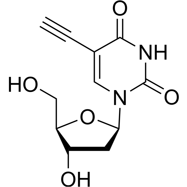
5-Ethynyl-2'-deoxyuridine (EdU), a thymidine analogue, is incorporated into cellular DNA during DNA replication and the subsequent reaction of EdU with a fluorescent azide in a “Click” reaction. EdU staining is a fast, sensitive and reproducible method to study cell proliferation . 5-Ethynyl-2'-deoxyuridine is an alkyl chain-based PROTAC linker that can be used in the synthesis of PROTACs . 5-Ethynyl-2'-deoxyuridine is a click chemistry reagent, it contains an Alkyne group and can undergo copper-catalyzed azide-alkyne cycloaddition (CuAAc) with molecules containing Azide groups.
loading...
    loading...
30
30 Cited Publications
Cat. No.: HY-K1071

MCE Cell Cycle and Apoptosis Analysis Kit (PI staining) provides a convenient method to detect cell cycle and cell apoptosis.


loading...
    loading...
23
23 Cited Publications
Cat. No.: HY-D1614
CAS No.: 216434-81-0
Target:  

Fluorescent Dye

Research Areas:  

Others

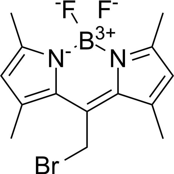
BODIPY493/503 methyl bromide is a BODIPY dye. BODIPY dye is a small molecule dye with strong ultraviolet absorption ability, its fluorescence peak is relatively sharp, and the quantum yield is high. They are relatively insensitive to the polarity and pH of the environment and are relatively stable under different physiological conditions. Due to its structural asymmetry, BODIPY derives a variety of structural products. BODIPY lipid droplet dyes can well pass through the cell membrane into the cell, and localize the neutral lipids in the cell to specifically stain the lipid droplets, which can be used for labeling of live cells and fixed cells . Maximum excitation/emission wavelength: 493/503 nm .
loading...
    loading...
18
18 Cited Publications
Cat. No.: HY-D1421
CAS No.: 257277-27-3
PKH67 is a fluorescent cell binding dye with green fluorescence. PKH67 can stain the cell membrane and the Ex/Em is 490/502 nm. PKH67 is often used in combination with the non-specific red fluorescent dye PKH26 (Ex/Em=551/567 nm) to label cells, detect cell proliferation in vitro, and trace cells in vitro and in vivo .
loading...
    loading...
16
16 Cited Publications
Cat. No.: HY-103311
CAS No.: 11103-72-3
Purity:  ≥97.0%
Synonyms: Ammoniated ruthenium oxychloride
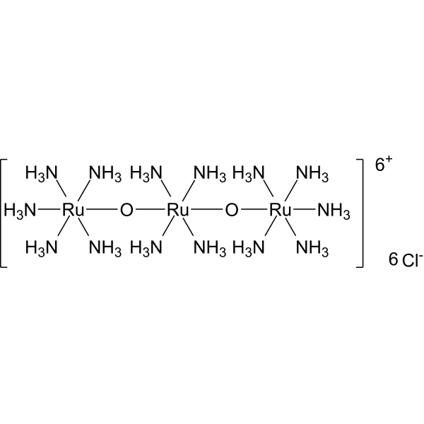
Ruthenium red (Ammoniated ruthenium oxychloride) is a polycationic dye widely used for electron microscopy (EM) of cells, tissues and vegetative bacteria. Ruthenium red strongly reacts with phospholipids and fatty acids and binds to acidic mucopolysaccharides. Ruthenium red is a L-type calcium current (ICa) blocker. When using Ruthenium Red to stain plant tissue sections affected by pathogens, the breakdown of mucilaginous substances by the pathogens prevents them from taking up the stain, whereas the cell walls of healthy tissue stain red .
loading...
    loading...
13
13 Cited Publications
Cat. No.: HY-K0315

MCE Hematoxylin-Eosin Staining Kit can be used for cell staining, the combination of hematoxylin and eosin results in a staining pattern where the cytoplasm appears red or pink and the nucleus takes on a blue or blue-violet.

loading...
    loading...
12
12 Cited Publications
Cat. No.: HY-D0714
CAS No.: 298-96-4
Synonyms: 2,3,5-Triphenyltetrazolium chloride; TPTZ; TTC
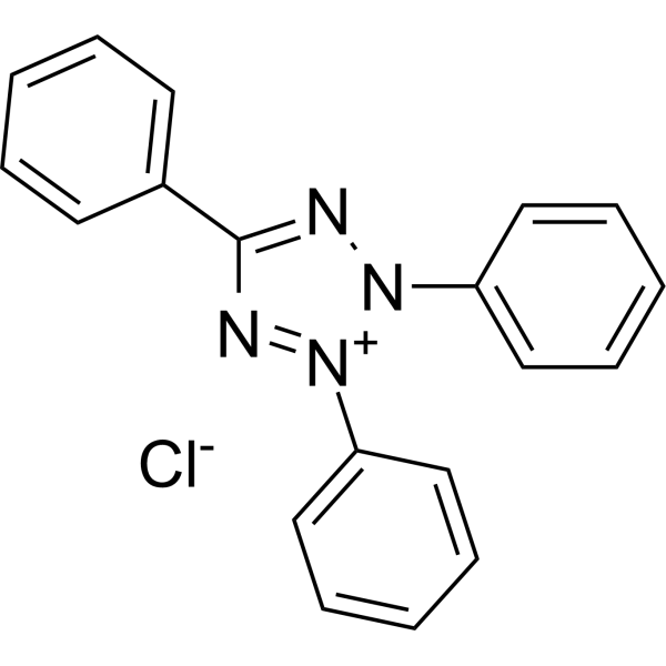
Tetrazolium Red (2,3,5-Triphenyltetrazolium chloride; TTC) is a not brain-penetrant, colorless, water-soluble dye that is reduced by mitochondrial enzymes to a deep red, water-insoluble compound (formazan) mainly in the mitochondria of living cells. Tetrazolium Red is used to observe the activity of dehydrogenase, and it turns colorless to red when exposed to hydrogen. Tetrazolium Red distinguishes between surviving and infarcted brain tissue after stroke. Tetrazolium Red has been used to stain heart tissue to measure the extent of acute lesions and also used to stain brain tissue to detect the size of the infarcted area. The absorption wavelength of Tetrazolium Red is 570 nm .
loading...
    loading...
12
12 Cited Publications
Cat. No.: HY-K1089

MCE Cell Senescence β-Galactosidase Staining Kit can be used to evaluate the senescence of cells or tissues.

loading...
    loading...
12
12 Cited Publications
Cat. No.: HY-K1094

MCE Viability/Cytotoxicity Assay Kit for Live & Dead Cells (Calcein/PI) enables dual fluorescent staining of both live and dead cells, making it suitable for assessing cell viability and cytotoxicity. The 500 T is defined as the base specification. All larger sizes correspond to incremental volumes of this base.

loading...
    loading...

