



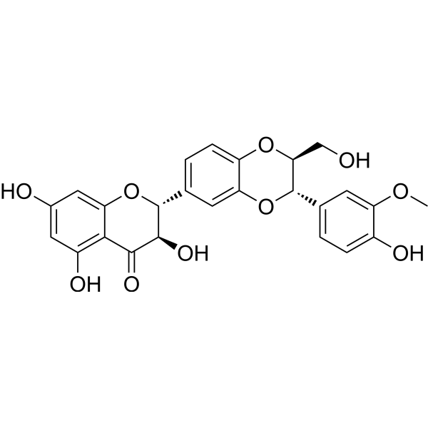
Silybin B

Based on 1 Customer Validation
Silybin B (Silibinin B) is an orally active amyloid-β aggregation inhibitor and ATR pathway activator that can cross the blood-brain barrier. Silybin B inhibits Aβ fibril formation and promotes amorphous aggregate formation, while activating the ATR-mediated DNA damage repair pathway and inhibiting JNK/p38 MAPK signaling. Silybin B can reduce Cisplatin (HY-17394)-induced neuronal DNA damage and apoptosis. Silybin B has anti-oxidative stress, cell cycle regulation and neuroprotective activities. Silybin B is mainly used in the study of Alzheimer's disease and Cisplatin chemotherapy-related neurotoxicity.
For research use only. We do not sell to patients.
- Purity : 99.90%
- CAS No.: 142797-34-0
- Formula: C25H22O10
- Molecular Weight:482.44
-
Storage:Powder -20°C, 3 years , 4°C, 2 years ; In solvent -80°C, 6 months , -20°C, 1 month

To place orders, for customer services and technical support, please contact: MedChemExpress USA
Tel: 609-228-6898 E-mail: [email protected] [email protected]


-
 Biological Activity
-
Chemical Information
-
Solvent & Solubility
- Protocols
- Purity & Documentation
- References
-
 Help & FAQs


-
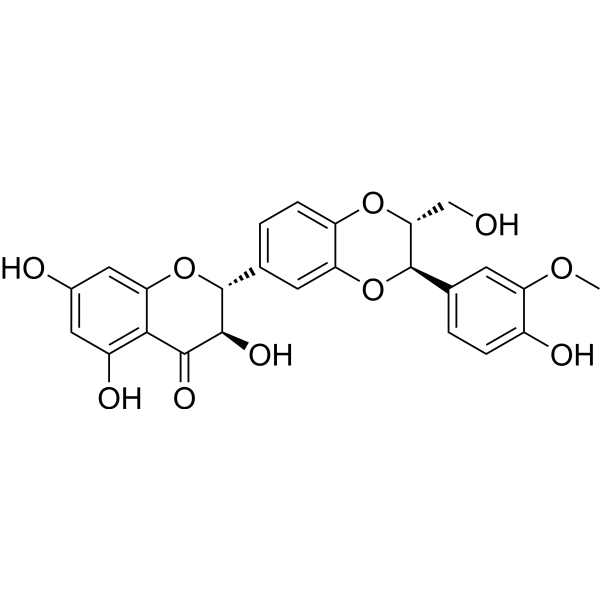
HY-13748
Silybin AAnticancer Agent -

HY-N7073
SilymarinMpro Inhibitor -

HY-N7046R
Silybin B (Standard)Anti-cancer Agent -
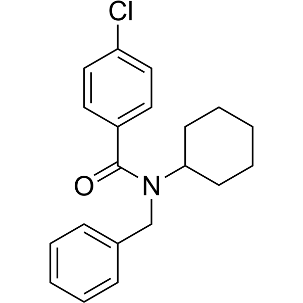
HY-19370
FPS-ZM1Amyloid-β Inhibitor -
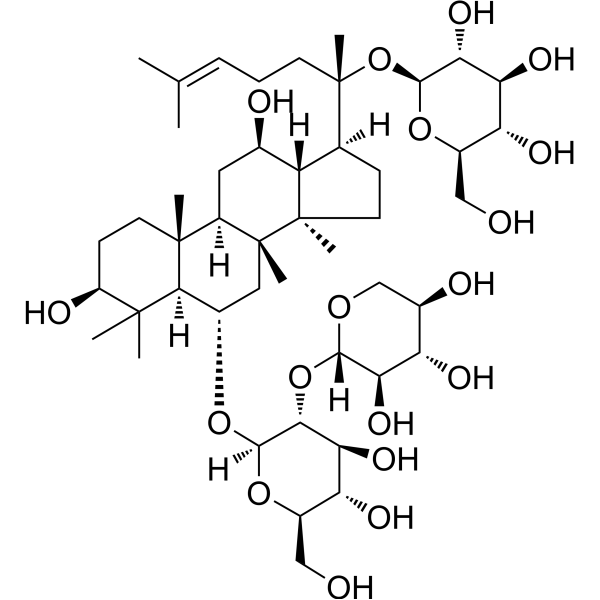
HY-N0615
Notoginsenoside R1Apoptosis Inhibitor -
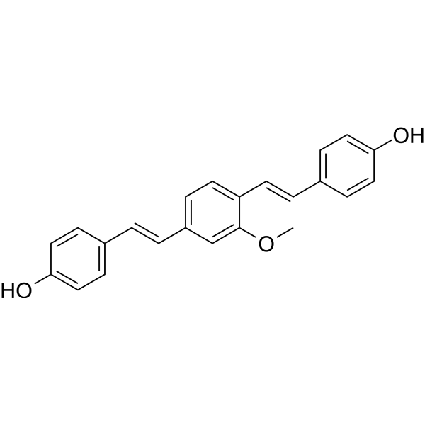
HY-103240
Methoxy-X04Fluorescent Dye -
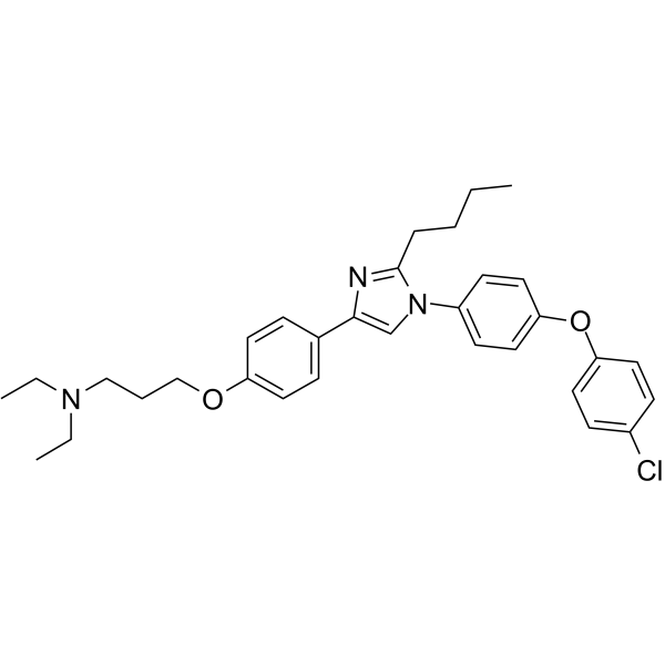
HY-50682
AzeliragonRAGE Inhibitor -
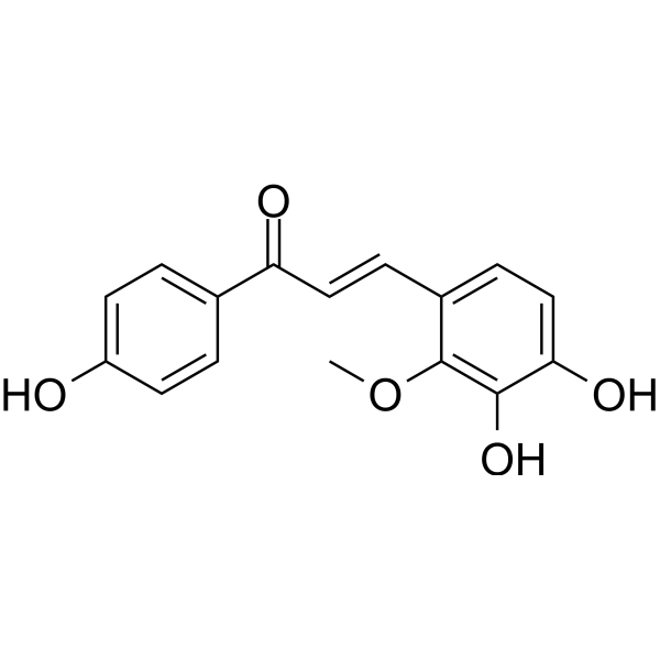
HY-N0373
Licochalcone BApoptosis Inducer -
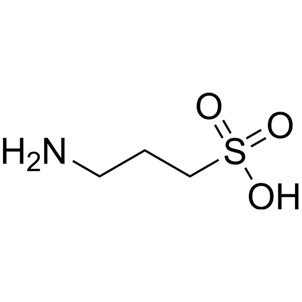
HY-14602
TramiprosateNeuroprotection Agent -
HY-N0249
Saikosaponin CMMP-2 Inducer -
HY-103242
CRANAD-2Fluorescent Probe -
HY-N0602
Ginsenoside Rg2NF-κB Inhibitor


Biological Activity
Description
Cellular Effect
|
Cell Line

|
Type | Value | Description | References |
|---|---|---|---|---|
| B16-4A5 | IC50 |
>100 μM
Compound: 6
|
Toxicity in mouse B16-4A5 cells assessed as inhibition of cell proliferation in presence of 1 mM theophylline after 72 hrs by WST8 dye reduction assay
Toxicity in mouse B16-4A5 cells assessed as inhibition of cell proliferation in presence of 1 mM theophylline after 72 hrs by WST8 dye reduction assay
|
[PMID: 20189399] |
| DU-145 | IC50 |
61.8 μM
Compound: 1
|
Antiproliferative activity against human DU145 cells after 72 hrs after ATPlite kit assay
Antiproliferative activity against human DU145 cells after 72 hrs after ATPlite kit assay
|
[PMID: 23260576] |
| HEK293 | IC50 |
5 μM
Compound: Silybin B
|
Inhibition of OATP1B3 (unknown origin) expressed in HEK293 cells assessed as reduction of [3H]estradiol-17beta-glucuronide uptake after 3 mins by beta-counting
Inhibition of OATP1B3 (unknown origin) expressed in HEK293 cells assessed as reduction of [3H]estradiol-17beta-glucuronide uptake after 3 mins by beta-counting
|
[PMID: 23401473] |
| HEK293 | IC50 |
8.5 μM
Compound: Silybin B
|
Inhibition of OATP1B1 (unknown origin) expressed in HEK293 cells assessed as reduction of [3H]estradiol-17beta-glucuronide uptake after 3 mins by beta-counting
Inhibition of OATP1B1 (unknown origin) expressed in HEK293 cells assessed as reduction of [3H]estradiol-17beta-glucuronide uptake after 3 mins by beta-counting
|
[PMID: 23401473] |
| Huh-7 | IC50 |
40 μM
Compound: 1
|
Antiviral activity against HCV JFH1 infected in human Huh7.5.1 cells assessed as reduction in viral proteins after 72 hrs post compound dose by Western blotting method
Antiviral activity against HCV JFH1 infected in human Huh7.5.1 cells assessed as reduction in viral proteins after 72 hrs post compound dose by Western blotting method
|
[PMID: 23260576] |
| Huh-7 | IC50 |
56.2 μM
Compound: 1
|
Antiproliferative activity against human Huh7.5.1 cells after 72 hrs after ATPlite kit assay
Antiproliferative activity against human Huh7.5.1 cells after 72 hrs after ATPlite kit assay
|
[PMID: 23260576] |
| Huh-7 | IC50 |
68.7 μM
Compound: 3
|
Cytotoxicity against human Huh7.5.1 cells after 72 hrs
Cytotoxicity against human Huh7.5.1 cells after 72 hrs
|
[PMID: 23673225] |
| Huh7.5.1 | CC50 |
68.7 μM
Compound: 4
|
Cytotoxicity against human Huh7.5.1 cells after 72 hrs by ATPlite assay
Cytotoxicity against human Huh7.5.1 cells after 72 hrs by ATPlite assay
|
[PMID: 30485098] |
| HUVEC | IC50 |
6.49 μM
Compound: 1b, (2R,3R,10S,11S)
|
Antiproliferative activity against HUVEC assessed as inhibition of cell growth after 3 days by MTT assay
Antiproliferative activity against HUVEC assessed as inhibition of cell growth after 3 days by MTT assay
|
[PMID: 21928794] |
| HUVEC | IC50 |
60.47 μM
Compound: 1b, (2R,3R,10S,11S)
|
Cytotoxicity against HUVEC assessed as cell viability after 16 hrs by MTT assay
Cytotoxicity against HUVEC assessed as cell viability after 16 hrs by MTT assay
|
[PMID: 21928794] |
| LNCaP | IC50 |
69.6 μM
Compound: 1
|
Antiproliferative activity against human LNCAP cells after 72 hrs after ATPlite kit assay
Antiproliferative activity against human LNCAP cells after 72 hrs after ATPlite kit assay
|
[PMID: 23260576] |
| MDCK-II | IC50 |
0.8 μM
Compound: Silybin B
|
Inhibition of OATP2B1 (unknown origin) expressed in MDCK2 cells assessed as reduction of [3H]estrone-3-sulfate uptake after 3 mins by beta-counting
Inhibition of OATP2B1 (unknown origin) expressed in MDCK2 cells assessed as reduction of [3H]estrone-3-sulfate uptake after 3 mins by beta-counting
|
[PMID: 23401473] |
| PC-3 | IC50 |
60.5 μM
Compound: 1
|
Antiproliferative activity against human PC3 cells after 72 hrs after ATPlite kit assay
Antiproliferative activity against human PC3 cells after 72 hrs after ATPlite kit assay
|
[PMID: 23260576] |
In Vitro
Silybin B (4 μM; 24 h) inhibits amyloid fibril formation, promoted amorphous aggregate formation, and reduces ThT fluorescence intensity in 10 μM Aβ40 solution[1].
Silybin B (20, 40, 80 μM; 48 h) dose-dependently antagonizes the cytotoxicity of 10 μM Cisplatin (HY-17394), and increases HT22 cell viability, reduces cell arrest in the G2/M phase, reduces cell apoptosis, and increases cleaved-PARP protein expression[2].
Silybin B (20, 40, 80 μM; 24 h) inhibits protein phosphorylation in the JNK/p38 MAPK signaling pathway in HT22 cells, upregulates p-ATR protein expression and downregulates γ-H2AX levels, and alleviates DNA damage[2].
MedChemExpress (MCE) has not independently confirmed the accuracy of these methods. They are for reference only.
-
Cell Line:Murine hippocampal neuronal HT22 cells
-
Concentration:20 μM, 40 μM, 80 μM
-
Incubation Time:48 h
-
Result:Dose-dependently increased cell viability in cisplatin (10 μM)-treated HT22 cells.
Compared to the cisplatin-alone group, cell viability increased from 30% to 60% (20 μM) and 70% (80 μM), indicating alleviation of cisplatin-induced cytotoxicity.
In Vivo
Silybin B (25, 50 mg/kg; gavage; once a day; 15 days) increases the antioxidant indexes of SOD, GSH, and T-AOC in the brain, reduces the level of LPO, and alleviates neuronal damage and apoptosis in the hippocampus in a Cisplatin (HY-17394)-induced mouse neurotoxicity model[2].
MedChemExpress (MCE) has not independently confirmed the accuracy of these methods. They are for reference only.
-
Animal Model:Male Balb/c mice (6-7 weeks old) with Cisplatin-induced neurotoxicity model[2]
-
Dosage:25 mg/kg, 50 mg/kg
-
Administration:Oral gavage, daily for 15 consecutive days, with a single intraperitoneal injection of Cisplatin (10 mg/kg) on day 10
-
Result:Significantly improved motor coordination and learning memory in Cisplatin-treated mice, as evidenced by reduced escape latency in the Morris water maze and decreased time to descend the pole test.
I ncreased the levels of superoxide dismutase (SOD), reduced glutathione (GSH), and total antioxidant capacity (T-AOC) in brain tissue, while decreased lipid peroxidation (LPO) levels.
- Neurotoxicity Study
- Detection of Apoptosis
- TUNEL staining for apoptotic DNA fragmentation
- Flow cytometric DNA-content cell-cycle staining
- Annexin V plus membrane-impermeant dye apoptosis staining
- ROS/oxidative-stress fluorescent staining
- BrdU Incorporation Assay
- Apoptosis Solutions
- Protocol for Cell Cycle
- Amyloid: Congo Red Amyloid Staining
- Genotoxicity/Mutagenicity Study
- Alzheimer’s Disease Modeling


Chemical Information
-
CAS No. 142797-34-0
-
Appearance Solid
-
Molecular Weight 482.44
-
Formula C25H22O10
-
Color White to off-white
-
SMILES
O=C1[C@H](O)[C@@H](C2=CC=C(O[C@@H](CO)[C@H](C3=CC=C(O)C(OC)=C3)O4)C4=C2)OC5=CC(O)=CC(O)=C15
-
Synonyms
Silibinin B
-
Structure Classification
-
Shipping
Room temperature in continental US; may vary elsewhere.
-
Storage
Powder -20°C 3 years 4°C 2 years In solvent -80°C 6 months -20°C 1 month


Solvent & Solubility
In Vitro:
DMSO : 250 mg/mL (518.20 mM; Need ultrasonic; Hygroscopic DMSO has a significant impact on the solubility of product, please use newly opened DMSO)
Please refer to the solubility information to select the appropriate solvent. Once prepared, please aliquot and store the solution to prevent product inactivation from repeated freeze-thaw cycles.
Storage method and period of stock solution: -80°C, 6 months; -20°C, 1 month. When stored at -80°C, please use it within 6 months. When stored at -20°C, please use it within 1 month.
Please refer to the solubility information to select the appropriate solvent. Once prepared, please aliquot and store the solution to prevent product inactivation from repeated freeze-thaw cycles.
Storage method and period of stock solution: -80°C, 6 months; -20°C, 1 month. When stored at -80°C, please use it within 6 months. When stored at -20°C, please use it within 1 month.
Concentration (start) × Volume (start) = Concentration (final) × Volume (final)
In Vivo:
Select the appropriate dissolution method based on your experimental animal and administration route.
- For the following dissolution methods, please ensure to first prepare a clear stock solution using an In Vitro approach and then sequentially add co-solvents:
- To ensure reliable experimental results, the clarified stock solution can be appropriately stored based on storage conditions. As for the working solution for In Vivo experiments, it is recommended to prepare freshly and use it on the same day.
- The percentages shown for the solvents indicate their volumetric ratio in the final prepared solution. If precipitation or phase separation occurs during preparation, heat and/or sonication can be used to aid dissolution.
Add each solvent one by one: 10% DMSO 40% PEG300 5% Tween-80 45% Saline
Solubility: ≥ 2.08 mg/mL (4.31 mM); Clear solution
This protocol yields a clear solution of ≥ 2.08 mg/mL (saturation unknown).
Taking 1 mL working solution as an example, add 100 μL DMSO stock solution (20.8 mg/mL) to 400 μL PEG300, and mix evenly; then add 50 μL Tween-80 and mix evenly; then add 450 μL Saline to adjust the volume to 1 mL.
Preparation of Saline: Dissolve 0.9 g sodium chloride in ddH₂O and dilute to 100 mL to obtain a clear Saline solution.
Add each solvent one by one: 10% DMSO 90% (20% SBE-β-CD in Saline)
Solubility: ≥ 2.08 mg/mL (4.31 mM); Clear solution
This protocol yields a clear solution of ≥ 2.08 mg/mL (saturation unknown).
Taking 1 mL working solution as an example, add 100 μL DMSO stock solution (20.8 mg/mL) to 900 μL 20% SBE-β-CD in Saline, and mix evenly.
Preparation of 20% SBE-β-CD in Saline (4°C, storage for one week): 2 g SBE-β-CD powder is dissolved in 10 mL Saline, completely dissolve until clear.
In Vivo Dissolution Calculator
Please enter the basic information of animal experiments:
-
-
-
-
Recommended: Prepare an additional quantity of animals to account for potential losses during experiments.
Please enter your animal formula composition:
-
%DMSO +
Recommended: Keep the proportion of DMSO in working solution below 2% if your animal is weak.
-
%+
-
+%Tween-80 + +
-
%Saline +
The co-solvents required include: DMSO, . All of co-solvents are available by MedChemExpress (MCE). , Tween 80. All of co-solvents are available by MedChemExpress (MCE).
Working solution concentration: 0.22 mg/mL
Method for preparing stock solution: mg drug dissolved in μL DMSO. Stock solution concentration: mg/mL.
1. Take μL DMSO stock solution;
2. Add μL .
μL , mix evenly;
3. Then add μL Tween 80, mix evenly;
4. Then add μL
Please ensure that the stock solution in the first step is dissolved to a clear state, and add co-solvents in sequence. You can use ultrasonic heating (ultrasonic cleaner, recommended frequency 20-40 kHz), vortexing, etc. to assist dissolution.


Purity & Documentation
-
Data Sheet (278 KB)
-
SDS (393 KB)
- English - EN (393 KB)
- Français - FR (393 KB)
- Deutsch - DE (393 KB)
- Norwegian - NO (393 KB)
- Español - ES (393 KB)
- Swedish - SV (393 KB)
- Italian - IT (393 KB)
- Korean - KR (393 KB)
- Portuguese - PT (393 KB)
-
Handling Instructions (2659 KB)
References
[1]. Sciacca MFM, et al. Inhibition of Aβ Amyloid Growth and Toxicity by Silybins: The Crucial Role of Stereochemistry. ACS Chem Neurosci. 2017 Aug 16;8(8):1767-1778. [Content Brief]
[2]. Wang XL, et al. Silybin B exerts protective effect on cisplatin-induced neurotoxicity by alleviating DNA damage and apoptosis. J Ethnopharmacol. 2022 Apr 24;288:114938. [Content Brief]


Complete Stock Solution Preparation Table
Please refer to the solubility information to select the appropriate solvent. Once prepared, please aliquot and store the solution to prevent product inactivation from repeated freeze-thaw cycles.
Storage method and period of stock solution: -80°C, 6 months; -20°C, 1 month. When stored at -80°C, please use it within 6 months. When stored at -20°C, please use it within 1 month.
| Optional Solvent | Concentration Solvent Mass | 1 mg | 5 mg | 10 mg | 25 mg |
|---|---|---|---|---|---|
| DMSO | 1 mM | 2.0728 mL | 10.3640 mL | 20.7280 mL | 51.8199 mL |
| 5 mM | 0.4146 mL | 2.0728 mL | 4.1456 mL | 10.3640 mL | |
| 10 mM | 0.2073 mL | 1.0364 mL | 2.0728 mL | 5.1820 mL | |
| 15 mM | 0.1382 mL | 0.6909 mL | 1.3819 mL | 3.4547 mL | |
| 20 mM | 0.1036 mL | 0.5182 mL | 1.0364 mL | 2.5910 mL | |
| 25 mM | 0.0829 mL | 0.4146 mL | 0.8291 mL | 2.0728 mL | |
| 30 mM | 0.0691 mL | 0.3455 mL | 0.6909 mL | 1.7273 mL | |
| 40 mM | 0.0518 mL | 0.2591 mL | 0.5182 mL | 1.2955 mL | |
| 50 mM | 0.0415 mL | 0.2073 mL | 0.4146 mL | 1.0364 mL | |
| 60 mM | 0.0345 mL | 0.1727 mL | 0.3455 mL | 0.8637 mL | |
| 80 mM | 0.0259 mL | 0.1295 mL | 0.2591 mL | 0.6477 mL | |
| 100 mM | 0.0207 mL | 0.1036 mL | 0.2073 mL | 0.5182 mL |


Silybin B Related Classifications
HY-N7046 Related Classifications
 Powered by Bioz