



Swertianolin
Based on 1 publication(s) in Google Scholar
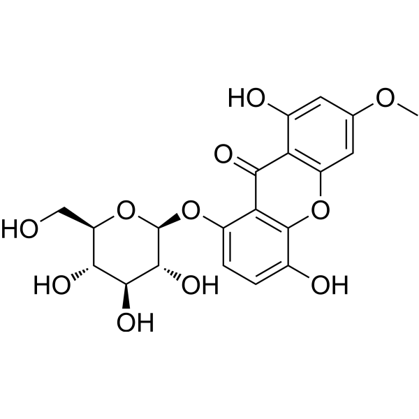
Swertianolin is a xanthone glycoside. Swertianolin can be isolated from plants of the Gentianaceae family (e.g., Gentiana campestris and Swertia punicea). Swertianolin inhibits p38 phosphorylation and NF-κB activation, and reduces NO and ROS production. Swertianolin exhibits anti-Helicobacter pylori activity with an IC50 of 6.1 μM and a minimum bactericidal concentration of 91.7 μM. Swertianolin modulates immune function in sepsis. Swertianolin is useful in the research of diseases such as Alzheimer's disease, hepatitis B, Helicobacter pylori infection, and sepsis.
For research use only. We do not sell to patients.
- Purity : 99.54%
- CAS No.: 23445-00-3
- Formula: C20H20O11
- Molecular Weight:436.37
-
Storage:
4°C, protect from light
* In solvent : -80°C, 6 months; -20°C, 1 month (protect from light)

To place orders, for customer services and technical support, please contact: MedChemExpress USA
Tel: 609-228-6898 E-mail: [email protected] [email protected]


-
 Biological Activity
-
Chemical Information
-
Solvent & Solubility
- Protocols
- Purity & Documentation
- References
-
 Help & FAQs


-

HY-N2192R
Swertianolin (Standard)Standard Of Swertianolin -
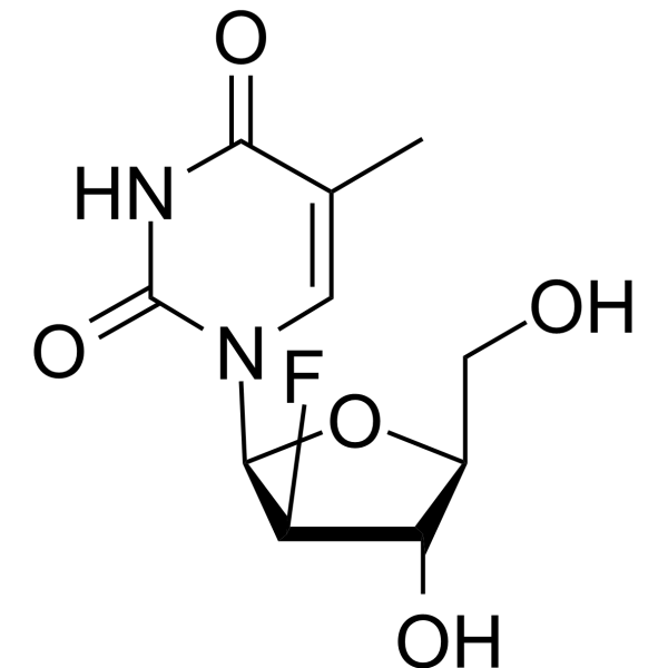
HY-13859
ClevudineHBV Inhibitor -
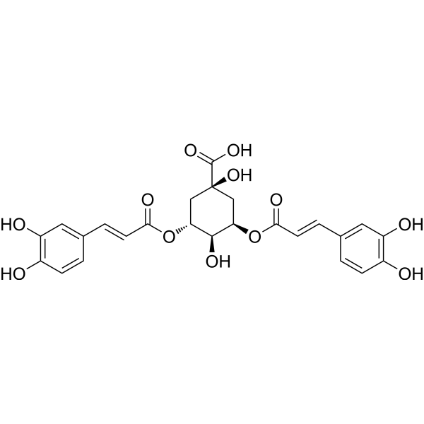
HY-N0056
Isochlorogenic acid AAntioxidant -
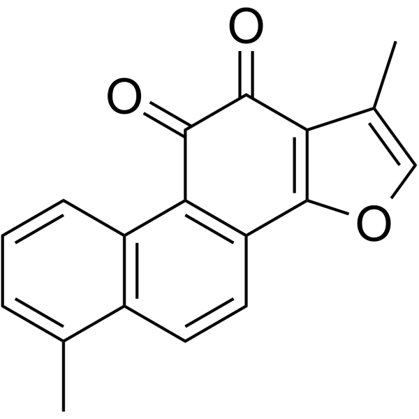
HY-N0134
Tanshinone IPhospholipase Inhibitor -
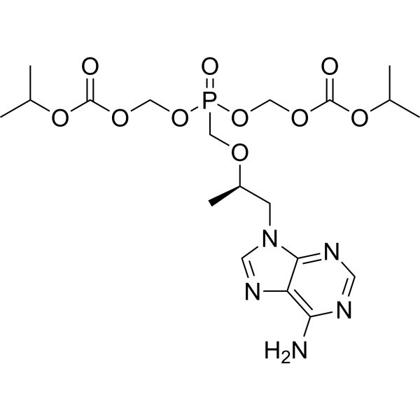
HY-13782A
Tenofovir DisoproxilReverse Transcriptase Inhibitor -
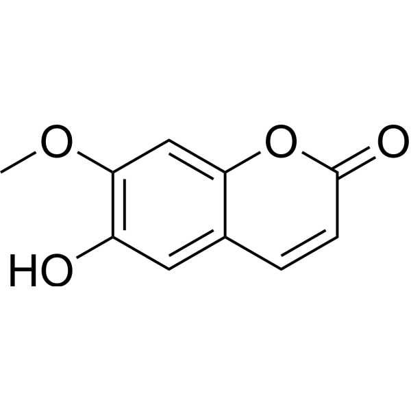
HY-N1365
IsoscopoletinHBV Inhibitor -
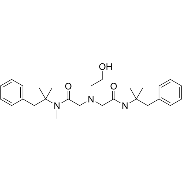
HY-B0955
OxethazaineAnalgesic Agent -
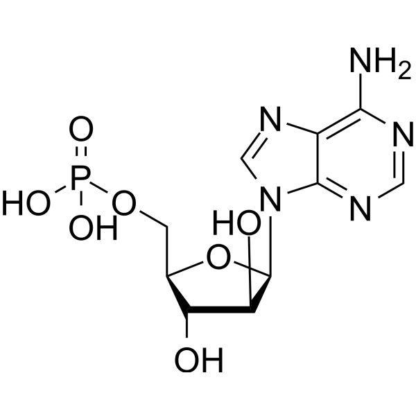
HY-B0277A
Vidarabine phosphateAntiviral Agent -

HY-N1365R
Isoscopoletin (Standard)HBV Inhibitor -

HY-N0134R
Tanshinone I (Standard)Phospholipase Inhibitor -

HY-13782AR
Tenofovir Disoproxil (Standard)Reverse Transcriptase Inhibitor -
HY-B0955R
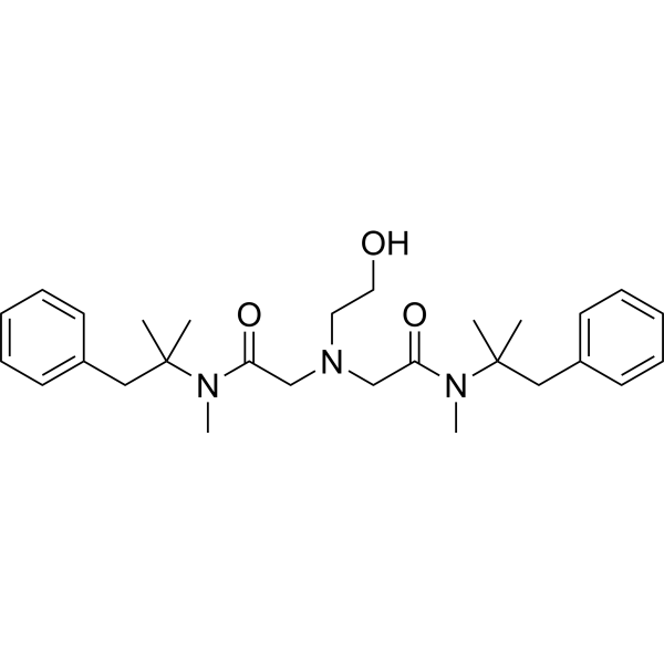
Oxethazaine (Standard)Analgesic Agent
Publications Citing Use of MedChemExpress (MCE) Swertianolin
More

Biological Activity
Description
IC50 & Target
|
AChE |
In Vitro
Swertianolin (1.6-25 μg/mL; 8 days) suppresses the expression of HBeAg in HepG₂ cells with an IC50 of 8.0 μg/mL[2].
Swertianolin (12.5-200 μM; 24-72 h) significantly reduces the secretion of IL-10, NO, ROS, and arginase in MDSCs, inhibits MDSC proliferation, promotes their differentiation into dendritic cells, and improves T-cell activity by blocking the immunosuppressive effect of MDSCs[3].
Swertianolin exhibits anti-Helicobacter pylori activity with an IC50 of 6.1 μM and a minimum bactericidal concentration of 91.7 μM[4].
MedChemExpress (MCE) has not independently confirmed the accuracy of these methods. They are for reference only. Further protocols information, click here.
In Vivo
MedChemExpress (MCE) has not independently confirmed the accuracy of these methods. They are for reference only.
-
Animal Model:8-week-old C57BL/6 mice (weight 25-30 g) with sepsis induced by cecal ligation and puncture (CLP) [5]
-
Dosage:12.5 μg/kg
-
Administration:intraperitoneal injection; 7 days (from the 5th day after CLP surgery)
-
Result:Reduced the proportion of MDSCs in the marrow (58.81% vs 87.89% in normal saline group) and spleen (28.60% vs 48.02% in normal saline group).
Increased CD4+ T cells in the spleen (28.99% vs 20.44% in normal saline group).
Decreased plasma concentrations of IL-10, NO, ROS, and arginase; reduced lung damage and improved survival rate after Legionella pneumophila challenge by inhibiting p38 phosphorylation and NF-κB activation.


Chemical Information
-
CAS No. 23445-00-3
-
Appearance Solid
-
Molecular Weight 436.37
-
Formula C20H20O11
-
Color White to yellow
-
SMILES
O=C1C2=C(OC3=C1C(O[C@H]4[C@@H]([C@H]([C@@H]([C@@H](CO)O4)O)O)O)=CC=C3O)C=C(OC)C=C2O
-
Structure Classification
-
Shipping
Room temperature in continental US; may vary elsewhere.
-
Storage
4°C, protect from light
* In solvent : -80°C, 6 months; -20°C, 1 month (protect from light)


Publications (1)
-
Journal Impact Factor
-
Most Recent
-
Vet Microbiol
The Chinese medicine monomer Schisandrin C inhibits PRRSV infection by regulating the OGT-PI3K/AKT/mTOR signaling pathway. [Abstract]2026 May:316:110992. PMID: 41865607


Solvent & Solubility
In Vitro:
DMSO : 25 mg/mL (57.29 mM; ultrasonic and warming and heat to 60°C; Hygroscopic DMSO has a significant impact on the solubility of product, please use newly opened DMSO)
Please refer to the solubility information to select the appropriate solvent. Once prepared, please aliquot and store the solution to prevent product inactivation from repeated freeze-thaw cycles.
Storage method and period of stock solution: -80°C, 6 months; -20°C, 1 month (protect from light). When stored at -80°C, please use it within 6 months. When stored at -20°C, please use it within 1 month.
Please refer to the solubility information to select the appropriate solvent. Once prepared, please aliquot and store the solution to prevent product inactivation from repeated freeze-thaw cycles.
Storage method and period of stock solution: -80°C, 6 months; -20°C, 1 month (protect from light). When stored at -80°C, please use it within 6 months. When stored at -20°C, please use it within 1 month.
Concentration (start) × Volume (start) = Concentration (final) × Volume (final)
In Vivo:
Select the appropriate dissolution method based on your experimental animal and administration route.
- For the following dissolution methods, please ensure to first prepare a clear stock solution using an In Vitro approach and then sequentially add co-solvents:
- To ensure reliable experimental results, the clarified stock solution can be appropriately stored based on storage conditions. As for the working solution for In Vivo experiments, it is recommended to prepare freshly and use it on the same day.
- The percentages shown for the solvents indicate their volumetric ratio in the final prepared solution. If precipitation or phase separation occurs during preparation, heat and/or sonication can be used to aid dissolution.
Add each solvent one by one: 10% DMSO 40% PEG300 5% Tween-80 45% Saline
Solubility: ≥ 2.5 mg/mL (5.73 mM); Clear solution
This protocol yields a clear solution of ≥ 2.5 mg/mL (saturation unknown).
Taking 1 mL working solution as an example, add 100 μL DMSO stock solution (25.0 mg/mL) to 400 μL PEG300, and mix evenly; then add 50 μL Tween-80 and mix evenly; then add 450 μL Saline to adjust the volume to 1 mL.
Preparation of Saline: Dissolve 0.9 g sodium chloride in ddH₂O and dilute to 100 mL to obtain a clear Saline solution.
In Vivo Dissolution Calculator
Please enter the basic information of animal experiments:
-
-
-
-
Recommended: Prepare an additional quantity of animals to account for potential losses during experiments.
Please enter your animal formula composition:
-
%DMSO +
Recommended: Keep the proportion of DMSO in working solution below 2% if your animal is weak.
-
%+
-
+%Tween-80 + +
-
%Saline +
The co-solvents required include: DMSO, . All of co-solvents are available by MedChemExpress (MCE). , Tween 80. All of co-solvents are available by MedChemExpress (MCE).
Working solution concentration: 0.22 mg/mL
Method for preparing stock solution: mg drug dissolved in μL DMSO. Stock solution concentration: mg/mL. * In solvent : -80°C, 6 months; -20°C, 1 month (protect from light)
1. Take μL DMSO stock solution;
2. Add μL .
μL , mix evenly;
3. Then add μL Tween 80, mix evenly;
4. Then add μL
Please ensure that the stock solution in the first step is dissolved to a clear state, and add co-solvents in sequence. You can use ultrasonic heating (ultrasonic cleaner, recommended frequency 20-40 kHz), vortexing, etc. to assist dissolution.

Protocols
-
Kinase activity and phosphorylation assays
Kinase activity assays measure the ability of kinases to transfer phosphate groups from ATP to specific substrates, while phosphorylation assays detect the presence and levels of phosphorylated proteins. Common methods include radiolabeled ATP incorporation (e. g. ,), ADP release detection via bioluminescence (e. g. ,[3]), enzyme-linked immunosorbent assays (ELISA) for phospho-specific epitopes (e. g. ,[6]), and microtiter-based formats for high-throughput screening (e. g. ,[8]). The ADP-Glo assay quantifies kinase activity by measuring ADP produced during phosphorylation using a luciferase-based system. Radiometric assays involve autoradiography or scintillation counting after incorporation of 32P-labeled ATP into substrate proteins. ELISA-based approaches rely on phospho-specific antibodies to detect activated kinases in cell lysates or purified samples.
-
Western Blot
Western blotting (WB) is a commonly used experimental method in molecular biology, biochemistry, and immunogenetics for identifying and quantifying target proteins. It combines gel electrophoresis with immunoassay, enabling researchers to analyze protein expression, post-translational modifications, and molecular weight.
-
Research Protocol for Infectious Diseases
Infectious-disease experiments test how pathogens interact with host barriers, innate immune receptors, inflammatory signaling, pathogen replication, and tissue injury; pattern-recognition receptors such as TLRs, RIG-I-like receptors, NOD-like receptors, and inflammasomes detect microbial molecules and activate NF-κB, interferon, and cytokine responses. The central hypothesis is that infection severity reflects the balance between pathogen burden and host response: protective inflammation restricts pathogen growth, whereas excessive or mislocalized inflammation contributes to tissue damage and disease phenotype. Unresolved questions include which host pathways are protective versus pathogenic, why some infection models fail to translate to human disease, and which combined readouts best predict clinically relevant infection outcomes.
-
Cell Cytotoxicity Assay
Cytotoxicity assays are usually based on the assessment of cell membrane damage, which can also be indirectly detected by measuring cell viability. Detection methods include MTT assay, CKK-8 assay, LDH assay and ATP assay, etc.
-
LPS-Induced Endotoxemia/Systemic Inflammation
Lipopolysaccharide (LPS)-induced endotoxemia is a widely used in vivo model of acute systemic inflammation in which LPS, a Gram-negative bacterial endotoxin, activates innate immune signaling primarily through TLR4, leading to rapid and transient induction of pro-inflammatory cytokines such as TNF-α, IL-6, and IL-1β in circulation and tissues. This cytokine surge is commonly used as a measurable readout of systemic inflammatory activation and immune dysregulation, and is typically assessed within hours after intraperitoneal LPS administration in mouse models of endotoxemia. The model captures key features of systemic inflammatory response syndrome, including cytokine release, immune cell activation, and downstream tissue responses, and has been used to evaluate anti-inflammatory interventions such as cytokine modulation, lipid mediators, and immune cell-targeting therapies.
-
ROS/oxidative-stress fluorescent staining
ROS/oxidative-stress fluorescent staining uses cell-permeant fluorogenic probes that become fluorescent after oxidation inside cells or tissues; commonly used examples include DCFH-DA/DCFDA for broad cellular oxidant detection, DHE for superoxide-related signal detection, MitoSOX for mitochondrial superoxide-related signal detection, and CellROX probes for oxidative-stress-associated fluorescence readouts. The assay detects probe oxidation rather than a single ROS species unless the probe and analysis method have been chemically validated for that species. DCFH-DA enters cells, is deacetylated by intracellular esterases to DCFH, and produces fluorescent DCF after oxidation, so the readout is used as an operational measure of total cellular oxidative stress rather than a species-specific ROS measurement. DHE and MitoSOX can report superoxide-related oxidation, but red fluorescence alone can include non-specific ethidium-like oxidation products; HPLC or optimized spectral approaches are
-
Protocol for Kinase activity and phosphorylation assays
Kinase activity assays measure transfer of phosphate from ATP to a protein or peptide substrate, generating phosphorylated substrate, ADP, or incorporated radiolabeled phosphate as the readout; phosphorylation assays measure site-specific phosphorylation in cells or tissues as a proxy for kinase-pathway activation, inhibition, or substrate regulation. Phosphorylation can be detected by phospho-specific Western blot, immunoprecipitation kinase assay, phospho-immunofluorescence, phospho-flow cytometry, luminescent ADP detection, radiolabeled ATP incorporation, or reporter-based pathway assays, and these readouts can be applied to cancer cells, primary neurons, mouse tumors, organoids, inflammatory macrophages, ferroptosis studies, and mitophagy studies when the kinase target is biologically relevant.
-
Research Protocol for Inflammation-related Diseases
The NLRP3 inflammasome is a cytosolic innate immune signaling platform that integrates priming signals and danger-signal activation to promote caspase-1 activation, maturation of IL-1β and IL-18, and gasdermin D-mediated pyroptotic cell death. The core experimental logic is to determine whether inflammatory disease phenotypes are driven by increased NLRP3 expression, ASC-containing inflammasome assembly, caspase-1 cleavage, GSDMD cleavage, and extracellular release of IL-1β/IL-18 rather than by nonspecific cell injury alone. The pathway is strongly linked to inflammation-related disease phenotypes because monosodium urate crystals activate NALP3/NLRP3 inflammasome signaling in gout-like crystal inflammation, cholesterol crystals activate NLRP3 inflammasomes in atherogenesis models, and DSS-induced intestinal inflammation has been reported to involve NLRP3 inflammasome activity. However, experimental colitis studies also show context-dependent protective effects of NLRP3 inflammasome co
-
Alzheimer’s Disease Modeling
Alzheimer’s Disease (AD) is a neurodegenerative disorder characterized by a progressive decline in cognitive functions and loss of specific types of neurons and synapses. Alzheimer's symptoms can be simulated in mice by injecting drugs (such as Aβ) or genetically modified.


Purity & Documentation
-
Data Sheet (279 KB)
-
SDS (392 KB)
- English - EN (392 KB)
- Français - FR (392 KB)
- Deutsch - DE (392 KB)
- Norwegian - NO (392 KB)
- Español - ES (392 KB)
- Swedish - SV (392 KB)
- Italian - IT (392 KB)
- Korean - KR (392 KB)
- Portuguese - PT (392 KB)
-
Handling Instructions (2659 KB)
References
[1]. Urbain A, et al. Xanthones from Gentiana campestris as new acetylcholinesterase inhibitors. Planta Med. 2004 Oct;70(10):1011-4. [Content Brief]
[3]. Ren Z, et al. Swertianolin ameliorates immune dysfunction in sepsis via blocking the immunosuppressive function of myeloid- derived suppressor cells. Eur J Histochem. 2021 Sep 1;65(3):3292. [Content Brief]
[4]. Suzuki R, et al. Anti-Helicobacter pylori activity of swertianolin, isolated from swertia herb. J Nat Med. 2023 Sep;77(4):1005-1008. [Content Brief]
[5]. Tang H, et al. Swertianolin regulates immunosuppression of myeloid suppressor cells in septic mice by inhibiting NF-κB and P38 signaling. Transpl Immunol. 2025 May;90:102217. [Content Brief]


Complete Stock Solution Preparation Table
Please refer to the solubility information to select the appropriate solvent. Once prepared, please aliquot and store the solution to prevent product inactivation from repeated freeze-thaw cycles.
Storage method and period of stock solution: -80°C, 6 months; -20°C, 1 month (protect from light). When stored at -80°C, please use it within 6 months. When stored at -20°C, please use it within 1 month.
| Optional Solvent | Concentration Solvent Mass | 1 mg | 5 mg | 10 mg | 25 mg |
|---|---|---|---|---|---|
| DMSO | 1 mM | 2.2916 mL | 11.4582 mL | 22.9163 mL | 57.2908 mL |
| 5 mM | 0.4583 mL | 2.2916 mL | 4.5833 mL | 11.4582 mL | |
| 10 mM | 0.2292 mL | 1.1458 mL | 2.2916 mL | 5.7291 mL | |
| 15 mM | 0.1528 mL | 0.7639 mL | 1.5278 mL | 3.8194 mL | |
| 20 mM | 0.1146 mL | 0.5729 mL | 1.1458 mL | 2.8645 mL | |
| 25 mM | 0.0917 mL | 0.4583 mL | 0.9167 mL | 2.2916 mL | |
| 30 mM | 0.0764 mL | 0.3819 mL | 0.7639 mL | 1.9097 mL | |
| 40 mM | 0.0573 mL | 0.2865 mL | 0.5729 mL | 1.4323 mL | |
| 50 mM | 0.0458 mL | 0.2292 mL | 0.4583 mL | 1.1458 mL |


Swertianolin Related Classifications
HY-N2192 Related Classifications
 Powered by Bioz