



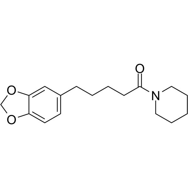
Tetrahydropiperine

Based on 1 Customer Validation
Tetrahydropiperine is an orally effective, selective inhibitor of NF-κB and MAPKs, and an activator of the PI3K/Akt/mTOR pathway. Tetrahydropiperine reduces the production of pro-inflammatory cytokines such as TNF-α, IL-6, and nitric oxide (NO) by inhibiting the nuclear translocation of NF-κB and the phosphorylation of MAPKs such as ERK, JNK, and p38. At the same time, Tetrahydropiperine inhibits excessive autophagy by activating the PI3K/Akt/mTOR pathway, protecting neurons from oxidative damage. Tetrahydropiperine has anti-inflammatory, anti-apoptotic, and neuroprotective effects, and is mainly used in the study of inflammatory diseases (such as endotoxemia, arthritis) and neurological diseases such as ischemic stroke.
For research use only. We do not sell to patients.
- Purity : 99.26%
- CAS No.: 23434-88-0
- Formula: C17H23NO3
- Molecular Weight:289.37
-
Storage:
4°C, protect from light
* In solvent : -80°C, 6 months; -20°C, 1 month (protect from light)

To place orders, for customer services and technical support, please contact: MedChemExpress USA
Tel: 609-228-6898 E-mail: [email protected] [email protected]


-
 Biological Activity
-
Chemical Information
-
Solvent & Solubility
- Protocols
- Purity & Documentation
- References
-
 Help & FAQs




Biological Activity
Description
IC50 & Target
[2]|
CYP1A1 23 μM (IC50) |
In Vitro
Tetrahydropiperine (1, 5, 10 μM; pretreatment for 1 h + LPS stimulation for 24 h) can significantly inhibit the production of NO, TNF-α, IL-6 and IL-1β induced by 1 μg/mL LPS in RAW 264.7 cells, and down-regulate the expression of COX-2 and i-NOS[1].
Tetrahydropiperine (10, 20, 40 μg/mL; 24 h) can improve the survival rate of PC-12 cells in the oxygen glucose deprivation (OGD) model, reduce cell morphological damage, and up-regulate the expression of proteins related to the PI3K/Akt/mTOR pathway[2].
MedChemExpress (MCE) has not independently confirmed the accuracy of these methods. They are for reference only.
-
Cell Line:RAW 264.7 cells
-
Concentration:5, 10 μM
-
Incubation Time:1 h pretreatment + 24 h LPS stimulation
-
Result:Significantly attenuated the expression of COX-2 and i-NOS proteins in LPS-stimulated cells, and inhibited the phosphorylation of NF-κB p65 and IκBα, indicating downregulation of the NF-κB signaling pathway.
In Vivo
MedChemExpress (MCE) has not independently confirmed the accuracy of these methods. They are for reference only.
-
Animal Model:Sprague-Dawley rats (male, 200±20 g, 6-8 weeks) permanent middle cerebral artery occlusion (pMCAO) model[2]
-
Dosage:10 mg/kg, 20 mg/kg, 30 mg/kg
-
Administration:Oral gavage daily, starting 1 h after pMCAO surgery, 14 days
-
Result:Dose-dependently improved neurological function, decreased cerebral infarct volume (notably 20 mg/kg and 30 mg/kg groups), and enhanced neuronal survival as shown by Nissl staining.
Western blot analysis revealed upregulation of PI3K, p-Akt/Akt, and p-mTOR/mTOR proteins, and downregulation of autophagy-related proteins Atg7 and Beclin1, indicating inhibition of excessive autophagy via the PI3K/Akt/mTOR pathway.
- Western Blot
- ROS/oxidative-stress fluorescent staining
- Research Protocol for Inflammation-related Diseases
- Kinase activity and phosphorylation assays
- Cytoplasmic-Nuclear Fractionated Protein Extraction
- Collagen-Induced Arthritis
- Detection of Apoptosis
- LPS-Induced Endotoxemia/Systemic Inflammation
- Autophagy
- Apoptosis Solutions
- Protocol for Kinase activity and phosphorylation assays
- Lysosome and acidic-vesicle live-cell staining
- Macroautophagy Solutions


Chemical Information
-
CAS No. 23434-88-0
-
Appearance Solid
-
Molecular Weight 289.37
-
Formula C17H23NO3
-
Color White to off-white
-
SMILES
O=C(N1CCCCC1)CCCCC2=CC=C(OCO3)C3=C2
-
Structure Classification
-
Initial Source
-
Shipping
Room temperature in continental US; may vary elsewhere.
-
Storage
4°C, protect from light
* In solvent : -80°C, 6 months; -20°C, 1 month (protect from light)


Solvent & Solubility
In Vitro:
DMSO : 100 mg/mL (345.58 mM; Need ultrasonic; Hygroscopic DMSO has a significant impact on the solubility of product, please use newly opened DMSO)
Please refer to the solubility information to select the appropriate solvent. Once prepared, please aliquot and store the solution to prevent product inactivation from repeated freeze-thaw cycles.
Storage method and period of stock solution: -80°C, 6 months; -20°C, 1 month (protect from light). When stored at -80°C, please use it within 6 months. When stored at -20°C, please use it within 1 month.
Please refer to the solubility information to select the appropriate solvent. Once prepared, please aliquot and store the solution to prevent product inactivation from repeated freeze-thaw cycles.
Storage method and period of stock solution: -80°C, 6 months; -20°C, 1 month (protect from light). When stored at -80°C, please use it within 6 months. When stored at -20°C, please use it within 1 month.
Concentration (start) × Volume (start) = Concentration (final) × Volume (final)
In Vivo:
Select the appropriate dissolution method based on your experimental animal and administration route.
- For the following dissolution methods, please ensure to first prepare a clear stock solution using an In Vitro approach and then sequentially add co-solvents:
- To ensure reliable experimental results, the clarified stock solution can be appropriately stored based on storage conditions. As for the working solution for In Vivo experiments, it is recommended to prepare freshly and use it on the same day.
- The percentages shown for the solvents indicate their volumetric ratio in the final prepared solution. If precipitation or phase separation occurs during preparation, heat and/or sonication can be used to aid dissolution.
Add each solvent one by one: 10% DMSO 90% (20% SBE-β-CD in Saline)
Solubility: ≥ 2.5 mg/mL (8.64 mM); Clear solution
This protocol yields a clear solution of ≥ 2.5 mg/mL (saturation unknown).
Taking 1 mL working solution as an example, add 100 μL DMSO stock solution (25.0 mg/mL) to 900 μL 20% SBE-β-CD in Saline, and mix evenly.
Preparation of 20% SBE-β-CD in Saline (4°C, storage for one week): 2 g SBE-β-CD powder is dissolved in 10 mL Saline, completely dissolve until clear.
In Vivo Dissolution Calculator
Please enter the basic information of animal experiments:
-
-
-
-
Recommended: Prepare an additional quantity of animals to account for potential losses during experiments.
Please enter your animal formula composition:
-
%DMSO +
Recommended: Keep the proportion of DMSO in working solution below 2% if your animal is weak.
-
%+
-
+%Tween-80 + +
-
%Saline +
The co-solvents required include: DMSO, . All of co-solvents are available by MedChemExpress (MCE). , Tween 80. All of co-solvents are available by MedChemExpress (MCE).
Working solution concentration: 0.22 mg/mL
Method for preparing stock solution: mg drug dissolved in μL DMSO. Stock solution concentration: mg/mL. * In solvent : -80°C, 6 months; -20°C, 1 month (protect from light)
1. Take μL DMSO stock solution;
2. Add μL .
μL , mix evenly;
3. Then add μL Tween 80, mix evenly;
4. Then add μL
Please ensure that the stock solution in the first step is dissolved to a clear state, and add co-solvents in sequence. You can use ultrasonic heating (ultrasonic cleaner, recommended frequency 20-40 kHz), vortexing, etc. to assist dissolution.


Purity & Documentation
-
Data Sheet (277 KB)
-
SDS (393 KB)
- English - EN (393 KB)
- Français - FR (393 KB)
- Deutsch - DE (393 KB)
- Norwegian - NO (393 KB)
- Español - ES (393 KB)
- Swedish - SV (393 KB)
- Italian - IT (393 KB)
- Korean - KR (393 KB)
- Portuguese - PT (393 KB)
-
Handling Instructions (2659 KB)
References
[1]. Kumar D, et al. Synthesis, characterization, and anti-inflammatory activity of tetrahydropiperine, piperic acid, and tetrahydropiperic acid via down regulation of NF-κB pathway. Cytokine. 2024 Jun;178:156578. [Content Brief]
[2]. Ren H, et al. Tetrahydropiperine, a natural alkaloid with neuroprotective effects in ischemic stroke. J Chem Neuroanat. 2024 Mar;136:102397. [Content Brief]
[3]. Koul S, et al. Structure-activity relationship of piperine and its synthetic analogues for their inhibitory potentials of rat hepatic microsomal constitutive and inducible cytochromeP450 activities. Bioorg Med Chem. 2000 Jan;8(1):251-68. [Content Brief]


Complete Stock Solution Preparation Table
Please refer to the solubility information to select the appropriate solvent. Once prepared, please aliquot and store the solution to prevent product inactivation from repeated freeze-thaw cycles.
Storage method and period of stock solution: -80°C, 6 months; -20°C, 1 month (protect from light). When stored at -80°C, please use it within 6 months. When stored at -20°C, please use it within 1 month.
| Optional Solvent | Concentration Solvent Mass | 1 mg | 5 mg | 10 mg | 25 mg |
|---|---|---|---|---|---|
| DMSO | 1 mM | 3.4558 mL | 17.2789 mL | 34.5578 mL | 86.3946 mL |
| 5 mM | 0.6912 mL | 3.4558 mL | 6.9116 mL | 17.2789 mL | |
| 10 mM | 0.3456 mL | 1.7279 mL | 3.4558 mL | 8.6395 mL | |
| 15 mM | 0.2304 mL | 1.1519 mL | 2.3039 mL | 5.7596 mL | |
| 20 mM | 0.1728 mL | 0.8639 mL | 1.7279 mL | 4.3197 mL | |
| 25 mM | 0.1382 mL | 0.6912 mL | 1.3823 mL | 3.4558 mL | |
| 30 mM | 0.1152 mL | 0.5760 mL | 1.1519 mL | 2.8798 mL | |
| 40 mM | 0.0864 mL | 0.4320 mL | 0.8639 mL | 2.1599 mL | |
| 50 mM | 0.0691 mL | 0.3456 mL | 0.6912 mL | 1.7279 mL | |
| 60 mM | 0.0576 mL | 0.2880 mL | 0.5760 mL | 1.4399 mL | |
| 80 mM | 0.0432 mL | 0.2160 mL | 0.4320 mL | 1.0799 mL | |
| 100 mM | 0.0346 mL | 0.1728 mL | 0.3456 mL | 0.8639 mL |


Tetrahydropiperine Related Classifications
HY-N4205 Related Classifications
 Powered by Bioz