



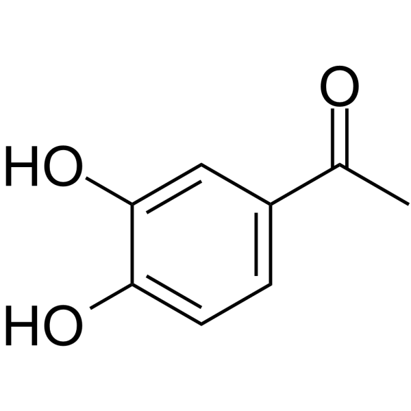
3',4'-Dihydroxyacetophenone

Based on 1 Customer Validation
3',4'-Dihydroxyacetophenone (3,4-DHAP) is a phenolic compound with oral bioavailability, possessing potent antioxidant, anti-inflammatory, anticancer and cardiovascular protective activities. 3',4'-Dihydroxyacetophenone inhibits mushroom Tyrosinase activity with an IC50 of 10 μM, thereby suppressing melanogenesis. 3',4'-Dihydroxyacetophenone inhibits platelet aggregation in platelet-rich plasma. 3',4'-Dihydroxyacetophenone reduces ROS levels in human umbilical vein endothelial cells treated with high glucose, upregulates the expression of Nrf2, HO-1 and PARP-1 in cells, and promotes the nuclear translocation of Nrf2. 3',4'-Dihydroxyacetophenone induces autophagy and apoptosis. 3',4'-Dihydroxyacetophenone inhibits seed germination/growth in most plants. 3',4'-Dihydroxyacetophenone can be used in the research of cancer, neurodegenerative diseases, non-alcoholic steatohepatitis, diabetes, obesity, skin pigmentation disorders, and cardiovascular and cerebrovascular diseases.
For research use only. We do not sell to patients.
- Purity : 99.87%
- CAS No.: 1197-09-7
- Formula: C8H8O3
- Molecular Weight:152.15
-
Storage:Powder -20°C, 3 years , 4°C, 2 years ; In solvent -80°C, 6 months , -20°C, 1 month

To place orders, for customer services and technical support, please contact: MedChemExpress USA
Tel: 609-228-6898 E-mail: [email protected] [email protected]


-
 Biological Activity
-
Chemical Information
-
Solvent & Solubility
- Protocols
- Purity & Documentation
- References
-
 Help & FAQs


-

HY-N1775R
3',4'-Dihydroxyacetophenone (Standard)Tyrosinase Inhibitor -
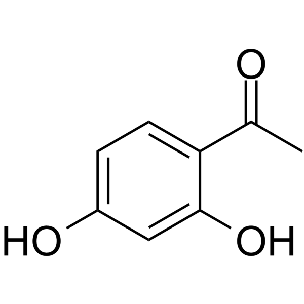
HY-Y0694
2',4'-DihydroxyacetophenonePlant Metabolite -
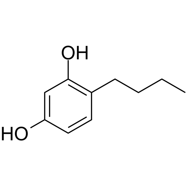
HY-107369
4-ButylresorcinolTyrosinase Inhibitor -
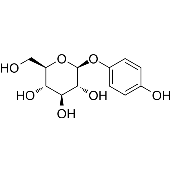
HY-N0192
ArbutinEndogenous Metabolite -
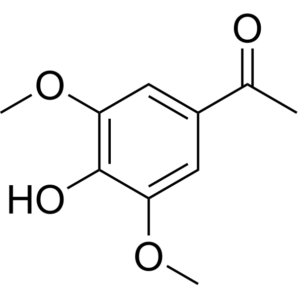
HY-W009884
Acetosyringone -
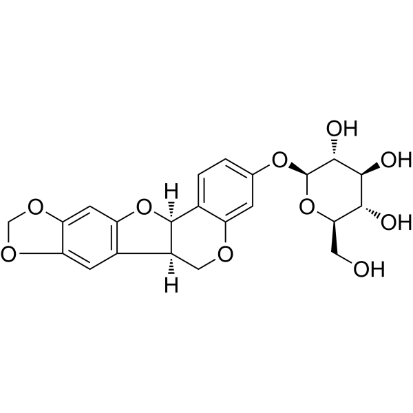
HY-N0616
TrifolirhizinPterocarpan Flavonoid -

HY-N0192R
Arbutin (Standard)Endogenous Metabolite -
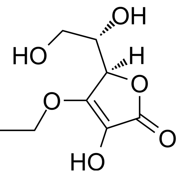
HY-W003607
3-O-Ethyl-L-ascorbic acidVitamin C Derivative -
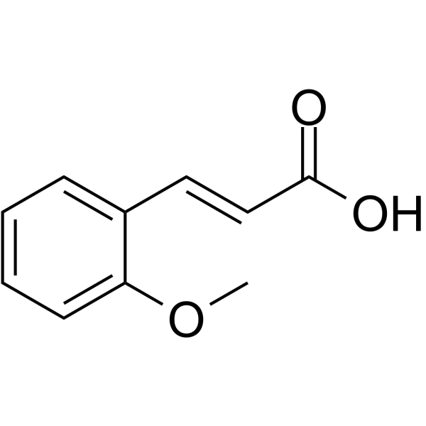
HY-N1386
2-Methoxycinnamic acidTyrosinase Inhibitor -
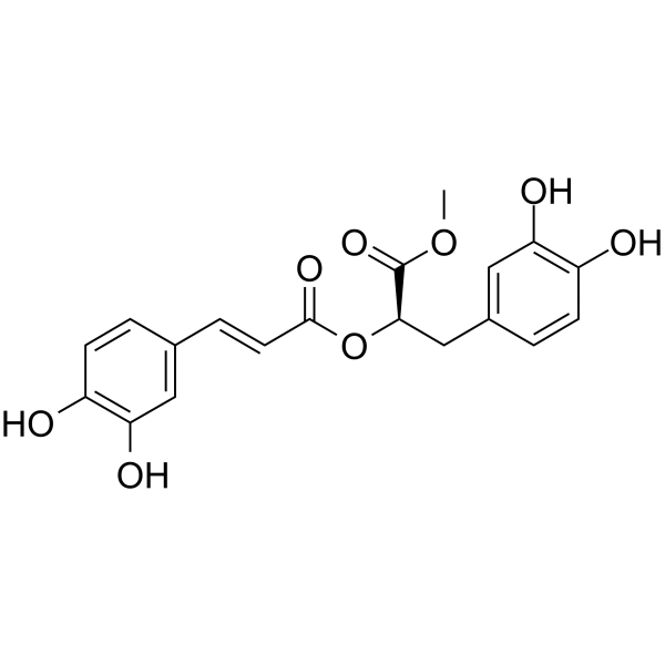
HY-N3266
Methyl rosmarinateHydroxycinnamic Acid -
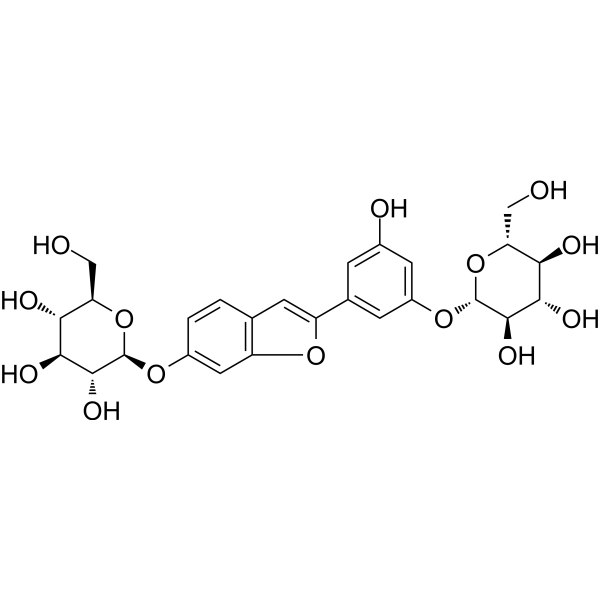
HY-N3518
Mulberroside FTyrosinase Inhibitor -
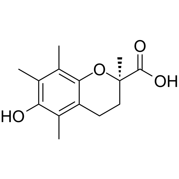
HY-101445A
(R)-TroloxTyrosinase Inhibitor


Biological Activity
Description
IC50 & Target
IC50: 10 μM (Tyrosinase)[1].
In Vitro
3',4'-Dihydroxyacetophenone (3,4-DHAP) potently inhibits mushroom tyrosinase activity with an IC50 of 10 μM[2].
3',4'-Dihydroxyacetophenone (3,4-DHAP) (10-200 μM; 24 h) dose-dependently inhibits tyrosinase activity in α-MSH-stimulated B16 melanoma cells, with significant inhibition starting at 10 μM after 24 h of treatment[2].
3',4'-Dihydroxyacetophenone (3 min pre-incubation; 5 min aggregation monitoring) potently inhibits ADP-stimulated platelet aggregation in rabbit PRP with an IC50 of 99.22 µmol/l[3].
3',4'-Dihydroxyacetophenone (1-100 µmol/l; 6 h) increases viability of human umbilical vein endothelial cells at 10 µmol/l, while higher and lower concentrations reduce viability, and 10 µmol/l protects cells against high glucose-induced viability loss[5].
3',4'-Dihydroxyacetophenone (10 µmol/l; 6 h) significantly reduces reactive oxygen species levels in high glucose-treated human umbilical vein endothelial cells[5].
3',4'-Dihydroxyacetophenone (10 µmol/l; 6 h) significantly increases the LC3-II/LC3-I protein ratio, indicating enhanced autophagy, in high glucose-treated human umbilical vein endothelial cells[5].
3',4'-Dihydroxyacetophenone (3,4-DHAP) (0.1-10 mM; up to 21 days) dose-dependently inhibits Picea schrenkiana seed germination rate and vigor, with significant inhibition starting at 2.5 mM for germination rate and 1 mM for germination vigor, and complete elimination of vigor at 10 mM[6].
3',4'-Dihydroxyacetophenone (3,4-DHAP) (0.1-10 mM; up to 14 days) does not inhibit Oryza sativa seed germination rate, but significantly promotes germination vigor at 1, 2.5, and 10 mM[6].
3',4'-Dihydroxyacetophenone (3,4-DHAP) (0.1-10 mM; up to 30 days) dose-dependently inhibits Picea schrenkiana seedling shoot length, root length, and fresh weight, with significant inhibition starting at 5 mM for shoot length, 2.5 mM for root length and fresh weight[6].
3',4'-Dihydroxyacetophenone (3,4-DHAP) (0.1-10 mM; up to 10 days) dose-dependently inhibits Triticum aestivum seedling shoot length and root length (significant at 5 mM and 2.5 mM, respectively) and exhibits hormetic effects on fresh weight, promoting growth at 0.5 mM and inhibiting it at 2.5 mM and higher[6].
3',4'-Dihydroxyacetophenone (3,4-DHAP) (0.1-10 mM; up to 10 days) dose-dependently inhibits seedling root growth of Latuca sativa, Oryza sativa, Raphanus sativus, Cucumis sativus, and Phaseolus radiatus, with significant inhibition starting at concentrations ranging from 0.5 mM to 2.5 mM; it also promotes Raphanus sativus shoot growth at 10 mM[6].
MedChemExpress (MCE) has not independently confirmed the accuracy of these methods. They are for reference only. Further protocols information, click here.
-
Cell Line:α-MSH-stimulated B16 melanoma cells
-
Concentration:10, 50, and 250 μM
-
Incubation Time:24 h
-
Result:Suppressed the amount of tyrosinase protein in α-MSH-stimulated B16 cells.
-
Cell Line:human umbilical vein endothelial cells (HUVECs)
-
Concentration:1, 10, 20, 50, 100 µM
-
Incubation Time:6 h (pretreated prior to 12 h high glucose exposure; variable for viability assessment)
-
Result:Increased HUVEC viability significantly compared to the control group at 10 µmol/l.
Reduced cell viability at 1, 20, 50 µM and 100 µM.
Increased cell viability significantly compared to the high glucose group at 10 µM.
-
Cell Line:human umbilical vein endothelial cells (HUVECs)
-
Concentration:10 µM
-
Incubation Time:6 h (pretreated prior to 12 h high glucose exposure)
-
Result:Increased total Nrf2 protein expression significantly compared to the high glucose group.\n
Increased nuclear Nrf2 protein expression significantly compared to the high glucose group.\n
Increased HO-1 protein expression significantly compared to the high glucose group.\n
Increased the LC3-II/LC3-I protein ratio significantly compared to the high glucose group.\n
Increased PARP-1 protein expression significantly compared to the high glucose group.
-
Cell Line:human umbilical vein endothelial cells (HUVECs)
-
Concentration:10 µM
-
Incubation Time:6 h (pretreated prior to 12 h high glucose exposure)
-
Result:Increased Nrf2 mRNA expression significantly compared to the high glucose group.
Increased HO-1 mRNA expression significantly compared to the high glucose group.
-
Cell Line:human umbilical vein endothelial cells (HUVECs)
-
Concentration:10 µM
-
Incubation Time:6 h (pretreated prior to 12 h high glucose exposure)
-
Result:Increased nuclear Nrf2 fluorescence intensity significantly compared to the high glucose group.
Parmacokinetics
| Species | Dose | Route | T1/2 | CL/F | AUC0-t | AUC0-∞ | MRT0-t | T1/2 (Absorption) | Bioavailability |
|---|---|---|---|---|---|---|---|---|---|
| Rat[3] | 0.20 mM | i.v. | 0.15 h | 2.30 L/h/kg | 66.32 μM/L·h | 87.48 μM/L·h | 0.22 h | / | / |
| Rat[3] | 0.39 mM | i.m. | 0.16 h | 3.121 L/h/kg | 124.22 μM/L·h | 129.94 μM/L·h | 0.46 h | 0.14 h | 93.81 % |
| Rat[3] | 0.79 mM | i.g. | 0.16 h | 17.39 L/h/kg | 47.78 μM/L·h | 48.64 μM/L·h | 0.38 h | 0.06 h | 18.01 % |
In Vivo
3',4'-Dihydroxyacetophenone (10 mg/kg; p.o.; daily; 8 weeks) improves endothelial function in rats with high-fat diet-induced obesity, as evidenced by enhanced endothelium-dependent vasodilation, upregulated activity of the eNOS-NO pathway, reduced oxidative stress and inflammatory responses, and improved lipid metabolism markers[4].
MedChemExpress (MCE) has not independently confirmed the accuracy of these methods. They are for reference only.
-
Animal Model:Wistar rats (male/female, 250-300 g)[3]
-
Dosage:0.79 mmol/kg
-
Administration:i.g.; single dose
-
Result:Significantly inhibited platelet aggregation compared to vehicle controls at 10 minutes post-administration (P=0.003).
Showed significantly reduced antiplatelet aggregation activity at 40 minutes post-administration compared to 10 minutes post-administration (P=0.003).
-
Animal Model:Wistar (male, 6 weeks old, high-fat diet-induced obesity)[4]
-
Dosage:10 mg/kg
-
Administration:p.o.; daily; 8 weeks
-
Result:Reduced plasma triglycerides, free fatty acids, serum tumor necrosis factor-α, and malondialdehyde.
Increased serum adiponectin.
Enhanced acetylcholine-induced endothelium-dependent vasodilatation.
Increased eNOS activity and NO production in endothelial cells.
Reduced NF-kB positive cells in the aorta.
Decreased superoxide anion production in the aorta.
Did not reduce body weight, visceral fat mass, or plasma insulin levels in obese rats.


Chemical Information
-
CAS No. 1197-09-7
-
Appearance Solid
-
Molecular Weight 152.15
-
Formula C8H8O3
-
Color Light brown to brown
-
SMILES
CC(C1=CC=C(O)C(O)=C1)=O
-
Synonyms
3,4-DHAP
-
Structure Classification
-
Initial Source
-
Shipping
Room temperature in continental US; may vary elsewhere.
-
Storage
Powder -20°C 3 years 4°C 2 years In solvent -80°C 6 months -20°C 1 month


Solvent & Solubility
In Vitro:
DMSO : 50 mg/mL (328.62 mM; Need ultrasonic; Hygroscopic DMSO has a significant impact on the solubility of product, please use newly opened DMSO)
Please refer to the solubility information to select the appropriate solvent. Once prepared, please aliquot and store the solution to prevent product inactivation from repeated freeze-thaw cycles.
Storage method and period of stock solution: -80°C, 6 months; -20°C, 1 month. When stored at -80°C, please use it within 6 months. When stored at -20°C, please use it within 1 month.
Please refer to the solubility information to select the appropriate solvent. Once prepared, please aliquot and store the solution to prevent product inactivation from repeated freeze-thaw cycles.
Storage method and period of stock solution: -80°C, 6 months; -20°C, 1 month. When stored at -80°C, please use it within 6 months. When stored at -20°C, please use it within 1 month.
Concentration (start) × Volume (start) = Concentration (final) × Volume (final)
In Vivo:
Select the appropriate dissolution method based on your experimental animal and administration route.
- For the following dissolution methods, please ensure to first prepare a clear stock solution using an In Vitro approach and then sequentially add co-solvents:
- To ensure reliable experimental results, the clarified stock solution can be appropriately stored based on storage conditions. As for the working solution for In Vivo experiments, it is recommended to prepare freshly and use it on the same day.
- The percentages shown for the solvents indicate their volumetric ratio in the final prepared solution. If precipitation or phase separation occurs during preparation, heat and/or sonication can be used to aid dissolution.
Add each solvent one by one: 10% DMSO 40% PEG300 5% Tween-80 45% Saline
Solubility: ≥ 2.5 mg/mL (16.43 mM); Clear solution
This protocol yields a clear solution of ≥ 2.5 mg/mL (saturation unknown).
Taking 1 mL working solution as an example, add 100 μL DMSO stock solution (25.0 mg/mL) to 400 μL PEG300, and mix evenly; then add 50 μL Tween-80 and mix evenly; then add 450 μL Saline to adjust the volume to 1 mL.
Preparation of Saline: Dissolve 0.9 g sodium chloride in ddH₂O and dilute to 100 mL to obtain a clear Saline solution.
Add each solvent one by one: 10% DMSO 90% (20% SBE-β-CD in Saline)
Solubility: ≥ 2.5 mg/mL (16.43 mM); Clear solution
This protocol yields a clear solution of ≥ 2.5 mg/mL (saturation unknown).
Taking 1 mL working solution as an example, add 100 μL DMSO stock solution (25.0 mg/mL) to 900 μL 20% SBE-β-CD in Saline, and mix evenly.
Preparation of 20% SBE-β-CD in Saline (4°C, storage for one week): 2 g SBE-β-CD powder is dissolved in 10 mL Saline, completely dissolve until clear.
In Vivo Dissolution Calculator
Please enter the basic information of animal experiments:
-
-
-
-
Recommended: Prepare an additional quantity of animals to account for potential losses during experiments.
Please enter your animal formula composition:
-
%DMSO +
Recommended: Keep the proportion of DMSO in working solution below 2% if your animal is weak.
-
%+
-
+%Tween-80 + +
-
%Saline +
The co-solvents required include: DMSO, . All of co-solvents are available by MedChemExpress (MCE). , Tween 80. All of co-solvents are available by MedChemExpress (MCE).
Working solution concentration: 0.22 mg/mL
Method for preparing stock solution: mg drug dissolved in μL DMSO. Stock solution concentration: mg/mL.
1. Take μL DMSO stock solution;
2. Add μL .
μL , mix evenly;
3. Then add μL Tween 80, mix evenly;
4. Then add μL
Please ensure that the stock solution in the first step is dissolved to a clear state, and add co-solvents in sequence. You can use ultrasonic heating (ultrasonic cleaner, recommended frequency 20-40 kHz), vortexing, etc. to assist dissolution.

Protocols
-
RNA extraction experimental
By lysing cells, releasing RNA, and removing impurities such as proteins and DNA, high-purity RNA products are finally obtained. The commonly used traditional method is the guanidine isothiocyanate/phenol/chloroform method (Trizol), which is suitable for a variety of animal materials including animal tissues, microorganisms, cultured cells, etc., and most plant materials.
-
Annexin V plus membrane-impermeant dye apoptosis staining
Annexin V-based apoptosis assays rely on the detection of phosphatidylserine (PS) externalization from the inner leaflet of the plasma membrane to the outer leaflet, an early biochemical hallmark of apoptosis. Fluorescently labeled Annexin V binds PS in a calcium-dependent manner, enabling identification of early apoptotic cells by flow cytometry or fluorescence microscopy. When combined with a membrane-impermeant DNA-binding dye (e. g. , propidium iodide), this approach allows discrimination between viable (Annexin V−/dye−), early apoptotic (Annexin V+/dye−), and late apoptotic or necrotic (Annexin V+/dye+) cell populations by assessing membrane integrity and PS exposure.
-
ROS/oxidative-stress fluorescent staining
ROS/oxidative-stress fluorescent staining uses cell-permeant fluorogenic probes that become fluorescent after oxidation inside cells or tissues; commonly used examples include DCFH-DA/DCFDA for broad cellular oxidant detection, DHE for superoxide-related signal detection, MitoSOX for mitochondrial superoxide-related signal detection, and CellROX probes for oxidative-stress-associated fluorescence readouts. The assay detects probe oxidation rather than a single ROS species unless the probe and analysis method have been chemically validated for that species. DCFH-DA enters cells, is deacetylated by intracellular esterases to DCFH, and produces fluorescent DCF after oxidation, so the readout is used as an operational measure of total cellular oxidative stress rather than a species-specific ROS measurement. DHE and MitoSOX can report superoxide-related oxidation, but red fluorescence alone can include non-specific ethidium-like oxidation products; HPLC or optimized spectral approaches are
-
Research Protocol for Inflammation-related Diseases
The NLRP3 inflammasome is a cytosolic innate immune signaling platform that integrates priming signals and danger-signal activation to promote caspase-1 activation, maturation of IL-1β and IL-18, and gasdermin D-mediated pyroptotic cell death. The core experimental logic is to determine whether inflammatory disease phenotypes are driven by increased NLRP3 expression, ASC-containing inflammasome assembly, caspase-1 cleavage, GSDMD cleavage, and extracellular release of IL-1β/IL-18 rather than by nonspecific cell injury alone. The pathway is strongly linked to inflammation-related disease phenotypes because monosodium urate crystals activate NALP3/NLRP3 inflammasome signaling in gout-like crystal inflammation, cholesterol crystals activate NLRP3 inflammasomes in atherogenesis models, and DSS-induced intestinal inflammation has been reported to involve NLRP3 inflammasome activity. However, experimental colitis studies also show context-dependent protective effects of NLRP3 inflammasome co
-
Research Protocol for Metabolic Diseases
AMP-activated protein kinase, AMPK, is a conserved cellular energy sensor that responds to reduced cellular energy status and coordinates metabolism by increasing ATP-generating catabolic pathways while suppressing ATP-consuming anabolic processes. In metabolic disease research, the AMPK pathway is experimentally relevant because it regulates hepatic lipid synthesis, fatty acid oxidation, glucose production, skeletal-muscle glucose disposal, mTORC1-linked biosynthesis, autophagy, mitochondrial homeostasis, and whole-body energy balance. The central pathway logic is that energy stress, metformin, exercise-like stimulation, or direct AMPK activators increase AMPKα Thr172 phosphorylation and downstream substrate phosphorylation, including ACC and RAPTOR. Phosphorylation of ACC suppresses lipogenesis and supports fatty acid oxidation, whereas phosphorylation of RAPTOR suppresses mTORC1 signaling and links cellular energy status to growth and protein synthesis control. The pathway is linked
-
Cytoplasmic-Nuclear Fractionated Protein Extraction
Cytoplasmic-nuclear fractionated protein extraction separates soluble cytoplasmic proteins from nuclear-enriched proteins by mild plasma-membrane permeabilization, differential centrifugation, washing of nuclei, and extraction of nuclear proteins for downstream immunoblotting or related molecular analysis. The readout is the relative abundance of a protein in cytoplasmic and nuclear fractions, commonly assessed by western blotting together with compartment markers such as tubulin or pyruvate kinase for cytoplasm and lamin, nucleoporin, hnRNP, H2AX, or Lamin B for nuclear fractions.
-
Apoptosis
Apoptosis, also called programmed cell death, is generally characterized by distinct morphological characteristics.
-
TUNEL staining for apoptotic DNA fragmentation
TUNEL staining detects DNA strand breaks by using terminal deoxynucleotidyl transferase to add labeled nucleotides to exposed 3′-OH DNA termini, generating either microscopic staining in fixed cells or tissue sections, or fluorescence/cytometric signal in cell suspensions. TUNEL positivity reflects DNA fragmentation but should not be interpreted alone as definitive apoptosis, because TUNEL can also label necrotic, autolytic, mechanically damaged, or DNA-repair-associated DNA breaks.
-
Cell Cytotoxicity Assay
Cytotoxicity assays are usually based on the assessment of cell membrane damage, which can also be indirectly detected by measuring cell viability. Detection methods include MTT assay, CKK-8 assay, LDH assay and ATP assay, etc.
-
Protocol for Pharmacokinetic Study
Pharmacokinetic studies quantify how an organism handles a drug over time through absorption, distribution, metabolism, and excretion, and the core experimental readout is the concentration-time profile of parent drug and, when relevant, metabolites in biological matrices such as plasma, whole blood, urine, bile, or tissue. Pharmacokinetic analysis links dose, route, exposure, clearance, half-life, distribution, bioavailability, and systemic exposure to drug efficacy and toxicity hypotheses rather than measuring a signaling pathway directly. The literature links pharmacokinetics to drug-development phenotypes by showing that drug metabolism and pharmacokinetics influence compound progression, exposure-response interpretation, safety margins, dosing strategy, and failure risk during discovery and development. DMPK science contributes to compound optimization by integrating physicochemical properties, in vitro metabolism, transporter behavior, in vivo exposure, and pharmacodynamic contex
-
Endothelial Tube Formation Assay
Endothelial tube formation assay evaluates the ability of endothelial cells to attach, migrate, align, and organize into capillary-like networks when cultured on gelled basement membrane extract or Matrigel; the readout is the morphology and quantity of tube-like networks, which reflects an in vitro endothelial morphogenesis step related to angiogenesis. Basement membrane extract/Matrigel provides laminin-rich extracellular matrix cues that support endothelial differentiation into capillary-like structures, but it can contain biologically active growth factors, so growth-factor-reduced matrix is preferred when testing defined angiogenic stimulators or inhibitors.
-
Autophagy
Autophagy is a process in which eukaryotic cells use lysosomes to degrade their own cytoplasmic proteins and damaged organelles under the regulation of autophagy related gene (Atg). Microtubule-associated proteins light chain 3 (LC3) is recognized as autophagy marker, which transfers from cytoplasmic LC3 (LC3-I) to membrane type (LC3-II). LC3-II/I ratio could be detected by Western Blot and fluorescence microscopy.
-
Apoptosis Solutions
Apoptosis is a regulated, generally non-lytic cell-death pathway that removes unwanted, damaged, infected, or abnormal cells through coordinated morphological changes, caspase activation, DNA fragmentation, and membrane remodeling. The intrinsic apoptosis pathway is controlled mainly by mitochondrial outer membrane permeabilization, BCL-2 family proteins, cytochrome c release, apoptosome formation, caspase-9 activation, and downstream executioner caspase-3/7 activation. The extrinsic apoptosis pathway is initiated by death receptors such as Fas, TNFR, and TRAIL receptors, which recruit adaptor proteins and activate caspase-8 before engaging executioner caspases or mitochondrial amplification through BID cleavage. Apoptosis is linked to many phenotypes, including cancer cell killing, tissue homeostasis, immune regulation, neurodegeneration, infection response, and treatment-induced cytotoxicity; unresolved questions include how apoptosis interacts with necroptosis, pyroptosis, ferroptos
-
Lysosome and acidic-vesicle live-cell staining
Lysosome and acidic-vesicle live-cell staining detects acidic intracellular compartments by using membrane-permeant acidotropic probes that accumulate in low-pH vesicles, including lysosomes, late endosomes, autolysosomes, and acidic phagosomes. LysoTracker staining is commonly used as an intensity-based readout of acidic lysosomal compartment abundance or enlargement, while acridine orange produces green fluorescence in less concentrated compartments and red fluorescence after concentration-dependent accumulation in acidic vesicular organelles. Loss or reduction of acridine-orange red signal can be used as a readout of lysosomal membrane permeabilization or reduced acidic-vesicle integrity. This protocol is designed for live cultured cells and can be adapted for fluorescence microscopy, high-content imaging, plate-reader readout, or flow cytometry when the selected literature supports the readout. Because these dyes report acidotropic accumulation rather than lysosome identity alone,
-
Macroautophagy Solutions
Macroautophagy is a conserved lysosome-dependent degradation pathway in which cytoplasmic material is sequestered into double-membrane autophagosomes and delivered to lysosomes for degradation and recycling. The pathway supports cellular homeostasis during nutrient limitation, organelle stress, protein-aggregate accumulation, infection, differentiation, and tissue remodeling by coupling cargo sequestration, autophagosome maturation, lysosomal fusion, and degradation of cargo-derived macromolecules. The core molecular sequence includes initiation by nutrient- and stress-regulated autophagy machinery, autophagosome nucleation, LC3/ATG8-family conjugation to autophagosomal membranes, cargo selection through receptors such as SQSTM1/p62, autophagosome-lysosome fusion, and lysosomal degradation. LC3 was identified as a mammalian homolog of yeast Atg8 that localizes to autophagosomal membranes after processing, and p62/SQSTM1 was shown to connect ubiquitinated cargo with autophagic degradati


Purity & Documentation
-
Data Sheet (286 KB)
-
SDS (393 KB)
- English - EN (393 KB)
- Français - FR (393 KB)
- Deutsch - DE (393 KB)
- Norwegian - NO (393 KB)
- Español - ES (393 KB)
- Swedish - SV (393 KB)
- Italian - IT (393 KB)
- Korean - KR (393 KB)
- Portuguese - PT (393 KB)
-
Handling Instructions (2659 KB)
References
[1]. Wang W, et al. Synthesis and Health Effects of Phenolic Compounds: A Focus on Tyrosol, Hydroxytyrosol, and 3,4-Dihydroxyacetophenone. Antioxidants (Basel). 2025;14(4):476. Published 2025 Apr 16. [Content Brief]
[2]. Kim YJ, et al. Antimelanogenic activity of 3,4-dihydroxyacetophenone: inhibition of tyrosinase and MITF. Biosci Biotechnol Biochem. 2006;70(2):532-534. [Content Brief]
[3]. Sun N, et al. Synthesis, oral bioavailability evaluation and antiplatelet aggregation activity of three derivatives of 3,4‑dihydroxyacetophenone. Int J Mol Med. 2020;45(3):919-930. [Content Brief]
[4]. Hui Z, et al. Effect of 3,4-dihydroxyacetophenone on endothelial dysfunction in obese rats. Pharm Biol. 2015;53(8):1149-1154. [Content Brief]
[5]. Cao D, et al. 3,4‑Dihydroxyacetophenone attenuates oxidative stress‑induced damage to HUVECs via regulation of the Nrf2/HO‑1 pathway. Mol Med Rep. 2022;25(6):199. [Content Brief]
[6]. Ruan X, et al. Autotoxicity and allelopathy of 3,4-dihydroxyacetophenone isolated from Picea schrenkiana needles. Molecules. 2011;16(10):8874-8893. Published 2011 Oct 24. [Content Brief]


Complete Stock Solution Preparation Table
Please refer to the solubility information to select the appropriate solvent. Once prepared, please aliquot and store the solution to prevent product inactivation from repeated freeze-thaw cycles.
Storage method and period of stock solution: -80°C, 6 months; -20°C, 1 month. When stored at -80°C, please use it within 6 months. When stored at -20°C, please use it within 1 month.
| Optional Solvent | Concentration Solvent Mass | 1 mg | 5 mg | 10 mg | 25 mg |
|---|---|---|---|---|---|
| DMSO | 1 mM | 6.5725 mL | 32.8623 mL | 65.7246 mL | 164.3115 mL |
| 5 mM | 1.3145 mL | 6.5725 mL | 13.1449 mL | 32.8623 mL | |
| 10 mM | 0.6572 mL | 3.2862 mL | 6.5725 mL | 16.4312 mL | |
| 15 mM | 0.4382 mL | 2.1908 mL | 4.3816 mL | 10.9541 mL | |
| 20 mM | 0.3286 mL | 1.6431 mL | 3.2862 mL | 8.2156 mL | |
| 25 mM | 0.2629 mL | 1.3145 mL | 2.6290 mL | 6.5725 mL | |
| 30 mM | 0.2191 mL | 1.0954 mL | 2.1908 mL | 5.4771 mL | |
| 40 mM | 0.1643 mL | 0.8216 mL | 1.6431 mL | 4.1078 mL | |
| 50 mM | 0.1314 mL | 0.6572 mL | 1.3145 mL | 3.2862 mL | |
| 60 mM | 0.1095 mL | 0.5477 mL | 1.0954 mL | 2.7385 mL | |
| 80 mM | 0.0822 mL | 0.4108 mL | 0.8216 mL | 2.0539 mL | |
| 100 mM | 0.0657 mL | 0.3286 mL | 0.6572 mL | 1.6431 mL |


3',4'-Dihydroxyacetophenone Related Classifications
HY-N1775 Related Classifications
 Powered by Bioz