



Daporinad
Based on 67 publication(s) in Google Scholar
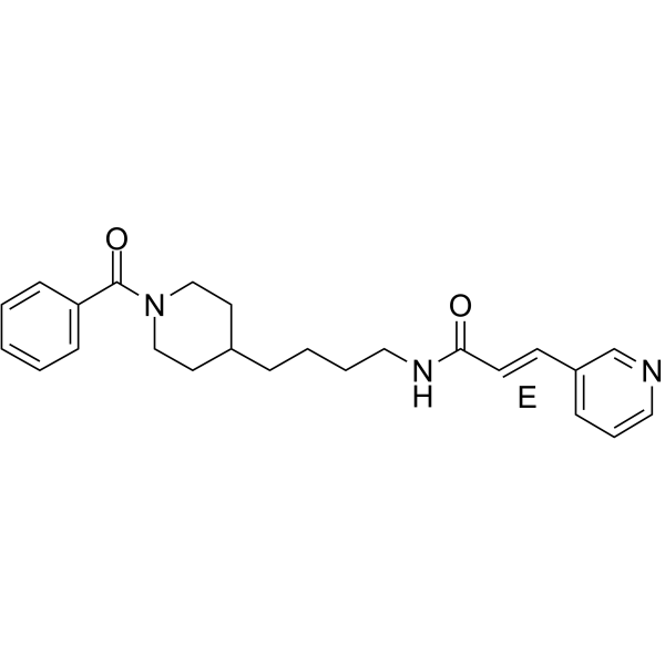
Daporinad (FK866) is a non-competitive inhibitor of nicotinamide phosphoribosyltransferase (Nampt), with a Ki value of 0.3 nM. Daporinad depletes NAD+ and ATP levels, inhibits mTORC1 and MAPK/ERK pathways, and activates TFEB to induce autophagy. Daporinad causes the depletion of the endoplasmic reticulum Ca²⁺ pool, ultimately weakening the mitogen-induced Ca²⁺ signal and the activation and function of T cells. Daporinad induces cell cycle arrest and apoptosis, and inhibits cell proliferation. Daporinad can be used for the study of myeloma, liver cancer, and immunosuppression.
For research use only. We do not sell to patients.
- Purity : 99.91%
- CAS No.: 658084-64-1
- Formula: C24H29N3O2
- Molecular Weight:391.51
-
Storage:Powder -20°C, 3 years , 4°C, 2 years ; In solvent -80°C, 2 years , -20°C, 1 year

To place orders, for customer services and technical support, please contact: MedChemExpress USA
Tel: 609-228-6898 E-mail: [email protected] [email protected]


-
 Biological Activity
-
Chemical Information
-
Solvent & Solubility
- Protocols
- Purity & Documentation
- References
-
 Help & FAQs


-
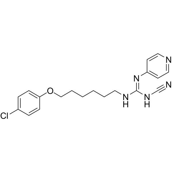
HY-10079
CHS-828NAMPT Inhibitor -
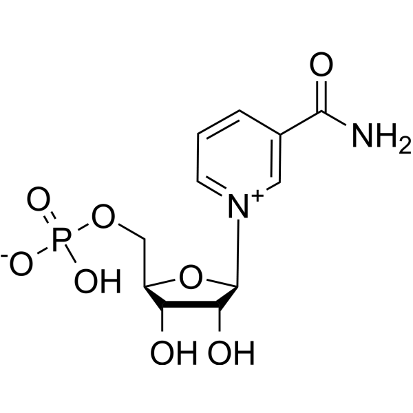
HY-F0004
FosribnicotinamideNAD+ Intermediate -
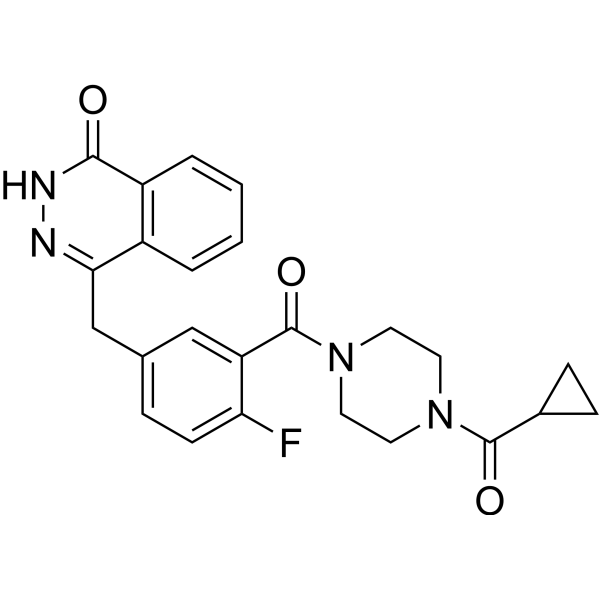
HY-10162
OlaparibPARP Inhibitor -
HY-B0150
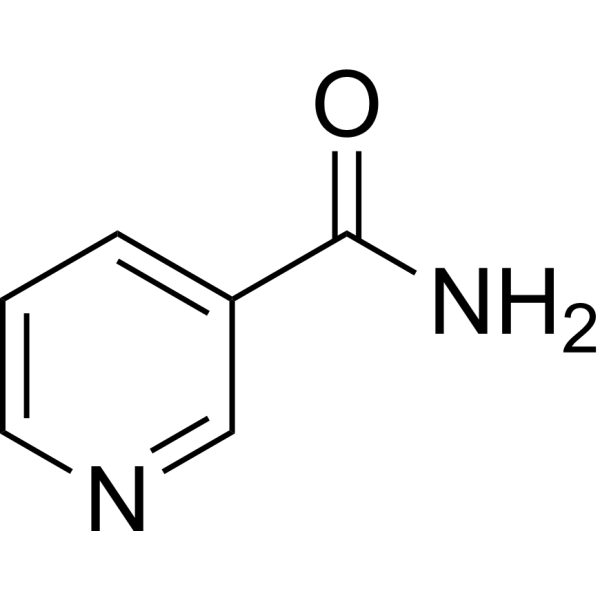
NicotinamideSIRT Inhibitor -
HY-123033A
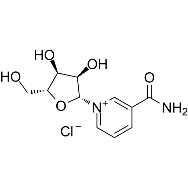
Nicotinamide riboside chlorideEndogenous Metabolite -

HY-10219
RapamycinmTOR Inhibitor -
HY-15136
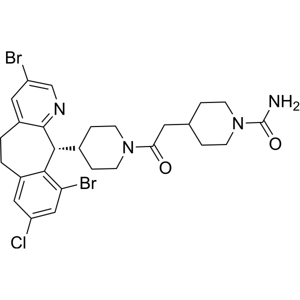
LonafarnibFarnesyltransferase Inhibitor -
HY-100229
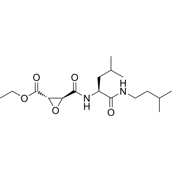
AloxistatinCysteine Protease Inhibitor -

HY-B0015
PaclitaxelAntitumor Agent -

HY-10227
BortezomibProteasome Inhibitor -

HY-14648
DexamethasoneGR Agonist -
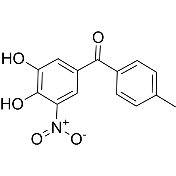
HY-17406
TolcaponeCOMT Inhibitor
Publications Citing Use of MedChemExpress (MCE) Daporinad
More- Cell Metab. 2024 Aug 6;36(8):1806-1822.e11. [Abstract]
- Nat Commun. 2024 Nov 12;15(1):9515. [Abstract]
- Exp Mol Med. 2025 Feb;57(1):72-85. [Abstract]
- J Adv Res. 2025 Dec 29:S2090-1232(25)01020-3. [Abstract]
- Redox Biol. 2024 Feb:69:103030. [Abstract]
- Adv Sci (Weinh). 2025 Mar;12(12):e2412282. [Abstract]
- Sci Adv. 2025 Mar 28;11(13):eado3923. [Abstract]
- Sci Adv. 2023 Apr 14;9(15):eadf8522. [Abstract]
- Cell Death Differ. 2024 Feb;31(2):239-253. [Abstract]
- Cell Death Dis. 2025 Apr 25;16(1):342. [Abstract]
- Acta Pharmacol Sin. 2023 Oct;44(10):2125-2138. [Abstract]
- Acta Pharmacol Sin. 2023 Sep;44(9):1906-1919. [Abstract]
- Cell Chem Biol. 2024 Aug 15;31(8):1529-1541.e12. [Abstract]
- Sci Signal. 2021 Jun 8;14(686):eabc7405. [Abstract]
- JCI Insight. 2025 Nov 24;10(22):e185914. [Abstract]
- J Agric Food Chem. 2024 Apr 15. [Abstract]
- Cells. 2023 Oct 2;12(19):2396. [Abstract]
- Commun Biol. 2025 Nov 26;8(1):1761. [Abstract]
- Clin Epigenetics. 2025 May 6;17(1):77. [Abstract]
- Int Immunopharmacol. 2023 Jul:120:110291. [Abstract]
- Biol Direct. 2025 Nov 27;20(1):113. [Abstract]
- Mol Neurobiol. 2023 Mar;60(3):1267-1280. [Abstract]
- Biol Res. 2025 May 4;58(1):25. [Abstract]
- Front Pharmacol. 2020 Jul 29:11:1136. [Abstract]
- Front Cell Dev Biol. 2022 Mar 24:10:853652. [Abstract]
- Chem Biol Interact. 2026 Feb 11:425:111900. [Abstract]
- Eur J Pharm Sci. 2025 Nov 1:214:107302. [Abstract]
- Molecules. 2022 Mar 21;27(6):2011. [Abstract]
- Biochim Biophys Acta Mol Basis Dis. 2025 Mar;1871(3):167662. [Abstract]
- Biochim Biophys Acta Mol Basis Dis. 2024 Oct;1870(7):167288. [Abstract]
- Mol Med Rep. 2017 Oct;16(4):5121-5128. [Abstract]
- PLoS Pathog. 2026 Apr 22;22(4):e1014165. [Abstract]
- Sci Rep. 2023 Feb 27;13(1):3334. [Abstract]
- PLoS Pathog. 2021 Mar 19;17(3):e1009436. [Abstract]
- Cancers (Basel). 2023 Apr 23;15(9):2427. [Abstract]
- J Inflamm Res. 2025 Jan 23:18:1091-1106. [Abstract]
- Clin Exp Med. 2025 Jul 10;25(1):243. [Abstract]
- Brain Res Bull. 2024 Nov:218:111114. [Abstract]
- J Biol Chem. 2022 Dec;298(12):102587. [Abstract]
- Adv Photonics Res. 2026 Apr 18;7(4):e202500258.
- J Cell Physiol. 2024 Jan;239(1):180-192. [Abstract]
- J Cell Physiol. 2019 Apr;234(4):4385-4395. [Abstract]
- Pancreatology. 2021 Aug;21(5):870-883. [Abstract]
- Mol Carcinog. 2024 Dec;63(12):2363-2381. [Abstract]
- PeerJ. 2021 May 14;9:e11401. [Abstract]
- Cancer Manag Res. 2021 Nov 30;13:8915-8928. [Abstract]
- Neurosci Lett. 2023 Sep 25:814:137471. [Abstract]
- Genes Genomics. 2022 Dec;44(12):1531-1541. [Abstract]
- Rapid Commun Mass Spectrom. 2021 Sep 30;35(18):e9150. [Abstract]
- bioRxiv. 2026 May 21.
- Res Sq. 2026 Mar 5.
- bioRxiv. 2025 Dec 25.
- bioRxiv. 2025 Oct 14.
- Res Sq. 2025 Sep 23.
- bioRxiv. 2025 Aug 02.
- bioRxiv. 2025 Jul 10:2025.07.08.663801. [Abstract]
- Res Sq. 2025 May 16:rs.3.rs-6590535. [Abstract]
- bioRxiv. 2025 March 26.
- bioRxiv. 2024 Nov 6:2024.11.04.621884. [Abstract]
- University of Minnesota. 2024.
- Research Square Preprint. 2024 Apr 12.
- Research Square Preprint. 2024 Mar 6.
- Research Square Preprint. 2023 Nov 14.
- Research Square Preprint. 2023 Sep 8.
- bioRxiv. 2019 Oct.
- bioRxiv. 2019 Oct 28.
- Patent. US20180263995A1.
 Customer Validation & Images

-
Histological Imaging/Staining

-
Flow Cytometry
-
Flow Cytometry
-
WB
-
WB


Biological Activity
Description
IC50 & Target
IC50: 0.09 nM (NMPRTase)
Cellular Effect
|
Cell Line

|
Type | Value | Description | References |
|---|---|---|---|---|
| A2780 | IC50 |
<3 nM
Compound: 1; FK866
|
Antiproliferative activity against human A2780 cells assessed as cell growth inhibition
Antiproliferative activity against human A2780 cells assessed as cell growth inhibition
|
[PMID: 37369332] |
| A2780 | IC50 |
0.001 μM
Compound: 1, APO866
|
Cytotoxicity against human A2780 cells after 72 hrs by SRB assay
Cytotoxicity against human A2780 cells after 72 hrs by SRB assay
|
[PMID: 23617784] |
| A2780 | IC50 |
0.001 μM
Compound: 2, APO-866
|
Antiproliferative activity against human A2780 cells assessed as growth inhibition after 72 hrs by SRB-based microplate reader analysis
Antiproliferative activity against human A2780 cells assessed as growth inhibition after 72 hrs by SRB-based microplate reader analysis
|
[PMID: 23859118] |
| A2780 | IC50 |
0.001 μM
Compound: 2, APO-866, FK866
|
Antiproliferative activity against human A2780 cells after 72 hrs by sulforhodamine B assay
Antiproliferative activity against human A2780 cells after 72 hrs by sulforhodamine B assay
|
[PMID: 24405419] |
| A2780 | IC50 |
0.001 μM
Compound: 8; APO866
|
Inhibition of NAMPT in human A2780 cells assessed as decrease in cell viability after 72 hrs by SRB assay
Inhibition of NAMPT in human A2780 cells assessed as decrease in cell viability after 72 hrs by SRB assay
|
[PMID: 27541271] |
| A2780 | IC50 |
0.28 nM
Compound: 1; FK886
|
Antiproliferative activity against human A2780 cells incubated for 72 hrs in carbon-dioxide atmosphere measured by cell counting kit-8 method
Antiproliferative activity against human A2780 cells incubated for 72 hrs in carbon-dioxide atmosphere measured by cell counting kit-8 method
|
[PMID: 36563407] |
| A2780 | IC50 |
1.6 nM
Compound: 1, APO866
|
Cytotoxicity against human A2780 cells assessed as growth inhibition after 72 hrs by WST-1 assay
Cytotoxicity against human A2780 cells assessed as growth inhibition after 72 hrs by WST-1 assay
|
[PMID: 24164086] |
| A2780 | IC50 |
4.2 nM
Compound: 1; FK866
|
Cytotoxicity against human A2780 cells assessed as reduction in cell viability
Cytotoxicity against human A2780 cells assessed as reduction in cell viability
|
[PMID: 28165742] |
| A2780 | IC50 |
5.7 nM
Compound: 1, APO866
|
Cytotoxicity against human A2780 cells by clonogenic assay
Cytotoxicity against human A2780 cells by clonogenic assay
|
[PMID: 24164086] |
| A-431 | IC50 |
6.1 nM
Compound: 1, APO866
|
Cytotoxicity against human A431 cells by clonogenic assay
Cytotoxicity against human A431 cells by clonogenic assay
|
[PMID: 24164086] |
| A549 | IC50 |
<0.16 μM
Compound: 1, FK-866
|
Cytotoxicity against human A549 cells after 6 days by SRB assay
Cytotoxicity against human A549 cells after 6 days by SRB assay
|
[PMID: 21330015] |
| A549 | IC50 |
0.028 μM
Compound: 1; FK866; AP0866
|
Antiproliferative activity against human A549 cells assessed as inhibition of cell growth incubated for 72 hrs by cell titer glo luminescent assay
Antiproliferative activity against human A549 cells assessed as inhibition of cell growth incubated for 72 hrs by cell titer glo luminescent assay
|
[PMID: 35640078] |
| A549 | IC50 |
3.7 μM
Compound: FK866
|
Cytotoxicity against human A549 cells assessed as inhibition of cell growth after 72 hrs by MTT assay
Cytotoxicity against human A549 cells assessed as inhibition of cell growth after 72 hrs by MTT assay
|
[PMID: 29348808] |
| A549 | IC50 |
34.53 μM
Compound: 1; FK866
|
Antiproliferative activity against NAMPT inhibitor-resistant A549 cells assessed as reduction in cell viability measured for 48 hrs in presence of epacadostat by CCK-8 assay
Antiproliferative activity against NAMPT inhibitor-resistant A549 cells assessed as reduction in cell viability measured for 48 hrs in presence of epacadostat by CCK-8 assay
|
[PMID: 36595482] |
| A549 | IC50 |
52.15 μM
Compound: 1; FK866
|
Antiproliferative activity against NAMPT inhibitor-resistant A549 cells assessed as reduction in cell viability measured for 48 hrs by CCK-8 assay
Antiproliferative activity against NAMPT inhibitor-resistant A549 cells assessed as reduction in cell viability measured for 48 hrs by CCK-8 assay
|
[PMID: 36595482] |
| B16 | IC50 |
219 nM
Compound: 1; FK866
|
Antitumor activity against mouse B16 cells assessed as inhibition of cell viability incubated for 72 hrs by SRB assay
Antitumor activity against mouse B16 cells assessed as inhibition of cell viability incubated for 72 hrs by SRB assay
|
[PMID: 38060985] |
| BXPC-3 | IC50 |
0.3 nM
Compound: 1; FK866, APO866, WK175
|
Antiproliferative activity against human BXPC-3 cells assessed as inhibition of cell growth measured after 72 hrs by XTT assay
Antiproliferative activity against human BXPC-3 cells assessed as inhibition of cell growth measured after 72 hrs by XTT assay
|
[PMID: 35724566] |
| CT26 | IC50 |
0.039 μM
Compound: FK866; AP0866
|
Cytotoxicity against mouse CT26 cells assessed as inhibition of cell growth incubated for 72 hrs by MTT assay
Cytotoxicity against mouse CT26 cells assessed as inhibition of cell growth incubated for 72 hrs by MTT assay
|
[PMID: 38722799] |
| CT26 | IC50 |
34.9 nM
Compound: 1; FK866
|
Antitumor activity against mouse CT26 cells assessed as inhibition of cell viability incubated for 72 hrs by SRB assay
Antitumor activity against mouse CT26 cells assessed as inhibition of cell viability incubated for 72 hrs by SRB assay
|
[PMID: 38060985] |
| DU-145 | IC50 |
5.12 nM
Compound: FK866, APO866; 1
|
Antiproliferative activity against human DU145 cells assessed as inhibition of cell growth after 72 hrs by CCK-8 assay
Antiproliferative activity against human DU145 cells assessed as inhibition of cell growth after 72 hrs by CCK-8 assay
|
[PMID: 30992165] |
| HCCLM3 | IC50 |
0.4 μM
Compound: FK866
|
Antiproliferative activity against human HCCLM3 cells incubated for 120 hrs in presence of JQ1 by MTT assay
Antiproliferative activity against human HCCLM3 cells incubated for 120 hrs in presence of JQ1 by MTT assay
|
[PMID: 38691889] |
| HCCLM3 | IC50 |
1.01 μM
Compound: FK866
|
Antiproliferative activity against human HCCLM3 cells incubated for 120 hrs by MTT assay
Antiproliferative activity against human HCCLM3 cells incubated for 120 hrs by MTT assay
|
[PMID: 38691889] |
| HCT-116 | IC50 |
<0.16 μM
Compound: 1, FK-866
|
Cytotoxicity against human HCT116 cells after 6 days by SRB assay
Cytotoxicity against human HCT116 cells after 6 days by SRB assay
|
[PMID: 21330015] |
| HCT-116 | IC50 |
1.6 μM
Compound: 2; FK228
|
Cytotoxicity in human HCT116 cells assessed as reduction in cell viability after 72 hrs by MTT assay
Cytotoxicity in human HCT116 cells assessed as reduction in cell viability after 72 hrs by MTT assay
|
[PMID: 28885834] |
| HCT-116 | IC50 |
1.6 μM
Compound: FK866
|
Cytotoxicity against human HCT116 cells assessed as inhibition of cell growth after 72 hrs by MTT assay
Cytotoxicity against human HCT116 cells assessed as inhibition of cell growth after 72 hrs by MTT assay
|
[PMID: 29348808] |
| HCT-116 | IC50 |
10.9 nM
Compound: 1, APO866
|
Cytotoxicity against human HCT116 cells assessed as growth inhibition after 72 hrs by WST-1 assay
Cytotoxicity against human HCT116 cells assessed as growth inhibition after 72 hrs by WST-1 assay
|
[PMID: 24164086] |
| HCT-116 | IC50 |
946 nM
Compound: 1, APO866
|
Cytotoxicity against APO866-resistant human HCT116 cells assessed as growth inhibition after 72 hrs by WST-1 assay
Cytotoxicity against APO866-resistant human HCT116 cells assessed as growth inhibition after 72 hrs by WST-1 assay
|
[PMID: 24164086] |
| HeLa | GI50 |
1.34 nM
Compound: 1; APO-866; FK866
|
Cytotoxicity against human HeLa cells assessed as cell growth inhibition after 72 hrs by CCK-8 assay
Cytotoxicity against human HeLa cells assessed as cell growth inhibition after 72 hrs by CCK-8 assay
|
[PMID: 27224875] |
| HeLa | IC50 |
3.75 nM
Compound: FK866, APO866; 1
|
Antiproliferative activity against human HeLa cells assessed as inhibition of cell growth after 72 hrs by CCK-8 assay
Antiproliferative activity against human HeLa cells assessed as inhibition of cell growth after 72 hrs by CCK-8 assay
|
[PMID: 30992165] |
| Hep 3B2 | IC50 |
1.55 μM
Compound: FK866
|
Synergistic antiproliferative activity against human Hep3B cells measured after 72 hrs in presence of JQ1
Synergistic antiproliferative activity against human Hep3B cells measured after 72 hrs in presence of JQ1
|
[PMID: 38691889] |
| Hep 3B2 | IC50 |
53.67 μM
Compound: FK866
|
Antiproliferative activity against human Hep3B cells incubated for 120 hrs by MTT assay
Antiproliferative activity against human Hep3B cells incubated for 120 hrs by MTT assay
|
[PMID: 38691889] |
| Hep 3B2 | IC50 |
53.67 μM
Compound: FK866
|
Antiproliferative activity against human Hep3B cells measured after 72 hrs
Antiproliferative activity against human Hep3B cells measured after 72 hrs
|
[PMID: 38691889] |
| HepG2 | IC50 |
0.89 μM
Compound: 2; FK228
|
Cytotoxicity in human HepG2 cells assessed as reduction in cell viability after 72 hrs by MTT assay
Cytotoxicity in human HepG2 cells assessed as reduction in cell viability after 72 hrs by MTT assay
|
[PMID: 28885834] |
| HepG2 | IC50 |
0.89 μM
Compound: FK866
|
Cytotoxicity against human HepG2 cells assessed as inhibition of cell growth after 72 hrs by MTT assay
Cytotoxicity against human HepG2 cells assessed as inhibition of cell growth after 72 hrs by MTT assay
|
[PMID: 29348808] |
| HepG2 | IC50 |
18.72 nM
Compound: 1; FK866
|
Cytotoxicity against human HepG2 cells assessed as reduction in cell viability measured after 72 hrs by SRB assay
Cytotoxicity against human HepG2 cells assessed as reduction in cell viability measured after 72 hrs by SRB assay
|
[PMID: 31818629] |
| HepG2 | IC50 |
2.2 nM
Compound: 1, APO866
|
Inhibition of NAMPT in human HepG2 cells using [14C]-nicotinamide/PRPP as substrate assessed as formation of [14C]-nicotinamide mononucleotide after 1 hr by liquid scintillation counting analysis
Inhibition of NAMPT in human HepG2 cells using [14C]-nicotinamide/PRPP as substrate assessed as formation of [14C]-nicotinamide mononucleotide after 1 hr by liquid scintillation counting analysis
|
[PMID: 24164086] |
| HL-60 | GI50 |
12.1 nM
Compound: FK866
|
Antiproliferative activity against p53-null human HL-60 cells assessed as inhibition of cell growth incubated for 48 hrs by resazurin dye based fluorescence analysis
Antiproliferative activity against p53-null human HL-60 cells assessed as inhibition of cell growth incubated for 48 hrs by resazurin dye based fluorescence analysis
|
[PMID: 38224650] |
| HT-1080 | IC50 |
<0.16 μM
Compound: 1, FK-866
|
Cytotoxicity against human HT1080 cells after 6 days by SRB assay
Cytotoxicity against human HT1080 cells after 6 days by SRB assay
|
[PMID: 21330015] |
| Huh-7 | IC50 |
0.67 μM
Compound: FK866
|
Antiproliferative activity against human Huh-7 cells incubated for 120 hrs in presence of JQ1 by MTT assay
Antiproliferative activity against human Huh-7 cells incubated for 120 hrs in presence of JQ1 by MTT assay
|
[PMID: 38691889] |
| Huh-7 | IC50 |
1.05 nM
Compound: FK866, APO866; 1
|
Antiproliferative activity against human HuH7 cells assessed as inhibition of cell growth after 72 hrs by CCK-8 assay
Antiproliferative activity against human HuH7 cells assessed as inhibition of cell growth after 72 hrs by CCK-8 assay
|
[PMID: 30992165] |
| Huh-7 | IC50 |
2.12 μM
Compound: FK866
|
Antiproliferative activity against human Huh-7 cells incubated for 120 hrs by MTT assay
Antiproliferative activity against human Huh-7 cells incubated for 120 hrs by MTT assay
|
[PMID: 38691889] |
| HUVEC | IC50 |
<0.001 nM
Compound: 1; FK866
|
Cytotoxicity activity against HUVEC cells assessed as cell growth inhibition
Cytotoxicity activity against HUVEC cells assessed as cell growth inhibition
|
[PMID: 37369332] |
| Jurkat | IC50 |
0.6 nM
Compound: 1; FK866, APO866, WK175
|
Antiproliferative activity against human Jurkat cells assessed as inhibition of cell growth measured after 72 hrs by XTT assay
Antiproliferative activity against human Jurkat cells assessed as inhibition of cell growth measured after 72 hrs by XTT assay
|
[PMID: 35724566] |
| Jurkat | IC50 |
0.73 nM
Compound: 1; FK866, APO866
|
Cytotoxicity against human Jurkat cells assessed as inhibition of cell viability at 72 hrs by SRB assay
Cytotoxicity against human Jurkat cells assessed as inhibition of cell viability at 72 hrs by SRB assay
|
[PMID: 36787658] |
| K562 | IC50 |
>20 μM
Compound: 1, FK-866
|
Cytotoxicity against human K562 cells after 6 days by SRB assay
Cytotoxicity against human K562 cells after 6 days by SRB assay
|
[PMID: 21330015] |
| K562 | IC50 |
0.96 nM
Compound: FK866, APO866; 1
|
Antiproliferative activity against human K562 cells assessed as inhibition of cell growth after 72 hrs by CCK-8 assay
Antiproliferative activity against human K562 cells assessed as inhibition of cell growth after 72 hrs by CCK-8 assay
|
[PMID: 30992165] |
| K562 | IC50 |
7.2 nM
Compound: 1, FK866, WK175, APO866
|
Cytotoxicity against human K562 cells after 96 hrs by MTT assay
Cytotoxicity against human K562 cells after 96 hrs by MTT assay
|
[PMID: 23679915] |
| LN-18 | IC50 |
33.6 nM
Compound: 1; FK866
|
Antitumor activity against human LN-18 cells assessed as inhibition of cell viability incubated for 72 hrs by SRB assay
Antitumor activity against human LN-18 cells assessed as inhibition of cell viability incubated for 72 hrs by SRB assay
|
[PMID: 38060985] |
| LX-2 | IC50 |
<0.032 μM
Compound: FK866
|
Antiproliferative activity against human LX2 cells incubated for 120 hrs in presence of JQ1 by MTT assay
Antiproliferative activity against human LX2 cells incubated for 120 hrs in presence of JQ1 by MTT assay
|
[PMID: 38691889] |
| LX-2 | IC50 |
0.21 μM
Compound: FK866
|
Antiproliferative activity against human LX2 cells incubated for 120 hrs by MTT assay
Antiproliferative activity against human LX2 cells incubated for 120 hrs by MTT assay
|
[PMID: 38691889] |
| MCF7 | GI50 |
0.29 nM
Compound: 1; APO-866; FK866
|
Cytotoxicity against human MCF7 cells assessed as cell growth inhibition after 72 hrs by CCK-8 assay
Cytotoxicity against human MCF7 cells assessed as cell growth inhibition after 72 hrs by CCK-8 assay
|
[PMID: 27224875] |
| MCF7 | IC50 |
0.41 nM
Compound: FK866, APO866; 1
|
Antiproliferative activity against human MCF7 cells assessed as inhibition of cell growth after 72 hrs by CCK-8 assay
Antiproliferative activity against human MCF7 cells assessed as inhibition of cell growth after 72 hrs by CCK-8 assay
|
[PMID: 30992165] |
| MCF7 | IC50 |
0.68 μM
Compound: 1, FK-866
|
Antitumor activity against human MCF7 cells at 10 uM after 6 days by SRB assay
Antitumor activity against human MCF7 cells at 10 uM after 6 days by SRB assay
|
[PMID: 21330015] |
| MCF7 | IC50 |
7.4 nM
Compound: 1, APO866
|
Cytotoxicity against human MCF-7 cells assessed as growth inhibition after 72 hrs by WST-1 assay
Cytotoxicity against human MCF-7 cells assessed as growth inhibition after 72 hrs by WST-1 assay
|
[PMID: 24164086] |
| MCF7 | IC50 |
8.4 nM
Compound: 1, APO866
|
Cytotoxicity against human MCF7 cells by clonogenic assay
Cytotoxicity against human MCF7 cells by clonogenic assay
|
[PMID: 24164086] |
| MDA-MB-231 | GI50 |
0.78 nM
Compound: 1; APO-866; FK866
|
Cytotoxicity against human MDA-MB-231 cells assessed as cell growth inhibition after 72 hrs by CCK-8 assay
Cytotoxicity against human MDA-MB-231 cells assessed as cell growth inhibition after 72 hrs by CCK-8 assay
|
[PMID: 27224875] |
| MDA-MB-231 | IC50 |
1.3 μM
Compound: 2; FK228
|
Cytotoxicity in human MDA-MB-231 cells assessed as reduction in cell viability after 72 hrs by MTT assay
Cytotoxicity in human MDA-MB-231 cells assessed as reduction in cell viability after 72 hrs by MTT assay
|
[PMID: 28885834] |
| MIA PaCa-2 | IC50 |
0.34 nM
Compound: 1; FK866, APO866
|
Antitumor activity in human MIA PaCa-2 cells assessed as intracellular NAD+ depletion incubated for 24 hrs by enzymatic cycling assay
Antitumor activity in human MIA PaCa-2 cells assessed as intracellular NAD+ depletion incubated for 24 hrs by enzymatic cycling assay
|
[PMID: 36787658] |
| MIA PaCa-2 | IC50 |
2.2 nM
Compound: 1; FK866, APO866, WK175
|
Antiproliferative activity against human MIA PaCa-2 cells assessed as inhibition of cell growth measured after 72 hrs by XTT assay
Antiproliferative activity against human MIA PaCa-2 cells assessed as inhibition of cell growth measured after 72 hrs by XTT assay
|
[PMID: 35724566] |
| MIA PaCa-2 | IC50 |
2.4 nM
Compound: 1; FK866, APO866
|
Cytotoxicity against human MIA PaCa-2 cells assessed as inhibition of cell viability at 72 hrs by SRB assay
Cytotoxicity against human MIA PaCa-2 cells assessed as inhibition of cell viability at 72 hrs by SRB assay
|
[PMID: 36787658] |
| ML-2 | IC50 |
0.24 nM
Compound: 1; FK866, APO866
|
Cytotoxicity against human ML-2 cells assessed as inhibition of cell viability at 72 hrs by SRB assay
Cytotoxicity against human ML-2 cells assessed as inhibition of cell viability at 72 hrs by SRB assay
|
[PMID: 36787658] |
| MV4-11 | GI50 |
7.95 nM
Compound: FK866
|
Antiproliferative activity against human MV4-11 cells harboring wild type p53 assessed as inhibition of cell growth incubated for 48 hrs by resazurin dye based fluorescence analysis
Antiproliferative activity against human MV4-11 cells harboring wild type p53 assessed as inhibition of cell growth incubated for 48 hrs by resazurin dye based fluorescence analysis
|
[PMID: 38224650] |
| MV4-11 | IC50 |
15.3 nM
Compound: FK866
|
Antiproliferative activity against human MV4-11 cells harboring wild type p53 assessed as inhibition of cell growth incubated for 48 hrs by resazurin dye based fluorescence analysis
Antiproliferative activity against human MV4-11 cells harboring wild type p53 assessed as inhibition of cell growth incubated for 48 hrs by resazurin dye based fluorescence analysis
|
[PMID: 38224650] |
| NAMALVA | IC50 |
0.37 nM
Compound: 1; FK866, APO866
|
Cytotoxicity against human NAMALVA cells assessed as inhibition of cell viability at 72 hrs by SRB assay
Cytotoxicity against human NAMALVA cells assessed as inhibition of cell viability at 72 hrs by SRB assay
|
[PMID: 36787658] |
| NB-4 | IC50 |
2 nM
Compound: 1; FK866, APO866
|
Cytotoxicity against human NB4 cells assessed as inhibition of cell viability at 72 hrs by SRB assay
Cytotoxicity against human NB4 cells assessed as inhibition of cell viability at 72 hrs by SRB assay
|
[PMID: 36787658] |
| NCI-H1975 | GI50 |
3.95 nM
Compound: 1; APO-866; FK866
|
Cytotoxicity against human NCI-H1975 cells assessed as cell growth inhibition after 72 hrs by CCK-8 assay
Cytotoxicity against human NCI-H1975 cells assessed as cell growth inhibition after 72 hrs by CCK-8 assay
|
[PMID: 27224875] |
| NCI-H1975 | IC50 |
4.76 nM
Compound: FK866, APO866; 1
|
Antiproliferative activity against human NCI-H1975 cells harboring EGFR L858R/T790M mutation assessed as inhibition of cell growth after 72 hrs by CCK-8 assay
Antiproliferative activity against human NCI-H1975 cells harboring EGFR L858R/T790M mutation assessed as inhibition of cell growth after 72 hrs by CCK-8 assay
|
[PMID: 30992165] |
| PANC-1 | IC50 |
0.6 nM
Compound: 1; FK866, APO866, WK175
|
Antiproliferative activity against human PANC-1 cells assessed as inhibition of cell growth measured after 72 hrs by XTT assay
Antiproliferative activity against human PANC-1 cells assessed as inhibition of cell growth measured after 72 hrs by XTT assay
|
[PMID: 35724566] |
| PC-3 | IC50 |
0.006 μM
Compound: 4; FK866
|
Antiproliferative activity against human PC3 cells assessed as reduction in cell viability by Celltiter-Glo assay
Antiproliferative activity against human PC3 cells assessed as reduction in cell viability by Celltiter-Glo assay
|
[PMID: 31303996] |
| PC-3 | IC50 |
3.8 nM
Compound: 1, APO866
|
Cytotoxicity against human PC3 cells by clonogenic assay
Cytotoxicity against human PC3 cells by clonogenic assay
|
[PMID: 24164086] |
| PC-3 | IC50 |
5.7 nM
Compound: 1; FK866
|
Antiproliferative activity against human PC3 cells assessed as reduction in cell viability incubated for 5 days by Cell-titer Glo reagent based assay
Antiproliferative activity against human PC3 cells assessed as reduction in cell viability incubated for 5 days by Cell-titer Glo reagent based assay
|
[PMID: 28610984] |
| RPMI-8226 | IC50 |
0.76 nM
Compound: 1; FK866, APO866
|
Cytotoxicity against human RPMI-8226 cells assessed as inhibition of cell viability at 72 hrs by SRB assay
Cytotoxicity against human RPMI-8226 cells assessed as inhibition of cell viability at 72 hrs by SRB assay
|
[PMID: 36787658] |
| SH-SY5Y | EC50 |
2.5 nM
Compound: 4; FK866
|
Cytotoxicity against human SH-SY5Y cells measured after 48 hrs by MTT assay
Cytotoxicity against human SH-SY5Y cells measured after 48 hrs by MTT assay
|
[PMID: 31400709] |
| SH-SY5Y | EC50 |
3.2 nM
Compound: FK866
|
Cytotoxicity against human SH-SY5Y cells assessed as reduction in cell viability after 48 hrs by MTT assay
Cytotoxicity against human SH-SY5Y cells assessed as reduction in cell viability after 48 hrs by MTT assay
|
10.1039/C5MD00261C |
| SH-SY5Y | EC50 |
3.4 nM
Compound: 1; FK866
|
Cytotoxicity against human SH-SY5Y cells assessed as reduction in cell viability after 56 hrs by MTT assay
Cytotoxicity against human SH-SY5Y cells assessed as reduction in cell viability after 56 hrs by MTT assay
|
[PMID: 28165742] |
| SH-SY5Y | IC50 |
0.5 nM
Compound: 1, FK-866, APO-866
|
Cytotoxicity against human SH-SY5Y cells assessed as reduction of total cellular NAD(P) level
Cytotoxicity against human SH-SY5Y cells assessed as reduction of total cellular NAD(P) level
|
[PMID: 19961183] |
| SH-SY5Y | IC50 |
1.7 nM
Compound: 1, FK-866, APO-866
|
Cytotoxicity against human SH-SY5Y cells assessed as cell viability after 48 hrs by MTT assay
Cytotoxicity against human SH-SY5Y cells assessed as cell viability after 48 hrs by MTT assay
|
[PMID: 19961183] |
| SH-SY5Y | IC50 |
30.1 pM
Compound: FK866
|
Inhibition of NAMPT in human SH-SY5Y cells assessed as NAD depletion incubated for 16 hrs
Inhibition of NAMPT in human SH-SY5Y cells assessed as NAD depletion incubated for 16 hrs
|
10.1039/C5MD00261C |
| SK-OV-3 | IC50 |
0.006 μM
Compound: FK866; AP0866
|
Cytotoxicity against human SK-OV-3 cells assessed as inhibition of cell growth incubated for 72 hrs by MTT assay
Cytotoxicity against human SK-OV-3 cells assessed as inhibition of cell growth incubated for 72 hrs by MTT assay
|
[PMID: 38722799] |
| SK-OV-3 | IC50 |
211 nM
Compound: 1, APO866
|
Cytotoxicity against human SKOV3 cells by clonogenic assay
Cytotoxicity against human SKOV3 cells by clonogenic assay
|
[PMID: 24164086] |
| SNU-638 | IC50 |
<0.16 μM
Compound: 1, FK-866
|
Cytotoxicity against human SNU638 cells after 6 days by SRB assay
Cytotoxicity against human SNU638 cells after 6 days by SRB assay
|
[PMID: 21330015] |
| U-87MG ATCC | IC50 |
0.41 μM
Compound: 7; FK866
|
Antiproliferative activity against human U-87 MG cells harboring IDH1 R132H mutant assessed as inhibition of cell growth incubated for 72 hrs by CCK8 assay
Antiproliferative activity against human U-87 MG cells harboring IDH1 R132H mutant assessed as inhibition of cell growth incubated for 72 hrs by CCK8 assay
|
[PMID: 38651495] |
| U-87MG ATCC | IC50 |
0.45 μM
Compound: 7; FK866
|
Synergistic antiproliferative activity against human U-87 MG cells harboring IDH1 R132H mutant assessed as inhibition of cell growth incubated for 48 hrs in presence of IDH305 by CCK8 assay
Synergistic antiproliferative activity against human U-87 MG cells harboring IDH1 R132H mutant assessed as inhibition of cell growth incubated for 48 hrs in presence of IDH305 by CCK8 assay
|
[PMID: 38651495] |
| U-87MG ATCC | IC50 |
0.56 μM
Compound: 7; FK866
|
Synergistic antiproliferative activity against human U-87 MG cells harboring IDH1 R132H mutant assessed as inhibition of cell growth incubated for 48 hrs in presence of AG-120 by CCK8 assay
Synergistic antiproliferative activity against human U-87 MG cells harboring IDH1 R132H mutant assessed as inhibition of cell growth incubated for 48 hrs in presence of AG-120 by CCK8 assay
|
[PMID: 38651495] |
| U-87MG ATCC | IC50 |
0.97 μM
Compound: 7; FK866
|
Antiproliferative activity against human U-87 MG cells harboring IDH1 R132H mutant assessed as inhibition of cell growth incubated for 48 hrs by CCK8 assay
Antiproliferative activity against human U-87 MG cells harboring IDH1 R132H mutant assessed as inhibition of cell growth incubated for 48 hrs by CCK8 assay
|
[PMID: 38651495] |
| U-87MG ATCC | IC50 |
1.04 μM
Compound: 7; FK866
|
Antiproliferative activity against human U-87 MG cells harboring IDH1 R132H mutant assessed as inhibition of cell growth incubated for 24 hrs by CCK8 assay
Antiproliferative activity against human U-87 MG cells harboring IDH1 R132H mutant assessed as inhibition of cell growth incubated for 24 hrs by CCK8 assay
|
[PMID: 38651495] |
| U-87MG ATCC | IC50 |
1.63 μM
Compound: 7; FK866
|
Antiproliferative activity against human U-87 MG cells harboring wildtype IDH1 assessed as inhibition of cell growth incubated for 72 hrs by CCK8 assay
Antiproliferative activity against human U-87 MG cells harboring wildtype IDH1 assessed as inhibition of cell growth incubated for 72 hrs by CCK8 assay
|
[PMID: 38651495] |
| U-87MG ATCC | IC50 |
1.69 μM
Compound: 7; FK866
|
Synergistic antiproliferative activity against human U-87 MG cells harboring wildtype IDH1 assessed as inhibition of cell growth incubated for 48 hrs in presence of IDH305 by CCK8 assay
Synergistic antiproliferative activity against human U-87 MG cells harboring wildtype IDH1 assessed as inhibition of cell growth incubated for 48 hrs in presence of IDH305 by CCK8 assay
|
[PMID: 38651495] |
| U-87MG ATCC | IC50 |
2.51 μM
Compound: 7; FK866
|
Synergistic antiproliferative activity against human U-87 MG cells harboring wildtype IDH1 assessed as inhibition of cell growth incubated for 48 hrs in presence of AG-120 by CCK8 assay
Synergistic antiproliferative activity against human U-87 MG cells harboring wildtype IDH1 assessed as inhibition of cell growth incubated for 48 hrs in presence of AG-120 by CCK8 assay
|
[PMID: 38651495] |
| U-87MG ATCC | IC50 |
2.55 μM
Compound: 7; FK866
|
Antiproliferative activity against human U-87 MG cells harboring wildtype IDH1 assessed as inhibition of cell growth incubated for 48 hrs by CCK8 assay
Antiproliferative activity against human U-87 MG cells harboring wildtype IDH1 assessed as inhibition of cell growth incubated for 48 hrs by CCK8 assay
|
[PMID: 38651495] |
| U-87MG ATCC | IC50 |
5.46 μM
Compound: 7; FK866
|
Antiproliferative activity against human U-87 MG cells harboring wildtype IDH1 assessed as inhibition of cell growth incubated for 24 hrs by CCK8 assay
Antiproliferative activity against human U-87 MG cells harboring wildtype IDH1 assessed as inhibition of cell growth incubated for 24 hrs by CCK8 assay
|
[PMID: 38651495] |
| U-937 | GI50 |
0.36 nM
Compound: 1; APO-866; FK866
|
Cytotoxicity against human U937 cells assessed as cell growth inhibition after 72 hrs by CCK-8 assay
Cytotoxicity against human U937 cells assessed as cell growth inhibition after 72 hrs by CCK-8 assay
|
[PMID: 27224875] |
In Vitro
Nampt inhibition with (E)-Daporinad (FK866) induces significant NAD+ intracellular reduction and selectively kills MM cells. (E)-Daporinad (FK866)-induced cell death is associated with inhibition of Nampt activity, rather than protein expression, and higher NAD+ baseline levels in MM cells than normal PBMCs confer (E)-Daporinad (FK866) sensitivity. (E)-Daporinad (FK866) abrogates the survival advantage conferred by the bone marrow microenvironment[1]. (E)-Daporinad (FK866) prevents the [Ca2+]i increase induced by different mitogens and reduces the Ca2+ content of TG-responsive Ca2+ stores in Jurkat and in activated PBLs. (E)-Daporinad (FK866) reduces the Ca2+ content of TG-responsive Ca2+ stores in Jurkat cells but not in Bcl2-Jurkat cells[2]. Inhibition of NAMPT by (E)-Daporinad (FK866), or inhibition of SIRT by nicotinamide decreases proliferation and triggered death of 293T cells involving the p53 acetylation pathway[3].
MedChemExpress (MCE) has not independently confirmed the accuracy of these methods. They are for reference only. Further protocols information, click here.
In Vivo
MedChemExpress (MCE) has not independently confirmed the accuracy of these methods. They are for reference only.
Clinical Trial
| NCT Number | Sponsor | Condition | Start Date |
Phase
|
|---|---|---|---|---|
| NCT01329991 | Plexxikon| | 2011-05 | PHASE1 |


Chemical Information
-
CAS No. 658084-64-1
-
Appearance Solid
-
Molecular Weight 391.51
-
Formula C24H29N3O2
-
Color White to light yellow
-
SMILES
O=C(C1=CC=CC=C1)N(CC2)CCC2CCCCNC(/C=C/C3=CC=CN=C3)=O
-
Synonyms
FK866; APO866
-
Shipping
Room temperature in continental US; may vary elsewhere.
-
Storage
Powder -20°C 3 years 4°C 2 years In solvent -80°C 2 years -20°C 1 year


Publications (67)
-
Journal Impact Factor
-
Most Recent
-
Cell Metab
Nicotinamide metabolism face-off between macrophages and fibroblasts manipulates the microenvironment in gastric cancer. [Abstract]2024 Aug 6;36(8):1806-1822.e11. PMID: 38897198
Daporinad purchased from MedChemExpress. Usage Cited in: Cell Metab. 2024 Aug 6;36(8):1806-1822.e11. [Abstract]
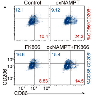
Flow cytometry analysis of M1 (CD86) and M2 (CD206) markers in human peripheral blood-derived macrophages or NAMPT-overexpressing (oxNAMPT) macrophages both treated with FK866 (10 nM, 48 h).
-
Nat Commun
2024 Nov 12;15(1):9515. PMID: 39532854 -
Exp Mol Med
CD300E+ macrophages facilitate liver regeneration after splenectomy in decompensated cirrhotic patients. [Abstract]2025 Feb;57(1):72-85. PMID: 39741181 -
J Adv Res
Single-Cell RNA sequencing identifies NAMPT as a potential therapeutic target in autoimmune uveitis. [Abstract]2025 Dec 29:S2090-1232(25)01020-3. PMID: 41475661
Daporinad purchased from MedChemExpress. Usage Cited in: J Adv Res. 2025 Dec 29:S2090-1232(25)01020-3. [Abstract]
Representative ocular fundus images and clinical scores from the Normal, EAU, and NAMPTi (FK866; 10 mg/kg; ip; daily for 14 days ) groups at day 14 post-immunization. White arrows highlight inflammatory infiltrates and vascular abnormalities. B. Representative histopathological images and histological scores from the Normal, EAU and NAMPTi groups at day 14 post-immunization. Black arrows highlight inflammatory cell infiltration, retinal detachment, and retinal folding.
Daporinad purchased from MedChemExpress. Usage Cited in: J Adv Res. 2025 Dec 29:S2090-1232(25)01020-3. [Abstract]
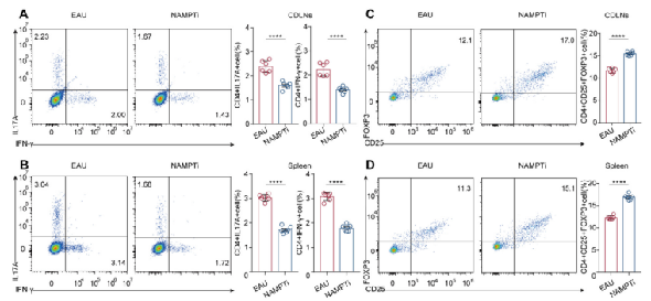
NAMPT inhibition ameliorates EAU by modulating the Teff/Treg balance and suppressing Hif1a in CD4+ T cells in vivo. A-B. Frequencies of CD4+IL 17A+ (Th17) cells and CD4+ IFN γ+(Th1) cells in both CDLNs (A) and spleen (B) of EAU and NAMPTi (FK866; 10 mg/kg; ip; daily for 14 days ) treated EAU mice on day 14 post-immunization. C-D. Frequencies of CD4+-CD25+Foxp3+ (Treg) cells in both CDLNs (C) and spleen (D).
-
Redox Biol
SIRT1 activated by AROS sensitizes glioma cells to ferroptosis via induction of NAD+ depletion-dependent activation of ATF3. [Abstract]2024 Feb:69:103030. PMID: 38181705 -
Adv Sci (Weinh)
Unraveling the Microenvironment and the Pathogenic Axis of HIF-1α-Visfatin-Fibrosis in Autoimmune Pancreatitis Using a Single-Cell Atlas. [Abstract]2025 Mar;12(12):e2412282. PMID: 39887620 -
Sci Adv
A dual chemodrug-loaded hyaluronan nanogel for differentiation induction therapy of refractory AML via disrupting lysosomal homeostasis. [Abstract]2025 Mar 28;11(13):eado3923. PMID: 40153509 -
Sci Adv
M2 macrophages drive leukemic transformation by imposing resistance to phagocytosis and improving mitochondrial metabolism. [Abstract]2023 Apr 14;9(15):eadf8522. PMID: 37058562 -
Cell Death Differ
TREM2 macrophage promotes cardiac repair in myocardial infarction by reprogramming metabolism via SLC25A53. [Abstract]2024 Feb;31(2):239-253. PMID: 38182899 -
Cell Death Dis
Targeting metabolic vulnerability by combining NAMPT inhibitors and disulfiram for treatment of recurrent ovarian cancer. [Abstract]2025 Apr 25;16(1):342. PMID: 40280967 -
Acta Pharmacol Sin
Activated SIRT1 contributes to DPT-induced glioma cell parthanatos by upregulation of NOX2 and NAT10. [Abstract]2023 Oct;44(10):2125-2138. PMID: 37277492 -
Acta Pharmacol Sin
TAX1BP1 contributes to deoxypodophyllotoxin-induced glioma cell parthanatos via inducing nuclear translocation of AIF by activation of mitochondrial respiratory chain complex I. [Abstract]2023 Sep;44(9):1906-1919. PMID: 37186123 -
Cell Chem Biol
AspSnFR: A genetically encoded biosensor for real-time monitoring of aspartate in live cells. [Abstract]2024 Aug 15;31(8):1529-1541.e12. PMID: 38806058 -
Sci Signal
Metabolic perturbations sensitize triple-negative breast cancers to apoptosis induced by BH3 mimetics. [Abstract]2021 Jun 8;14(686):eabc7405. PMID: 34103421 -
JCI Insight
2025 Nov 24;10(22):e185914. PMID: 41277555 -
J Agric Food Chem
Palmatine Attenuated Lipopolysaccharide-Induced Acute Lung Injury by Inhibiting M1 Phenotype Macrophage Polarization via NAMPT/TLR2/CCR1 Signaling. [Abstract]2024 Apr 15. PMID: 38619332 -
Cells
2023 Oct 2;12(19):2396. PMID: 37830610 -
Commun Biol
Nicotinamide mononucleotide treatment improves spermatogenesis in obese mice by reducing lysine acetylation of lactate dehydrogenase C. [Abstract]2025 Nov 26;8(1):1761. PMID: 41298813 -
Clin Epigenetics
Investigating the mechanisms by which low NAT1 expression in tumor cells contributes to chemo-resistance in colorectal cancer. [Abstract]2025 May 6;17(1):77. PMID: 40329330 -
Int Immunopharmacol
Cisatracurium besylate rescues Mycobacterium Tuberculosis-infected macrophages from necroptosis and enhances the bactericidal effect of isoniazid. [Abstract]2023 Jul:120:110291. PMID: 37182451 -
Biol Direct
NAMPT modulates muscle fiber type transition in PAD myopathy via the cGMP-PKG signaling pathway. [Abstract]2025 Nov 27;20(1):113. PMID: 41310770 -
Mol Neurobiol
The Depletion of NAMPT Disturbs Mitochondrial Homeostasis and Causes Neuronal Degeneration in Mouse Hippocampus. [Abstract]2023 Mar;60(3):1267-1280. PMID: 36441480 -
Biol Res
NAMPT regulates mitochondria and oxidative stress level for mouse early embryo development. [Abstract]2025 May 4;58(1):25. PMID: 40320561 -
Front Pharmacol
Trimetazidine Inhibits Renal Tubular Epithelial Cells to Mesenchymal Transition in Diabetic Rats via Upregulation of Sirt1. [Abstract]2020 Jul 29:11:1136. PMID: 32848753 -
Front Cell Dev Biol
AZD5153, a Bivalent BRD4 Inhibitor, Suppresses Hepatocarcinogenesis by Altering BRD4 Chromosomal Landscape and Modulating the Transcriptome of HCC Cells. [Abstract]2022 Mar 24:10:853652. PMID: 35399501 -
Chem Biol Interact
Melatonin suppresses glycolysis and coordinately disrupts DNA repair via targeting the YAP1-NAMPT signaling in breast cancer. [Abstract]2026 Feb 11:425:111900. PMID: 41485639 -
Eur J Pharm Sci
Activation of nicotinamide phosphoribosyltransferase protects against unilateral renal ischemia-reperfusion injury via the NAD+/SIRT1/PGC-1α signaling pathway and modulation of NFκB/TNF-α/IL-6. [Abstract]2025 Nov 1:214:107302. PMID: 41033563 -
Molecules
Quantitative Analysis of Daporinad (FK866) and Its In Vitro and In Vivo Metabolite Identification Using Liquid Chromatography-Quadrupole-Time-of-Flight Mass Spectrometry. [Abstract]2022 Mar 21;27(6):2011. PMID: 35335372 -
Biochim Biophys Acta Mol Basis Dis
Chlorophyllides repress gain-of-function p53 mutated HNSCC cell proliferation via activation of p73 and repression of p53 aggregation in vitro and in vivo. [Abstract]2025 Mar;1871(3):167662. PMID: 39788216 -
Biochim Biophys Acta Mol Basis Dis
Effect of alternating nicotinamide phosphoribosyltransferase expression levels on mitophagy in Alzheimer's disease mouse models. [Abstract]2024 Oct;1870(7):167288. PMID: 38862096 -
Mol Med Rep
Emodin alleviates severe acute pancreatitis-associated acute lung injury by decreasing pre-B-cell colony-enhancing factor expression and promoting polymorphonuclear neutrophil apoptosis. [Abstract]2017 Oct;16(4):5121-5128. PMID: 28849044
Daporinad purchased from MedChemExpress. Usage Cited in: Mol Med Rep. 2017 Oct;16(4):5121-5128. [Abstract]
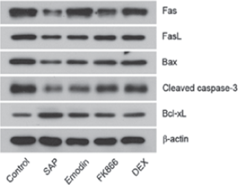
Effects of Emodin, FK866 and DEX on PMN apoptosis in SAP rats. Protein expression levels of Fas, FasL, Bax, cleaved caspase 3 and Bcl xL are detected by western blotting. β actin served as an internal control. Data are presented as the mean±standard deviation.
-
PLoS Pathog
Nicotinamide metabolism is essential for Hepatitis C Virus replication and the production of infectious Lipo-Viro-Particles. [Abstract]2026 Apr 22;22(4):e1014165. PMID: 42018568 -
Sci Rep
Inhibition of nicotinamide dinucleotide salvage pathway counters acquired and intrinsic poly(ADP-ribose) polymerase inhibitor resistance in high-grade serous ovarian cancer. [Abstract]2023 Feb 27;13(1):3334. PMID: 36849518 -
PLoS Pathog
2021 Mar 19;17(3):e1009436. PMID: 33740012 -
Cancers (Basel)
High-Dosage NMN Promotes Ferroptosis to Suppress Lung Adenocarcinoma Growth through the NAM-Mediated SIRT1-AMPK-ACC Pathway. [Abstract]2023 Apr 23;15(9):2427. PMID: 37173894 -
J Inflamm Res
Decrease of NAD+ Inhibits the Apoptosis of OLP T Cells via Inducing Mitochondrial Fission. [Abstract]2025 Jan 23:18:1091-1106. PMID: 39871961 -
Clin Exp Med
SPOP-dependent destabilization of SYT12 in a GSK-3β-dependent manner in papillary thyroid cancer cells. [Abstract]2025 Jul 10;25(1):243. PMID: 40640517 -
Brain Res Bull
Ablation of NAMPT in dopaminergic neurons leads to neurodegeneration and induces Parkinson's disease in mouse. [Abstract]2024 Nov:218:111114. PMID: 39489186 -
J Biol Chem
Glucose-6-phosphate dehydrogenase exerts anti-stress effects independently of its enzymatic activity. [Abstract]2022 Dec;298(12):102587. PMID: 36243112 -
-
J Cell Physiol
NAMPT regulates mitochondria function and lipid metabolism during porcine oocyte maturation. [Abstract]2024 Jan;239(1):180-192. PMID: 37992208 -
J Cell Physiol
Nicotinamide phosphoribosyl transferase regulates cell growth via the Sirt1/P53 signaling pathway and is a prognosis marker in colorectal cancer. [Abstract]2019 Apr;234(4):4385-4395. PMID: 30191976
Daporinad purchased from MedChemExpress. Usage Cited in: J Cell Physiol. 2019 Apr;234(4):4385-4395. [Abstract]
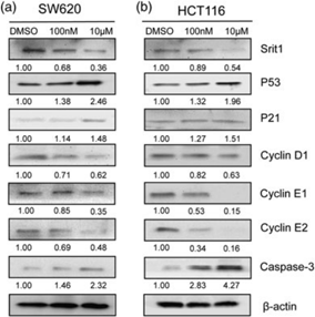
SW620 and HCT116 cells are seeded into a six-well plate for 72 hr, followed by treatment with different concentrations (100 nM and 10 μM) of FK866 for 24 hr.
-
Pancreatology
Inhibition of nicotinamide phosphoribosyltransferase protects against acute pancreatitis via modulating macrophage polarization and its related metabolites. [Abstract]2021 Aug;21(5):870-883. PMID: 33810973 -
Mol Carcinog
Adipocytes Promote Endometrial Cancer Progression Through Activation of the SIRT1-HMMR Signaling Axis. [Abstract]2024 Dec;63(12):2363-2381. PMID: 39254492 -
PeerJ
Oxidative stress induces cell death partially by decreasing both mRNA and protein levels of nicotinamide phosphoribosyltransferase in differentiated PC12 cells. [Abstract]2021 May 14;9:e11401. PMID: 34040894 -
Cancer Manag Res
Is the Fixed Periodic Treatment Effective for the Tumor System without Complete Information?. [Abstract]2021 Nov 30;13:8915-8928. PMID: 34876854 -
Neurosci Lett
Inhibition of neuronal Kv7 channels ameliorates MK-801-induced cognitive dysfunction in mice via up-regulating NAMPT expression. [Abstract]2023 Sep 25:814:137471. PMID: 37673371 -
Genes Genomics
FK866 inhibits colorectal cancer metastasis by reducing NAD+ levels in cancer-associated fibroblasts. [Abstract]2022 Dec;44(12):1531-1541. PMID: 36214983 -
Rapid Commun Mass Spectrom
Daporinad in vitro metabolite profiling via rat, dog, monkey and human liver microsomes by liquid chromatography/quadrupole-orbitrap mass spectrometry. [Abstract]2021 Sep 30;35(18):e9150. PMID: 34159659 -
-
-
-
-
-
-
bioRxiv
A Novel FNDC1-NAMPT-NAD axis is Implicated in Small and Large-vessel Arterial Disease and Drives Vascular Calcification. [Abstract]2025 Jul 10:2025.07.08.663801. PMID: 40672352 -
Res Sq
Novel mixed cancer-cell models designed to capture inter-patient tumor heterogeneity for accurate evaluation of drug combinations. [Abstract]2025 May 16:rs.3.rs-6590535. PMID: 40470249 -
-
bioRxiv
PAIRWISE: Deep Learning-based Prediction of Effective Personalized Drug Combinations in Cancer. [Abstract]2024 Nov 6:2024.11.04.621884. PMID: 39574568 -
-
-
-
-
-
-
-


Solvent & Solubility
In Vitro:
DMSO : ≥ 50 mg/mL (127.71 mM; Hygroscopic DMSO has a significant impact on the solubility of product, please use newly opened DMSO)
H2O : < 0.1 mg/mL (insoluble)
* "≥" means soluble, but saturation unknown.
Please refer to the solubility information to select the appropriate solvent. Once prepared, please aliquot and store the solution to prevent product inactivation from repeated freeze-thaw cycles.
Storage method and period of stock solution: -80°C, 2 years; -20°C, 1 year. When stored at -80°C, please use it within 2 years. When stored at -20°C, please use it within 1 year.
Please refer to the solubility information to select the appropriate solvent. Once prepared, please aliquot and store the solution to prevent product inactivation from repeated freeze-thaw cycles.
Storage method and period of stock solution: -80°C, 2 years; -20°C, 1 year. When stored at -80°C, please use it within 2 years. When stored at -20°C, please use it within 1 year.
Concentration (start) × Volume (start) = Concentration (final) × Volume (final)
In Vivo:
Select the appropriate dissolution method based on your experimental animal and administration route.
- For the following dissolution methods, please ensure to first prepare a clear stock solution using an In Vitro approach and then sequentially add co-solvents:
- To ensure reliable experimental results, the clarified stock solution can be appropriately stored based on storage conditions. As for the working solution for In Vivo experiments, it is recommended to prepare freshly and use it on the same day.
- The percentages shown for the solvents indicate their volumetric ratio in the final prepared solution. If precipitation or phase separation occurs during preparation, heat and/or sonication can be used to aid dissolution.
Add each solvent one by one: 10% DMSO 40% PEG300 5% Tween-80 45% Saline
Solubility: ≥ 2.5 mg/mL (6.39 mM); Clear solution
This protocol yields a clear solution of ≥ 2.5 mg/mL (saturation unknown).
Taking 1 mL working solution as an example, add 100 μL DMSO stock solution (25.0 mg/mL) to 400 μL PEG300, and mix evenly; then add 50 μL Tween-80 and mix evenly; then add 450 μL Saline to adjust the volume to 1 mL.
Preparation of Saline: Dissolve 0.9 g sodium chloride in ddH₂O and dilute to 100 mL to obtain a clear Saline solution.
Add each solvent one by one: 10% DMSO 90% (20% SBE-β-CD in Saline)
Solubility: ≥ 2.5 mg/mL (6.39 mM); Clear solution
This protocol yields a clear solution of ≥ 2.5 mg/mL (saturation unknown).
Taking 1 mL working solution as an example, add 100 μL DMSO stock solution (25.0 mg/mL) to 900 μL 20% SBE-β-CD in Saline, and mix evenly.
Preparation of 20% SBE-β-CD in Saline (4°C, storage for one week): 2 g SBE-β-CD powder is dissolved in 10 mL Saline, completely dissolve until clear.
For the following dissolution methods, please prepare the working solution directly:
It is recommended to prepare fresh solutions and use them promptly within a short period of time.
The percentages shown for the solvents indicate their volumetric ratio in the final prepared solution. If precipitation or phase separation occurs during preparation, heat and/or sonication can be used to aid dissolution.
In Vivo Dissolution Calculator
Please enter the basic information of animal experiments:
-
-
-
-
Recommended: Prepare an additional quantity of animals to account for potential losses during experiments.
Please enter your animal formula composition:
-
%DMSO +
Recommended: Keep the proportion of DMSO in working solution below 2% if your animal is weak.
-
%+
-
+%Tween-80 + +
-
%Saline +
The co-solvents required include: DMSO, . All of co-solvents are available by MedChemExpress (MCE). , Tween 80. All of co-solvents are available by MedChemExpress (MCE).
Working solution concentration: 0.22 mg/mL
Method for preparing stock solution: mg drug dissolved in μL DMSO. Stock solution concentration: mg/mL.
1. Take μL DMSO stock solution;
2. Add μL .
μL , mix evenly;
3. Then add μL Tween 80, mix evenly;
4. Then add μL
Please ensure that the stock solution in the first step is dissolved to a clear state, and add co-solvents in sequence. You can use ultrasonic heating (ultrasonic cleaner, recommended frequency 20-40 kHz), vortexing, etc. to assist dissolution.

Protocols
-
Annexin V plus membrane-impermeant dye apoptosis staining
Annexin V-based apoptosis assays rely on the detection of phosphatidylserine (PS) externalization from the inner leaflet of the plasma membrane to the outer leaflet, an early biochemical hallmark of apoptosis. Fluorescently labeled Annexin V binds PS in a calcium-dependent manner, enabling identification of early apoptotic cells by flow cytometry or fluorescence microscopy. When combined with a membrane-impermeant DNA-binding dye (e. g. , propidium iodide), this approach allows discrimination between viable (Annexin V−/dye−), early apoptotic (Annexin V+/dye−), and late apoptotic or necrotic (Annexin V+/dye+) cell populations by assessing membrane integrity and PS exposure.
-
BrdU Incorporation Assay
Bromodeoxyuridine (BrdU) incorporation assay is based on the principle that BrdU, a thymidine analog, is incorporated into newly synthesized DNA during the S phase of the cell cycle, thereby serving as a marker of DNA replication and cellular proliferation. Incorporated BrdU can be detected using anti-BrdU antibodies following DNA denaturation, enabling visualization or quantification of proliferating cells through immunochemical detection methods such as immunofluorescence or immunohistochemistry.
-
Somatic Cell Culture
A method of simulating the in vivo environment in vitro to maintain the cell growth, differentation and main functions.
-
CFSE Dye Dilution Proliferation Assay
The CFSE (carboxyfluorescein diacetate succinimidyl ester) dye dilution proliferation assay is based on the covalent labeling of intracellular proteins by a cell-permeant fluorescent dye that becomes fluorescent upon intracellular ester cleavage and then is stably retained within cells. As labeled cells divide, the dye is partitioned equally between daughter cells, resulting in a stepwise halving of fluorescence intensity that can be quantified by flow cytometry to determine the number of cell divisions undergone by each cell population. This fluorescence dilution approach enables quantitative tracking of lymphocyte proliferation at the single-cell level over multiple rounds of division. CFSE-based proliferation analysis has been widely applied to measure antigen-driven lymphocyte expansion in vitro, where discrete fluorescence peaks correspond to successive cell divisions and allow reconstruction of proliferative history within heterogeneous populations.
-
Dye-dilution cell tracking and proliferation staining
Dye-dilution cell tracking assays quantify cell proliferation by covalently labeling intracellular proteins with a stable fluorescent dye that is equally partitioned between daughter cells during mitosis, resulting in stepwise halving of fluorescence intensity with each cell division as measured by flow cytometry histograms. Carboxyfluorescein diacetate succinimidyl ester (CFSE) is a prototypical dye that diffuses into cells, is enzymatically converted into a fluorescent compound, and then covalently binds intracellular amine groups, producing long-lived fluorescence suitable for tracking multiple rounds of division in vitro and in vivo. Successive generations of dividing cells form discrete peaks of decreasing fluorescence intensity, enabling estimation of proliferation history, precursor frequency, and division index within heterogeneous populations. Alternative dyes such as CellTrace Violet (CTV) and far-red membrane dyes (e. g. , PKH26) follow the same dilution principle but differ
-
Protocol for Cell Cycle
Cell-cycle analysis by flow cytometry measures DNA content in single cells to estimate the fraction of cells in G0/G1, S, and G2/M phases. Propidium iodide intercalates into DNA, and after RNA removal with RNase, fluorescence intensity reflects cellular DNA content: 2N cells are assigned to G0/G1, cells between 2N and 4N to S phase, and 4N cells to G2/M. DNA-content analysis alone cannot reliably separate G0 from G1 or G2 from M. Ki-67 can distinguish quiescent G0 cells from cycling cells, EdU or BrdU incorporation marks active DNA synthesis in S phase, and phospho-histone H3 staining identifies mitotic cells within the 4N population.
-
CCK-8/WST-8 Cell Proliferation Assay
The CCK-8/WST-8 assay is based on the reduction of the water-soluble tetrazolium salt WST-8 to a water-soluble formazan product by cellular dehydrogenases in metabolically active cells, where the generated formazan amount is proportional to the number of living cells and is quantified by measuring absorbance in the visible range, providing a colorimetric readout for cell viability and proliferation assessment. This class of tetrazolium-based assays improves upon earlier MTT-based systems by producing a water-soluble formazan, eliminating the need for organic solubilization steps and enabling direct spectrophotometric measurement in culture medium.
-
Cell Counting-Based Growth Curve Assay
Cell counting-based growth curve assays quantify cell proliferation by directly measuring changes in viable cell number over time using manual or automated counting methods such as hemocytometer-based counting or instrument-assisted cell enumeration, enabling construction of growth curves that reflect population expansion dynamics in response to culture conditions. A widely used approach is trypan blue exclusion with hemocytometer counting, where membrane-compromised (non-viable) cells take up the dye, allowing discrimination between viable and non-viable cells while simultaneously enabling total cell number quantification. Repeated sampling across time points allows estimation of proliferation rate, growth phases, and comparative growth kinetics between experimental conditions.
-
Liver Cancer Modeling
Liver cancer can be classified into primary liver cancer and secondary liver cancer. Secondary liver cancer is the metastatic liver cancer. Primary liver cancer includes hepatocellular carcinoma (HCC), intrahepatic cholangiocarcinoma (ICC) and fibrolamellar HCC, of which HCC is the most common form, accounting for approximately 90% of primary liver cancers[1]. HCC mouse models include chemical agent-induced models, transplanted tumor models, and genetic engineered models.
-
Colony Formation (Clonogenic) Assay
The clonogenic (colony formation) assay measures the ability of a single cell to retain reproductive viability and form a macroscopic colony, typically defined as a cluster derived from one progenitor cell after a defined growth period. This assay is widely used to evaluate cell survival after exposure to ionizing radiation or cytotoxic treatments and is considered a standard method in radiation biology for generating dose-response relationships of reproductive cell death. Colony formation reflects long-term proliferative capacity rather than short-term metabolic activity, and survival is quantified by comparing treated versus untreated conditions based on colony number and derived survival fractions.
-
Cell Viability Determination by MTT Colorimetric Assay
The following protocol uses the MTT colorimetric assay as a classic literature-established method for assessing cell viability/metabolic activity in cultured mammalian cells. MTT[3-(4,5-dimethylthiazol-2-yl)-2,5-diphenyltetrazolium bromide] is reduced by metabolically active cells to a colored formazan product; the amount of formazan is quantified spectrophotometrically and provides an indirect measure of metabolically active viable cells. Importantly, MTT reduction reflects cellular oxidoreductase/metabolic activity rather than an absolute direct count of living cells, so changes in cellular metabolism can alter the signal independently of cell number.
-
EdU Incorporation Assay (Click Chemistry-Based DNA Synthesis Measurement)
The EdU incorporation assay measures DNA synthesis by adding the thymidine analog 5-ethynyl-2′-deoxyuridine to cells or tissues, where it is incorporated into newly synthesized DNA during S phase. Incorporated EdU is detected by copper-catalyzed azide-alkyne cycloaddition, in which a fluorescent azide covalently reacts with the ethynyl group on EdU, allowing S-phase cells to be detected by fluorescence microscopy, flow cytometry, or high-content imaging. EdU detection does not require DNA denaturation or anti-BrdU antibody access, which preserves sample structure and improves compatibility with immunostaining and multiparameter cytometry compared with BrdU-based detection. EdU can be cytotoxic in a cell-type- and exposure-dependent manner, so pulse duration, concentration, and continuous-labeling designs should be validated for each cell type.
-
Apoptosis
Apoptosis, also called programmed cell death, is generally characterized by distinct morphological characteristics.
-
TUNEL staining for apoptotic DNA fragmentation
TUNEL staining detects DNA strand breaks by using terminal deoxynucleotidyl transferase to add labeled nucleotides to exposed 3′-OH DNA termini, generating either microscopic staining in fixed cells or tissue sections, or fluorescence/cytometric signal in cell suspensions. TUNEL positivity reflects DNA fragmentation but should not be interpreted alone as definitive apoptosis, because TUNEL can also label necrotic, autolytic, mechanically damaged, or DNA-repair-associated DNA breaks.
-
Ki-67 Immunostaining Proliferation Assay
Ki-67 immunostaining measures the growth fraction of a cell population by detecting Ki-67, a nuclear antigen present in proliferating cells and absent in quiescent G0 cells. The readout is the percentage of Ki-67-positive nuclei among total counted cells, commonly called the Ki-67 labeling index or proliferation index.
-
Flow cytometric DNA-content cell-cycle staining
Flow cytometric DNA-content cell-cycle staining measures the fluorescence intensity of DNA-bound fluorochromes in single cells or nuclei to estimate DNA content distributions, allowing assignment of populations to G0/G1, S, and G2/M phases by DNA histogram deconvolution. Propidium iodide (PI) intercalates into DNA, and PI fluorescence is proportional to cellular DNA content when staining is performed under conditions that make DNA accessible and minimize non-DNA signal. Cells with G2/M DNA content are expected to show approximately twice the fluorescence intensity of G0/G1 cells, while S-phase cells occupy intermediate fluorescence values. PI-based DNA-content analysis can also detect cells with fractional DNA content, often reported as sub-G1, when DNA fragmentation and extraction during staining reduce retained DNA signal in apoptotic cells. DAPI is an alternative DNA fluorochrome for univariate DNA-content analysis, while bivariate approaches combining DNA content with proliferation
-
PCNA Immunodetection Proliferation Assay
PCNA immunodetection measures proliferative activity by detecting proliferating cell nuclear antigen, a nuclear protein associated with DNA polymerase δ function and DNA replication. The assay readout is the proportion of PCNA-positive nuclei among total counted cells, but PCNA labeling is not identical to BrdU labeling because PCNA can mark late G1/early S-associated replication competence and may persist beyond active DNA synthesis depending on fixation and extraction conditions.
-
Protocol for Cell Counting and Cell Density Analysis
Cell counting and cell-density analysis estimate the number of cells in a known volume or field area. Manual hemocytometer counting uses a chamber of defined geometry to convert counted cells into cells/mL, while automated counters and image-analysis workflows detect cell objects from optical, brightfield, fluorescence, impedance, or digital-image features. Trypan blue viability counting is based on dye exclusion: viable cells with intact membranes exclude dye, while non-viable cells with compromised membranes stain blue. The readout is total cell density, viable-cell density, dead-cell density, and percent viability. Cell density can also be estimated from microscopy images by counting objects per image area, from flow cytometry using calibrated volume or reference particles, or from in situ microscopy in bioreactors after calibration against reference methods such as hemocytometer or flow cytometry.
-
Autophagy
Autophagy is a process in which eukaryotic cells use lysosomes to degrade their own cytoplasmic proteins and damaged organelles under the regulation of autophagy related gene (Atg). Microtubule-associated proteins light chain 3 (LC3) is recognized as autophagy marker, which transfers from cytoplasmic LC3 (LC3-I) to membrane type (LC3-II). LC3-II/I ratio could be detected by Western Blot and fluorescence microscopy.
-
Apoptosis Solutions
Apoptosis is a regulated, generally non-lytic cell-death pathway that removes unwanted, damaged, infected, or abnormal cells through coordinated morphological changes, caspase activation, DNA fragmentation, and membrane remodeling. The intrinsic apoptosis pathway is controlled mainly by mitochondrial outer membrane permeabilization, BCL-2 family proteins, cytochrome c release, apoptosome formation, caspase-9 activation, and downstream executioner caspase-3/7 activation. The extrinsic apoptosis pathway is initiated by death receptors such as Fas, TNFR, and TRAIL receptors, which recruit adaptor proteins and activate caspase-8 before engaging executioner caspases or mitochondrial amplification through BID cleavage. Apoptosis is linked to many phenotypes, including cancer cell killing, tissue homeostasis, immune regulation, neurodegeneration, infection response, and treatment-induced cytotoxicity; unresolved questions include how apoptosis interacts with necroptosis, pyroptosis, ferroptos
-
Lysosome and acidic-vesicle live-cell staining
Lysosome and acidic-vesicle live-cell staining detects acidic intracellular compartments by using membrane-permeant acidotropic probes that accumulate in low-pH vesicles, including lysosomes, late endosomes, autolysosomes, and acidic phagosomes. LysoTracker staining is commonly used as an intensity-based readout of acidic lysosomal compartment abundance or enlargement, while acridine orange produces green fluorescence in less concentrated compartments and red fluorescence after concentration-dependent accumulation in acidic vesicular organelles. Loss or reduction of acridine-orange red signal can be used as a readout of lysosomal membrane permeabilization or reduced acidic-vesicle integrity. This protocol is designed for live cultured cells and can be adapted for fluorescence microscopy, high-content imaging, plate-reader readout, or flow cytometry when the selected literature supports the readout. Because these dyes report acidotropic accumulation rather than lysosome identity alone,
-
MTT Cell Proliferation Assay
The MTT assay is a colorimetric endpoint assay for estimating viable cell number, cell growth, cytotoxicity, or cell activation in cultured mammalian cells. Living cells reduce the yellow tetrazolium salt MTT into purple/blue formazan, while dead cells do not generate the same signal; the resulting color can be quantified with a multiwell spectrophotometer. MTT reduction is commonly interpreted as a readout of metabolic activity that often correlates with viable cell number, but it should not be treated as a direct cell-counting method unless the assay is optimized for the cell type and experimental condition. Studies show that MTT reduction can involve mitochondrial and non-mitochondrial reducing systems, and formazan may accumulate in intracellular lipid droplets rather than simply marking mitochondria.
-
Macroautophagy Solutions
Macroautophagy is a conserved lysosome-dependent degradation pathway in which cytoplasmic material is sequestered into double-membrane autophagosomes and delivered to lysosomes for degradation and recycling. The pathway supports cellular homeostasis during nutrient limitation, organelle stress, protein-aggregate accumulation, infection, differentiation, and tissue remodeling by coupling cargo sequestration, autophagosome maturation, lysosomal fusion, and degradation of cargo-derived macromolecules. The core molecular sequence includes initiation by nutrient- and stress-regulated autophagy machinery, autophagosome nucleation, LC3/ATG8-family conjugation to autophagosomal membranes, cargo selection through receptors such as SQSTM1/p62, autophagosome-lysosome fusion, and lysosomal degradation. LC3 was identified as a mammalian homolog of yeast Atg8 that localizes to autophagosomal membranes after processing, and p62/SQSTM1 was shown to connect ubiquitinated cargo with autophagic degradati


Purity & Documentation
-
Data Sheet (276 KB)
-
SDS (396 KB)
- English - EN (396 KB)
- Français - FR (396 KB)
- Deutsch - DE (396 KB)
- Norwegian - NO (396 KB)
- Español - ES (396 KB)
- Swedish - SV (396 KB)
- Italian - IT (396 KB)
- Korean - KR (396 KB)
- Portuguese - PT (396 KB)
-
Handling Instructions (2659 KB)
References
[1]. Cea M, et al. Targeting NAD+ salvage pathway induces autophagy in multiple myeloma cells via mTORC1 and extracellular signal-regulated kinase (ERK1/2) inhibition. Blood. 2012 Oct 25;120(17):3519-29. [Content Brief]
[2]. Magnone M, et al. NAD+ levels control Ca2+ store replenishment and mitogen-induced increase of cytosolic Ca2+ by Cyclic ADP-ribose-dependent TRPM2 channel gating in human T lymphocytes. J Biol Chem. 2012 Jun 15;287(25):21067-81. [Content Brief]
[3]. Thakur BK, et al. Inhibition of NAMPT pathway by FK866 activates the function of p53 in HEK293T cells. Biochem Biophys Res Commun. 2012 Aug 3;424(3):371-7. [Content Brief]
[4]. Hasmann M, Schemainda I. FK866, a highly specific noncompetitive inhibitor of nicotinamide phosphoribosyltransferase, represents a novel mechanism for induction of tumor cell apoptosis. Cancer Res. 2003 Nov 1;63(21):7436-42. [Content Brief]


Complete Stock Solution Preparation Table
Please refer to the solubility information to select the appropriate solvent. Once prepared, please aliquot and store the solution to prevent product inactivation from repeated freeze-thaw cycles.
Storage method and period of stock solution: -80°C, 2 years; -20°C, 1 year. When stored at -80°C, please use it within 2 years. When stored at -20°C, please use it within 1 year.
| Optional Solvent | Concentration Solvent Mass | 1 mg | 5 mg | 10 mg | 25 mg |
|---|---|---|---|---|---|
| DMSO | 1 mM | 2.5542 mL | 12.7711 mL | 25.5421 mL | 63.8553 mL |
| 5 mM | 0.5108 mL | 2.5542 mL | 5.1084 mL | 12.7711 mL | |
| 10 mM | 0.2554 mL | 1.2771 mL | 2.5542 mL | 6.3855 mL | |
| 15 mM | 0.1703 mL | 0.8514 mL | 1.7028 mL | 4.2570 mL | |
| 20 mM | 0.1277 mL | 0.6386 mL | 1.2771 mL | 3.1928 mL | |
| 25 mM | 0.1022 mL | 0.5108 mL | 1.0217 mL | 2.5542 mL | |
| 30 mM | 0.0851 mL | 0.4257 mL | 0.8514 mL | 2.1285 mL | |
| 40 mM | 0.0639 mL | 0.3193 mL | 0.6386 mL | 1.5964 mL | |
| 50 mM | 0.0511 mL | 0.2554 mL | 0.5108 mL | 1.2771 mL | |
| 60 mM | 0.0426 mL | 0.2129 mL | 0.4257 mL | 1.0643 mL | |
| 80 mM | 0.0319 mL | 0.1596 mL | 0.3193 mL | 0.7982 mL | |
| 100 mM | 0.0255 mL | 0.1277 mL | 0.2554 mL | 0.6386 mL |


Daporinad Related Classifications
HY-50876 Related Classifications
 Powered by Bioz