



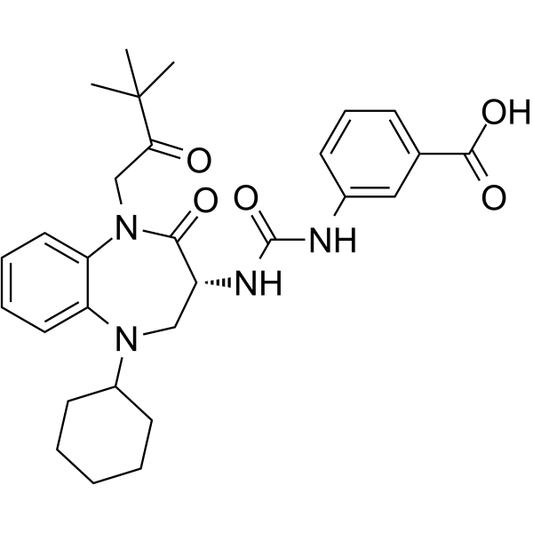
Nastorazepide
Based on 1 publication(s) in Google Scholar
Nastorazepide (Z-360) is an orally active 1,5-benzodiazepine derivative and gastrin/CCK-2 receptor antagonist. Nastorazepide inhibits the specific binding of [3H]CCK-8 to the human CCK-2 receptor with a Ki value of 0.47 nM. Nastorazepide inhibits IL-1β, ephrin B1, VEGF, and HIF-1alpha, reduces Akt and NR2B phosphorylation. Nastorazepide has antitumor activity against pancreatic cancer. Nastorazepide inhibits colorectal cancer liver metastasis and relieves pain.
For research use only. We do not sell to patients.
- Purity : 99.95%
- CAS No.: 209219-38-5
- Formula: C29H36N4O5
- Molecular Weight:520.62
-
Storage:Powder -20°C, 3 years , 4°C, 2 years ; In solvent -80°C, 2 years , -20°C, 1 year

To place orders, for customer services and technical support, please contact: MedChemExpress USA
Tel: 609-228-6898 E-mail: [email protected] [email protected]


-
 Biological Activity
-
Chemical Information
-
Solvent & Solubility
- Protocols
- Purity & Documentation
- References
-
 Help & FAQs


-
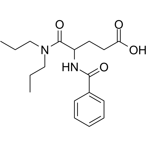
HY-B1330
ProglumideCCK-A/B Receptors Antagonist -
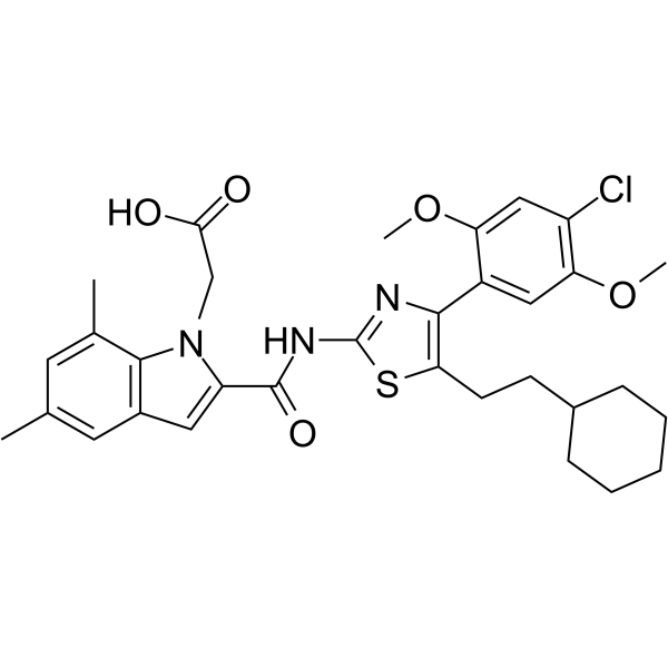
HY-11077
SR 146131Cholecystokinin Receptor Agonist -
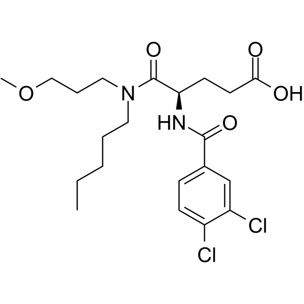
HY-128878
DexloxiglumideCCKR Inhibitor -
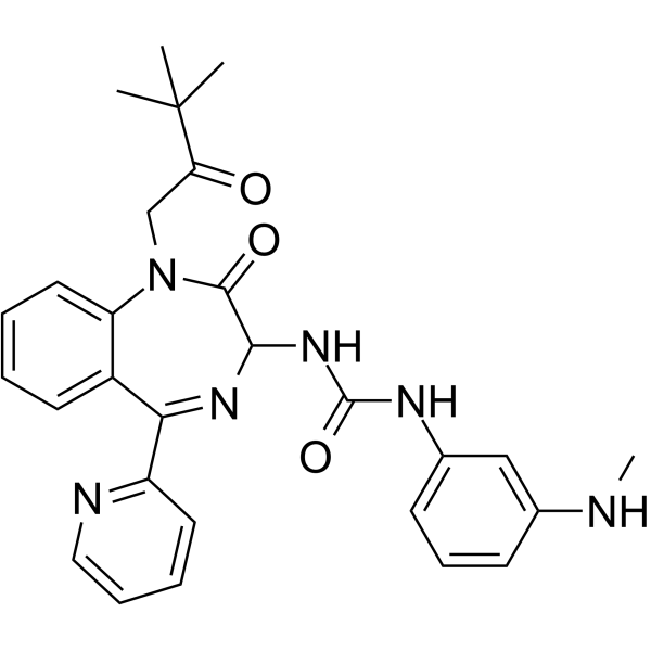
HY-U00360
(Rac)-Sograzepide -
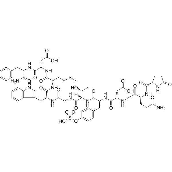
HY-A0190
CaeruleinCholecystokinin Receptor Agonist -
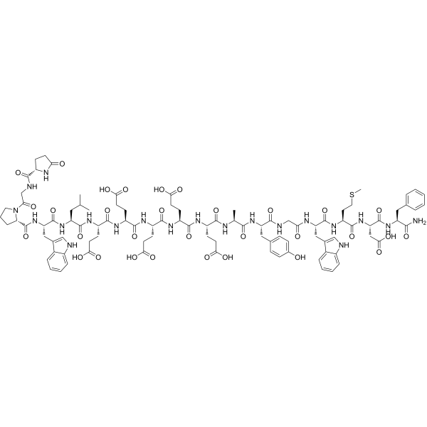
HY-P1097
Gastrin I, humanCCK2 Agonist -
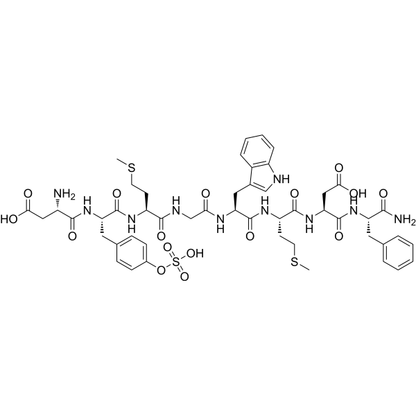
HY-P0093
SincalideAmino Acid Polypeptide Hormone Analogue -

HY-17617R
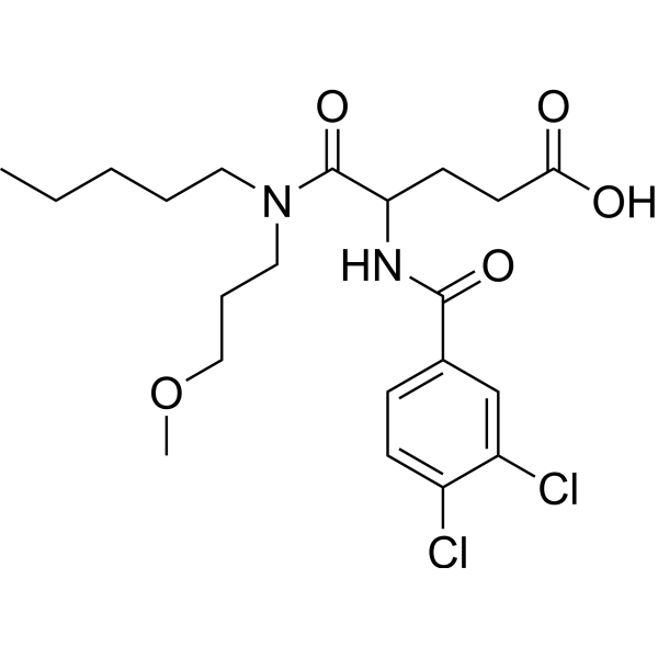
Nastorazepide (Standard)CCK-2 Receptor Antagonist -
HY-B2154
LoxiglumideCholecystokinin Receptor Antagonist -
![[Leu15]-Gastrin I (human)](https://file.medchemexpress.com/product_pic/hy-p2671.gif)
HY-P2671
[Leu15]-Gastrin I (human)Peptide Hormone -
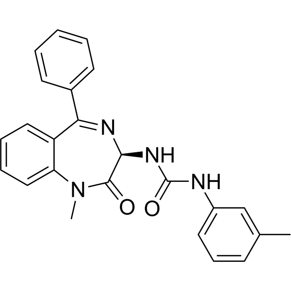
HY-106840
L-365260CCK-B Antagonist -
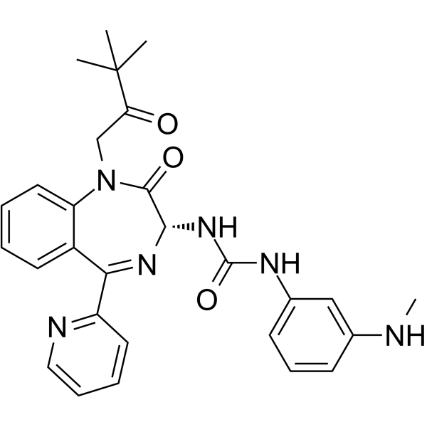
HY-14850
SograzepideGastrin/CCK-B Antagonist
Publications Citing Use of MedChemExpress (MCE) Nastorazepide
MoreAll VEGFR Isoforms
More

Biological Activity
Description
IC50 & Target
|
CCKBR |
In Vitro
Nastorazepide (Z-360) is an orally active 1,5-benzodiazepine derivative and gastrin/CCK-2 receptor antagonist. Nastorazepide inhibits the specific binding of [3H]CCK-8 to the human CCK-2 receptor with a Ki value of 0.47 nM. Nastorazepide inhibits IL-1β, ephrin B1, VEGF, and HIF-1alpha, reduces Akt and NR2B phosphorylation. Nastorazepide has antitumor activity against pancreatic cancer. Nastorazepide inhibits colorectal cancer liver metastasis and relieves pain[1][2][3][4][5][6][7].
MedChemExpress (MCE) has not independently confirmed the accuracy of these methods. They are for reference only. Further protocols information, click here.
In Vivo
Nastorazepide (3-100 mg/kg; p.o.; once daily) inhibits colorectal cancer liver metastasis in the C170HM2 mouse model, increases survival in the MGLVA1 ascites mouse model, and inhibits pancreatic tumor growth when combined with Gemcitabine in the PAN-1 orthotopic mouse model[3].
Nastorazepide (100 mg/kg; p.o.; once daily; 3 weeks) suppresses tumor growth in MIA PaCa-2-bearing mice via inhibition of Gastrin-induced anti-apoptotic effects[4].
Nastorazepide (30-300 mg/kg; p.o.; starting from day 7 and continuing until day 21 in the cancer pain model) can inhibit the late-phase nociceptive responses in the Formalin-induced pain model, produce an anti-allodynic effect in the cancer pain model[5].
Nastorazepide (100 mg/kg; p.o.; once daily; from day 7 to day 14) prevents up-regulation of ephrin B1 gene expression and phosphorylation of NR2B via suppression of IL-1β production in a cancer-induced pain model in mice[6].
MedChemExpress (MCE) has not independently confirmed the accuracy of these methods. They are for reference only.
-
Animal Model:BALB/c-nu/nuSlc nude mice (female, 7-week-old) with subcutaneous xenograft of MiaPaCa2 cells[2]
-
Dosage:10 mg/kg, 30 mg/kg, 100 mg/kg
-
Administration:Oral administration, once daily, for 21 days
-
Result:Significantly inhibited tumor growth of MiaPaCa2 subcutaneous xenografts in a dose-dependent manner.
Resulted in final tumor weight inhibition of 16.5%, 39.6%, and 41.7% at 10, 30, and 100 mg/kg, respectively.
-
Animal Model:
Clinical Trial
| NCT Number | Sponsor | Condition | Start Date |
Phase

|
|---|---|---|---|---|
| NCT01329991 | Plexxikon| | 2011-05 | PHASE1 |


Chemical Information
-
CAS No. 209219-38-5
-
Appearance Solid
-
Molecular Weight 520.62
-
Formula C29H36N4O5
-
Color White to off-white
-
SMILES
O=C(O)C1=CC=CC(NC(N[C@H]2C(N(CC(C(C)(C)C)=O)C3=CC=CC=C3N(C4CCCCC4)C2)=O)=O)=C1
-
Synonyms
Z-360
-
Shipping
Room temperature in continental US; may vary elsewhere.
-
Storage
Powder -20°C 3 years 4°C 2 years In solvent -80°C 2 years -20°C 1 year


Publications (1)
-
Journal Impact Factor
-
Most Recent


Solvent & Solubility
In Vitro:
DMSO : ≥ 32 mg/mL (61.47 mM; Hygroscopic DMSO has a significant impact on the solubility of product, please use newly opened DMSO)
* "≥" means soluble, but saturation unknown.
Please refer to the solubility information to select the appropriate solvent. Once prepared, please aliquot and store the solution to prevent product inactivation from repeated freeze-thaw cycles.
Storage method and period of stock solution: -80°C, 2 years; -20°C, 1 year. When stored at -80°C, please use it within 2 years. When stored at -20°C, please use it within 1 year.
Please refer to the solubility information to select the appropriate solvent. Once prepared, please aliquot and store the solution to prevent product inactivation from repeated freeze-thaw cycles.
Storage method and period of stock solution: -80°C, 2 years; -20°C, 1 year. When stored at -80°C, please use it within 2 years. When stored at -20°C, please use it within 1 year.
Concentration (start) × Volume (start) = Concentration (final) × Volume (final)
In Vivo:
Select the appropriate dissolution method based on your experimental animal and administration route.
- For the following dissolution methods, please ensure to first prepare a clear stock solution using an In Vitro approach and then sequentially add co-solvents:
- To ensure reliable experimental results, the clarified stock solution can be appropriately stored based on storage conditions. As for the working solution for In Vivo experiments, it is recommended to prepare freshly and use it on the same day.
- The percentages shown for the solvents indicate their volumetric ratio in the final prepared solution. If precipitation or phase separation occurs during preparation, heat and/or sonication can be used to aid dissolution.
Add each solvent one by one: 10% DMSO 90% (20% SBE-β-CD in Saline)
Solubility: 2.08 mg/mL (4.00 mM); Suspended solution; Need ultrasonic
This protocol yields a suspended solution of 2.08 mg/mL. Suspended solution can be used for oral and intraperitoneal injection.
Taking 1 mL working solution as an example, add 100 μL DMSO stock solution (20.8 mg/mL) to 900 μL 20% SBE-β-CD in Saline, and mix evenly.
Preparation of 20% SBE-β-CD in Saline (4°C, storage for one week): 2 g SBE-β-CD powder is dissolved in 10 mL Saline, completely dissolve until clear.
In Vivo Dissolution Calculator
Please enter the basic information of animal experiments:
-
-
-
-
Recommended: Prepare an additional quantity of animals to account for potential losses during experiments.
Please enter your animal formula composition:
-
%DMSO +
Recommended: Keep the proportion of DMSO in working solution below 2% if your animal is weak.
-
%+
-
+%Tween-80 + +
-
%Saline +
The co-solvents required include: DMSO, . All of co-solvents are available by MedChemExpress (MCE). , Tween 80. All of co-solvents are available by MedChemExpress (MCE).
Working solution concentration: 0.22 mg/mL
Method for preparing stock solution: mg drug dissolved in μL DMSO. Stock solution concentration: mg/mL.
1. Take μL DMSO stock solution;
2. Add μL .
μL , mix evenly;
3. Then add μL Tween 80, mix evenly;
4. Then add μL
Please ensure that the stock solution in the first step is dissolved to a clear state, and add co-solvents in sequence. You can use ultrasonic heating (ultrasonic cleaner, recommended frequency 20-40 kHz), vortexing, etc. to assist dissolution.

Protocols
-
Kinase activity and phosphorylation assays
Kinase activity assays measure the ability of kinases to transfer phosphate groups from ATP to specific substrates, while phosphorylation assays detect the presence and levels of phosphorylated proteins. Common methods include radiolabeled ATP incorporation (e. g. ,), ADP release detection via bioluminescence (e. g. ,[3]), enzyme-linked immunosorbent assays (ELISA) for phospho-specific epitopes (e. g. ,[6]), and microtiter-based formats for high-throughput screening (e. g. ,[8]). The ADP-Glo assay quantifies kinase activity by measuring ADP produced during phosphorylation using a luciferase-based system. Radiometric assays involve autoradiography or scintillation counting after incorporation of 32P-labeled ATP into substrate proteins. ELISA-based approaches rely on phospho-specific antibodies to detect activated kinases in cell lysates or purified samples.
-
Western Blot
Western blotting (WB) is a commonly used experimental method in molecular biology, biochemistry, and immunogenetics for identifying and quantifying target proteins. It combines gel electrophoresis with immunoassay, enabling researchers to analyze protein expression, post-translational modifications, and molecular weight.
-
CCK-8/WST-8 Cell Proliferation Assay
The CCK-8/WST-8 assay is based on the reduction of the water-soluble tetrazolium salt WST-8 to a water-soluble formazan product by cellular dehydrogenases in metabolically active cells, where the generated formazan amount is proportional to the number of living cells and is quantified by measuring absorbance in the visible range, providing a colorimetric readout for cell viability and proliferation assessment. This class of tetrazolium-based assays improves upon earlier MTT-based systems by producing a water-soluble formazan, eliminating the need for organic solubilization steps and enabling direct spectrophotometric measurement in culture medium.
-
Splenic/Portal-Vein Liver Metastasis Xenograft
Splenic and portal-vein liver metastasis xenograft models deliver tumor cells into the portal circulation so that cells reach the liver first and form hepatic metastatic lesions; splenic injection uses the spleen as an access route to the portal system, while direct portal-vein injection introduces cells into the portal vein without requiring splenectomy. The assay detects liver colonization, intrahepatic tumor growth, tumor distribution, treatment response, survival, and liver-metastasis microenvironment changes; readouts include bioluminescence or fluorescence imaging, gross liver nodule counts, liver weight or tumor burden, histology, and survival.
-
Protocol for Kinase activity and phosphorylation assays
Kinase activity assays measure transfer of phosphate from ATP to a protein or peptide substrate, generating phosphorylated substrate, ADP, or incorporated radiolabeled phosphate as the readout; phosphorylation assays measure site-specific phosphorylation in cells or tissues as a proxy for kinase-pathway activation, inhibition, or substrate regulation. Phosphorylation can be detected by phospho-specific Western blot, immunoprecipitation kinase assay, phospho-immunofluorescence, phospho-flow cytometry, luminescent ADP detection, radiolabeled ATP incorporation, or reporter-based pathway assays, and these readouts can be applied to cancer cells, primary neurons, mouse tumors, organoids, inflammatory macrophages, ferroptosis studies, and mitophagy studies when the kinase target is biologically relevant.
-
Subcutaneous Cell-Line-Derived Xenograft
Subcutaneous cell-line-derived xenograft (CDX) models are established by implanting cultured human cancer cell lines into immunodeficient mice, where the injected cells form localized tumors that can be monitored in vivo as a measure of tumorigenic potential, growth kinetics, and treatment response. These models are widely used in oncology research because they allow reproducible tumor formation and enable comparative assessment of tumor growth between different cell lines or genetic manipulations in a controlled in vivo microenvironment. Subcutaneous implantation of cancer cells in immunodeficient mice is a standard approach for evaluating tumor growth behavior and therapeutic response across multiple cancer types, including prostate, esophageal, pancreatic, and colon cancer models.
-
Orthotopic Cell-Line Xenograft
Orthotopic cell-line xenograft models involve implantation of human cancer cell lines into the anatomically corresponding organ of immunodeficient mice to reproduce tumor growth within a native microenvironment, enabling more clinically relevant tumor behavior compared with subcutaneous models. These models are widely used because orthotopic placement better recapitulates tumor progression, including invasion and metastatic spread, which are often underrepresented in heterotopic implantation systems. Compared with conventional xenografts, orthotopic implantation is described as more technically complex but provides improved simulation of tumor-microenvironment interactions and metastatic behavior, making it particularly valuable for translational oncology research. Surgical orthotopic implantation approaches have been emphasized as enabling faithful reproduction of clinical cancer features, including metastasis and disease progression patterns that align with the tumor’s organ of origi
-
Pyroptosis Solutions
Pyroptosis is a lytic inflammatory cell-death pathway executed by gasdermin pores, most classically through inflammasome-mediated activation of caspase-1, cleavage of gasdermin D, membrane pore formation, LDH release, and secretion of IL-1β and IL-18. The canonical pathway is commonly modeled by priming cells with an inflammatory signal such as LPS to induce pro-IL-1β and inflammasome components, followed by an activation signal such as ATP or nigericin to activate NLRP3, ASC speck formation, caspase-1 cleavage, GSDMD cleavage, cytokine release, and pyroptotic membrane rupture. The non-canonical pathway is triggered when cytosolic LPS activates mouse caspase-11 or human caspase-4/5, leading to GSDMD cleavage and pyroptosis, and this can secondarily activate NLRP3-dependent IL-1β release. Pyroptosis is linked to inflammatory injury, infection, cancer, liver disease, ocular disease, placental inflammation, and other disease phenotypes, but unresolved questions include which gasdermin fam


Purity & Documentation
-
Data Sheet (284 KB)
-
SDS (583 KB)
- English - EN (583 KB)
- Français - FR (583 KB)
- Deutsch - DE (583 KB)
- Norwegian - NO (583 KB)
- Español - ES (583 KB)
- Swedish - SV (583 KB)
- Italian - IT (583 KB)
- Korean - KR (583 KB)
- Portuguese - PT (583 KB)
-
Handling Instructions (2659 KB)
References
[1]. Kato H, et al. CCK-2/gastrin receptor signaling pathway is significant for gemcitabine-induced gene expression of VEGF in pancreatic carcinoma cells. Life Sci. 2011 Oct 24;89(17-18):603-8. [Content Brief]
[2]. Kawasaki D, et al. Effect of Z-360, a novel orally active CCK-2/gastrin receptor antagonist on tumor growth in human pancreatic adenocarcinoma cell lines in vivo and mode of action determinations in vitro. Cancer Chemother Pharmacol. 2008 Apr;61(5):883-92. [Content Brief]
[3]. Grabowska AM, et al. Pre-clinical evaluation of a new orally-active CCK-2R antagonist, Z-360, in gastrointestinal cancer models. Regul Pept. 2008 Feb 7;146(1-3):46-57. [Content Brief]
[4]. Shiomi Y, et al. Z-360 Suppresses Tumor Growth in MIA PaCa-2-bearing Mice via Inhibition of Gastrin-induced Anti-Apoptotic Effects. Anticancer Res. 2017 Aug;37(8):4127-4137. [Content Brief]
[5]. Yoshinaga K, et al. Pharmacological evaluation of analgesic effects of the cholecystokinin2 receptor antagonist Z-360 in mouse models of formalin- and cancer-induced pain. Biol Pharm Bull. 2010;33(2):244-8. [Content Brief]
[6]. Orikawa Y, et al. Z-360, a novel therapeutic agent for pancreatic cancer, prevents up-regulation of ephrin B1 gene expression and phosphorylation of NR2B via suppression of interleukin-1 β production in a cancer-induced pain model in mice. Mol Pain. 2010 Oct 28;6:72. [Content Brief]
[7]. Kobayashi N, et al. Z-360, a novel cholecystokinin-2/gastrin receptor antagonist, inhibits gemcitabine-induced expression of the vascular endothelial growth factor gene in human pancreatic cancer cells. Biol Pharm Bull. 2010;33(2):216-22. [Content Brief]


Complete Stock Solution Preparation Table
Please refer to the solubility information to select the appropriate solvent. Once prepared, please aliquot and store the solution to prevent product inactivation from repeated freeze-thaw cycles.
Storage method and period of stock solution: -80°C, 2 years; -20°C, 1 year. When stored at -80°C, please use it within 2 years. When stored at -20°C, please use it within 1 year.
| Optional Solvent | Concentration Solvent Mass | 1 mg | 5 mg | 10 mg | 25 mg |
|---|---|---|---|---|---|
| DMSO | 1 mM | 1.9208 mL | 9.6039 mL | 19.2079 mL | 48.0197 mL |
| 5 mM | 0.3842 mL | 1.9208 mL | 3.8416 mL | 9.6039 mL | |
| 10 mM | 0.1921 mL | 0.9604 mL | 1.9208 mL | 4.8020 mL | |
| 15 mM | 0.1281 mL | 0.6403 mL | 1.2805 mL | 3.2013 mL | |
| 20 mM | 0.0960 mL | 0.4802 mL | 0.9604 mL | 2.4010 mL | |
| 25 mM | 0.0768 mL | 0.3842 mL | 0.7683 mL | 1.9208 mL | |
| 30 mM | 0.0640 mL | 0.3201 mL | 0.6403 mL | 1.6007 mL | |
| 40 mM | 0.0480 mL | 0.2401 mL | 0.4802 mL | 1.2005 mL | |
| 50 mM | 0.0384 mL | 0.1921 mL | 0.3842 mL | 0.9604 mL | |
| 60 mM | 0.0320 mL | 0.1601 mL | 0.3201 mL | 0.8003 mL |


Nastorazepide Related Classifications
HY-17617 Related Classifications
 Powered by Bioz
Keywords