



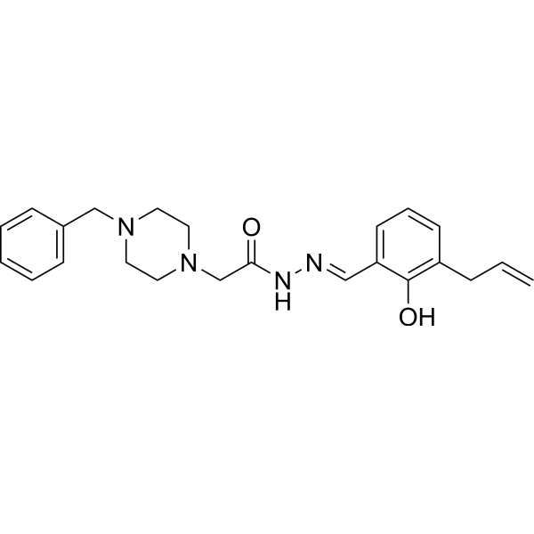
PAC-1
Based on 7 publication(s) in Google Scholar
PAC-1 is a procaspase-3 activator that induces apoptosis in cancer cells with an EC50 of 2.08 μM.
For research use only. We do not sell to patients.
- Purity : 99.88%
- CAS No.: 315183-21-2
- Formula: C23H28N4O2
- Molecular Weight:392.49
-
Storage:Powder -20°C, 3 years , 4°C, 2 years ; In solvent -80°C, 2 years , -20°C, 1 year

To place orders, for customer services and technical support, please contact: MedChemExpress USA
Tel: 609-228-6898 E-mail: [email protected] [email protected]


-
 Biological Activity
-
Chemical Information
-
Solvent & Solubility
- Protocols
- Purity & Documentation
- References
-
 Help & FAQs


-
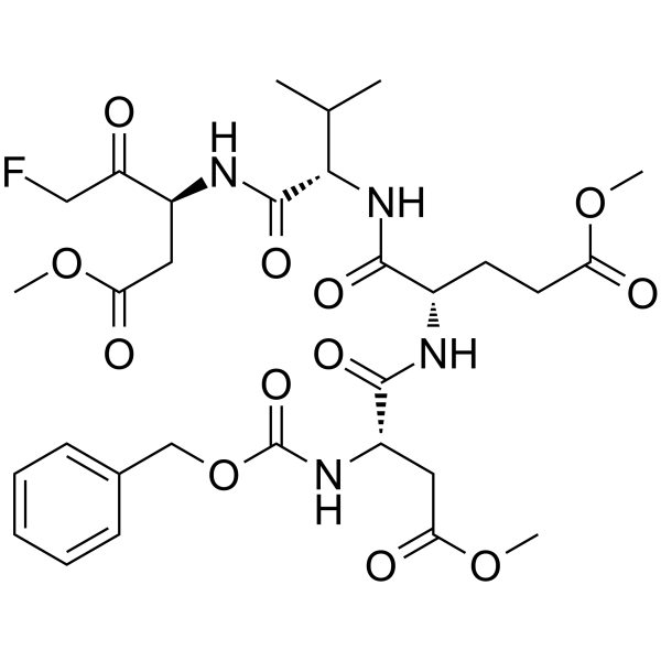
HY-12466
Z-DEVD-FMKCaspase-3 Inhibitor -
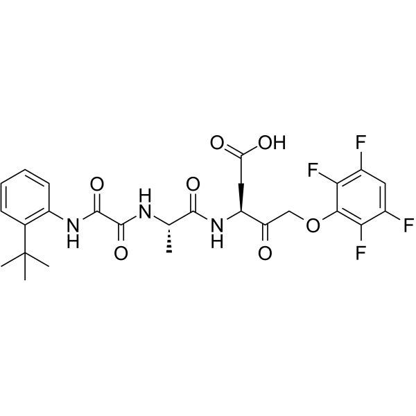
HY-10396
Emricasanpan-Caspase Inhibitor -
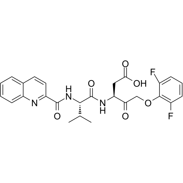
HY-12305
Q-VD-OPhPan-caspase Inhibitor -
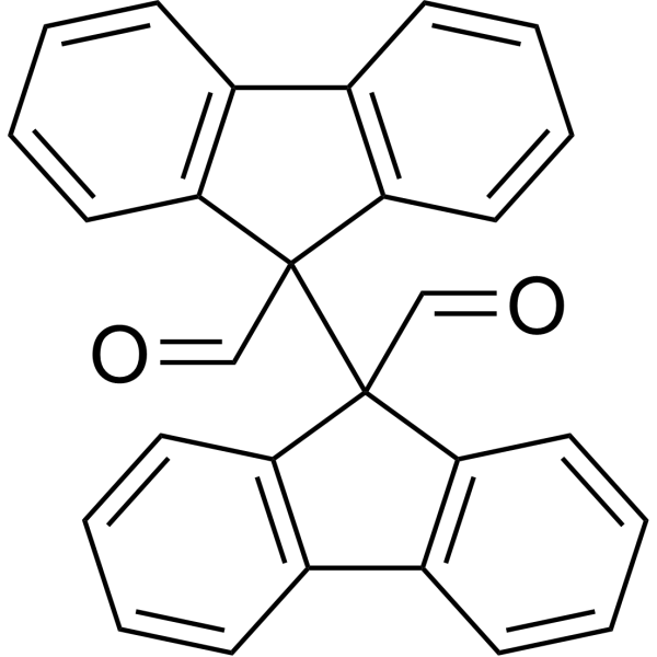
HY-121320
RaptinalCaspase-3 Activator -
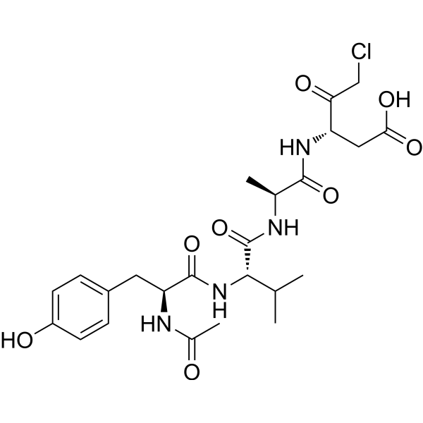
HY-16990
Ac-YVAD-cmkCaspase-1 Inhibitor -

HY-15763
ErastinFerroptosis Inducer -
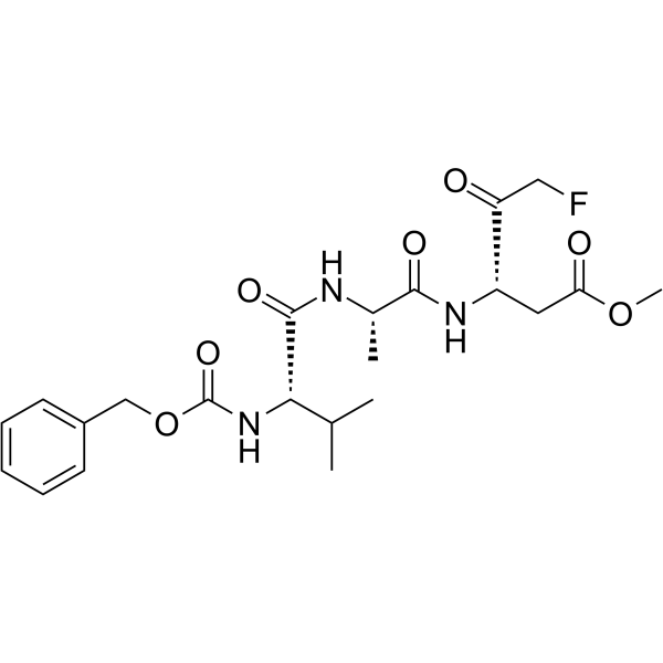
HY-16658
Z-VAD(OMe)-FMKPan-caspase Inhibitor -
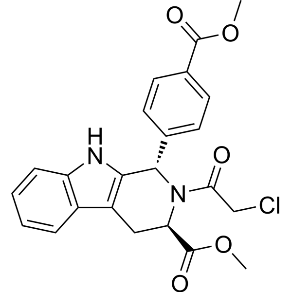
HY-100218A
RSL3GPX4 Inhibitor -
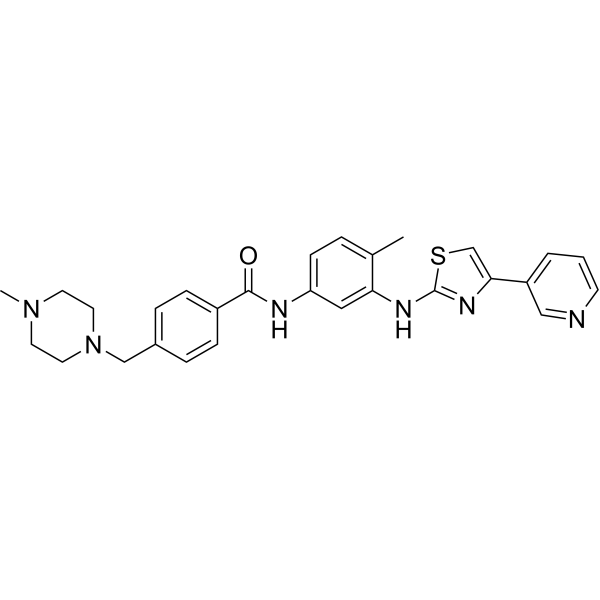
HY-10209
Masitinibc-Kit Inhibitor -
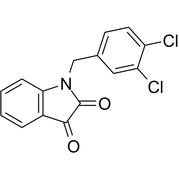
HY-18633
MDK83190Apoptosis Inducer -
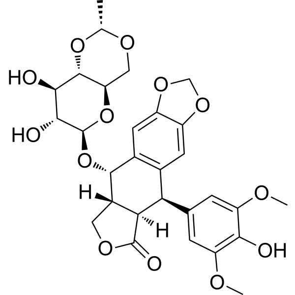
HY-13629
EtoposideTopoisomerase II Inhibitor -
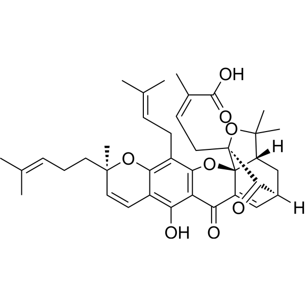
HY-N0087
Gambogic AcidBcl-2 Family Inhibitor
Publications Citing Use of MedChemExpress (MCE) PAC-1
More- Free Radic Biol Med. 2026 Feb 16:244:452-463.
- Mol Cancer Ther. 2020 Aug;19(8):1751-1760. [Abstract]
- Cell Mol Life Sci. 2024 Jul 30;81(1):325. [Abstract]
- Cancers (Basel). 2023 Apr 23;15(9):2427. [Abstract]
- Poult Sci. 2019 Dec 1;98(12):6367-6377. [Abstract]
- Vet Microbiol. 2021 Oct:261:109177. [Abstract]
- Oncotarget. 2017 Feb 14;8(7):12311-12322. [Abstract]
 Customer Validation & Images

-
Cell Imaging/Staining

-
Bio/Physico-chemical Assay
-
Microbiological Assay
-
Apoptosis Analysis

-
WB
All Caspase Isoforms
More

Biological Activity
Description
IC50 & Target
[1]|
Procaspase-3 2.08 μM (EC50) |
Cellular Effect
|
Cell Line

|
Type | Value | Description | References |
|---|---|---|---|---|
| A549 | IC50 |
2.51 μM
Compound: PAC-1
|
Cytotoxicity against human A549 cells after 72 hrs by MTT assay
Cytotoxicity against human A549 cells after 72 hrs by MTT assay
|
[PMID: 25874341] |
| B16-F10 | IC50 |
>100 μM
Compound: 1, PAC-1
|
Activation of procaspase-3-mediated human B16-F10 cell death after 24 hrs by MTS/PMS assay
Activation of procaspase-3-mediated human B16-F10 cell death after 24 hrs by MTS/PMS assay
|
[PMID: 16936720] |
| BT-549 | IC50 |
14.3 μM
Compound: 19
|
Cytotoxicity against Smac mimetic-resistant human BT549 cells assessed as growth inhibition after 24 hrs by CellTiter-Glo assay
Cytotoxicity against Smac mimetic-resistant human BT549 cells assessed as growth inhibition after 24 hrs by CellTiter-Glo assay
|
[PMID: 27578248] |
| Cancer cell lines | IC50 |
0.003 μM
Compound: 1, PAC-1
|
Cytotoxicity against human colon cancer cells after 24 hrs by MTS/PMS assay
Cytotoxicity against human colon cancer cells after 24 hrs by MTS/PMS assay
|
[PMID: 16936720] |
| Colon cell | IC50 |
5.02 μM
Compound: 1, PAC-1
|
Cytotoxicity against human colon cells after 24 hrs by MTS/PMS assay
Cytotoxicity against human colon cells after 24 hrs by MTS/PMS assay
|
[PMID: 16936720] |
| EL4 | IC50 |
6.5 μM
Compound: 1, PAC-1
|
Cytotoxicity against mouse EL4 cells assessed as cell death after 72 hrs by sulforhodamine B assay
Cytotoxicity against mouse EL4 cells assessed as cell death after 72 hrs by sulforhodamine B assay
|
[PMID: 25856364] |
| HL-60 | IC50 |
0.92 μM
Compound: 1, PAC-1
|
Activation of procaspase-3-mediated human HL60 cell death after 72 hrs by MTS/PMS assay
Activation of procaspase-3-mediated human HL60 cell death after 72 hrs by MTS/PMS assay
|
[PMID: 16936720] |
| HL-60 | IC50 |
0.92 μM
Compound: 38; PAC-1
|
Antitumor activity against human HL-60 cells assessed as cell growth inhibition
Antitumor activity against human HL-60 cells assessed as cell growth inhibition
|
[PMID: 36858050] |
| Hs-578T | IC50 |
>100 μM
Compound: 1, PAC-1
|
Activation of procaspase-3-mediated human Hs 578T cell death after 24 hrs by MTS/PMS assay
Activation of procaspase-3-mediated human Hs 578T cell death after 24 hrs by MTS/PMS assay
|
[PMID: 16936720] |
| HT-29 | IC50 |
0.83 μM
Compound: 7, PAC-1
|
Cytotoxicity against human HT-29 cells after 72 hrs by MTT assay
Cytotoxicity against human HT-29 cells after 72 hrs by MTT assay
|
[PMID: 23838381] |
| HT-29 | IC50 |
0.83 μM
Compound: 8; PAC-1
|
Cytotoxicity against human HT-29 cells after 72 hrs by MTT assay
Cytotoxicity against human HT-29 cells after 72 hrs by MTT assay
|
[PMID: 26897090] |
| HT-29 | IC50 |
0.97 μM
Compound: 5, PAC-1
|
Cytotoxicity against human HT-29 cells assessed as growth inhibition after 72 hrs by MTT assay
Cytotoxicity against human HT-29 cells assessed as growth inhibition after 72 hrs by MTT assay
|
[PMID: 23059545] |
| HT-29 | IC50 |
1.02 μM
Compound: PAC-1
|
Cytotoxicity against human HT-29 cells after 72 hrs by MTT assay
Cytotoxicity against human HT-29 cells after 72 hrs by MTT assay
|
[PMID: 25874341] |
| HT-29 | IC50 |
1.36 μM
Compound: PAC-1
|
Antiproliferative activity against human HT-29 cells after 72 hrs by MTT assay
Antiproliferative activity against human HT-29 cells after 72 hrs by MTT assay
|
[PMID: 25171780] |
| Jurkat | IC50 |
4.4 μM
Compound: 1, PAC-1
|
Cytotoxicity against human Jurkat cells assessed as cell death after 72 hrs by sulforhodamine B assay
Cytotoxicity against human Jurkat cells assessed as cell death after 72 hrs by sulforhodamine B assay
|
[PMID: 25856364] |
| KE-37 | IC50 |
241.66 nM
Compound: PAC-1
|
Anticancer activity against human KE-37 cells assessed as reduction in cell viability
Anticancer activity against human KE-37 cells assessed as reduction in cell viability
|
[PMID: 33774345] |
| MCF-10A | IC50 |
3.2 μM
Compound: 1, PAC-1
|
Activation of procaspase-3-mediated human MCF-10A cell death after 72 hrs by MTS/PMS assay
Activation of procaspase-3-mediated human MCF-10A cell death after 72 hrs by MTS/PMS assay
|
[PMID: 16936720] |
| MCF7 | IC50 |
>40 μM
Compound: PAC-1
|
Antiproliferative activity against human MCF7 cells after 24 hrs by CCK-8 assay
Antiproliferative activity against human MCF7 cells after 24 hrs by CCK-8 assay
|
[PMID: 31015910] |
| MCF7 | IC50 |
>75 μM
Compound: 1, PAC-1
|
Activation of procaspase-3 in human MCF-7 cells assessed as cell death after 72 hrs
Activation of procaspase-3 in human MCF-7 cells assessed as cell death after 72 hrs
|
[PMID: 16936720] |
| MCF7 | IC50 |
>75 μM
Compound: 38; PAC-1
|
Antitumor activity against human MCF7 cells assessed as cell growth inhibition
Antitumor activity against human MCF7 cells assessed as cell growth inhibition
|
[PMID: 36858050] |
| MDA-MB-231 | IC50 |
3.2 μM
Compound: PAC-1
|
Cytotoxicity against human MDA-MB-231 cells after 72 hrs by MTT assay
Cytotoxicity against human MDA-MB-231 cells after 72 hrs by MTT assay
|
[PMID: 25874341] |
| MDA-MB-231 | IC50 |
4.6 μM
Compound: 7, PAC-1
|
Cytotoxicity against human MDA-MB-231 cells after 72 hrs by MTT assay
Cytotoxicity against human MDA-MB-231 cells after 72 hrs by MTT assay
|
[PMID: 23838381] |
| MDA-MB-231 | IC50 |
4.6 μM
Compound: 8; PAC-1
|
Cytotoxicity against human MDA-MB-231 cells after 72 hrs by MTT assay
Cytotoxicity against human MDA-MB-231 cells after 72 hrs by MTT assay
|
[PMID: 26897090] |
| MDA-MB-231 | IC50 |
4.7 μM
Compound: 19
|
Cytotoxicity against Smac mimetic-sensitive human MDA-MB-231 cells assessed as growth inhibition after 24 hrs by CellTiter-Glo assay
Cytotoxicity against Smac mimetic-sensitive human MDA-MB-231 cells assessed as growth inhibition after 24 hrs by CellTiter-Glo assay
|
[PMID: 27578248] |
| MDA-MB-231 | IC50 |
4.8 μM
Compound: PAC-1
|
Antiproliferative activity against human MDA-MB-435 cells after 72 hrs by MTT assay
Antiproliferative activity against human MDA-MB-435 cells after 72 hrs by MTT assay
|
[PMID: 25171780] |
| MDA-MB-231 | IC50 |
6.11 μM
Compound: 5, PAC-1
|
Cytotoxicity against human MDA-MB-231 cells assessed as growth inhibition after 72 hrs by MTT assay
Cytotoxicity against human MDA-MB-231 cells assessed as growth inhibition after 72 hrs by MTT assay
|
[PMID: 23059545] |
| MDA-MB-453 | IC50 |
2 μM
Compound: 19
|
Cytotoxicity against Smac mimetic-resistant human MDA-MB-453 cells assessed as growth inhibition after 24 hrs by CellTiter-Glo assay
Cytotoxicity against Smac mimetic-resistant human MDA-MB-453 cells assessed as growth inhibition after 24 hrs by CellTiter-Glo assay
|
[PMID: 27578248] |
| MKN-45 | IC50 |
0.82 μM
Compound: 7, PAC-1
|
Cytotoxicity against human MKN45 cells after 72 hrs by MTT assay
Cytotoxicity against human MKN45 cells after 72 hrs by MTT assay
|
[PMID: 23838381] |
| MKN-45 | IC50 |
0.82 μM
Compound: 8; PAC-1
|
Cytotoxicity against human MKN45 cells after 72 hrs by MTT assay
Cytotoxicity against human MKN45 cells after 72 hrs by MTT assay
|
[PMID: 26897090] |
| MKN-45 | IC50 |
2.61 μM
Compound: PAC-1
|
Antiproliferative activity against human MKN45 cells after 72 hrs by MTT assay
Antiproliferative activity against human MKN45 cells after 72 hrs by MTT assay
|
[PMID: 25171780] |
| NCI-H226 | IC50 |
0.35 μM
Compound: 1, PAC-1
|
Activation of procaspase-3-mediated human NCI-H226 cell death after 24 hrs by MTS/PMS assay
Activation of procaspase-3-mediated human NCI-H226 cell death after 24 hrs by MTS/PMS assay
|
[PMID: 16936720] |
| NCI-H226 | IC50 |
0.35 μM
Compound: 38; PAC-1
|
Antitumor activity against human NCI-H226 cells assessed as cell growth inhibition
Antitumor activity against human NCI-H226 cells assessed as cell growth inhibition
|
[PMID: 36858050] |
| NCI-H226 | IC50 |
1.02 μM
Compound: PAC-1
|
Antiproliferative activity against human NCI-H226 cells after 72 hrs by MTT assay
Antiproliferative activity against human NCI-H226 cells after 72 hrs by MTT assay
|
[PMID: 25171780] |
| NCI-H446 | IC50 |
2326.69 μM
Compound: PAC-1
|
Anticancer activity against human NCI-H446 cells assessed as reduction in cell viability
Anticancer activity against human NCI-H446 cells assessed as reduction in cell viability
|
[PMID: 33774345] |
| NCI-H460 | IC50 |
1.25 μM
Compound: PAC-1
|
Cytotoxicity against human H460 cells after 72 hrs by MTT assay
Cytotoxicity against human H460 cells after 72 hrs by MTT assay
|
[PMID: 25874341] |
| NCI-H460 | IC50 |
3.57 μM
Compound: 5, PAC-1
|
Cytotoxicity against human H460 cells assessed as growth inhibition after 72 hrs by MTT assay
Cytotoxicity against human H460 cells assessed as growth inhibition after 72 hrs by MTT assay
|
[PMID: 23059545] |
| PBL | IC50 |
123.81 μM
Compound: 8; PAC-1
|
Cytotoxicity against human PBL cells after 72 hrs by MTT assay
Cytotoxicity against human PBL cells after 72 hrs by MTT assay
|
[PMID: 26897090] |
| PC-12 | EC50 |
122 μM
Compound: 2, PAC-1
|
Induction of apoptosis in rat PC12 cells after 24 hrs by trypan blue exclusion assay
Induction of apoptosis in rat PC12 cells after 24 hrs by trypan blue exclusion assay
|
[PMID: 23859779] |
| SK-N-SH | IC50 |
3.06 μM
Compound: PAC-1
|
Antiproliferative activity against human SK-N-SH cells after 72 hrs by MTT assay
Antiproliferative activity against human SK-N-SH cells after 72 hrs by MTT assay
|
[PMID: 25171780] |
| U-937 | IC50 |
10.2 μM
Compound: 1, PAC-1
|
Cytotoxicity against human U937 cells assessed as cell death after 72 hrs by sulforhodamine B assay
Cytotoxicity against human U937 cells assessed as cell death after 72 hrs by sulforhodamine B assay
|
[PMID: 25856364] |
| U-937 | IC50 |
4.8 μM
Compound: 1, PAC-1
|
Cytotoxicity against human U937 cells after 72 hrs by sulforhodamine B assay
Cytotoxicity against human U937 cells after 72 hrs by sulforhodamine B assay
|
[PMID: 19708658] |
| U-937 | IC50 |
6 μM
Compound: PAC-1
|
Antiproliferative activity against human procaspase-3 over-expressing U937 cells after 24 hrs by CCK-8 assay
Antiproliferative activity against human procaspase-3 over-expressing U937 cells after 24 hrs by CCK-8 assay
|
[PMID: 31015910] |
| UACC-62 | IC50 |
>100 μM
Compound: 1, PAC-1
|
Activation of procaspase-3-mediated human UACC62 cell death after 24 hrs by MTS/PMS assay
Activation of procaspase-3-mediated human UACC62 cell death after 24 hrs by MTS/PMS assay
|
[PMID: 16936720] |
In Vitro
PAC-1 activates procaspase-3 with an EC50 of 2.08 μM. PAC-1 exhibits an enhanced zinc chelating ability (EC50= 7.08 μM). PAC-1 induces leukemia cell death with IC50 of 4.03 μM, which is consistent with the values reported by other investigators. PAC-1 treatment also results in death of other malignant cells in a concentration-dependent manner with IC50s ranging from 4.03 to 53.44μM. The overall mean IC50 in the fifteen malignant cell lines is 0.88 mM for WF-210 and 19.40 μM for PAC-1. In contrast, the sensitivity of the normal human cells (PBL, L-02, HUVEC and MCF 10A) to WF-210 is 2.6-fold lower (mean IC50=412.34 μM) than PAC-1 (mean IC50=158.29 μM)[1]. Procaspase-activating compound-1 (PAC-1) is the first direct caspase-activating compound discovered. PAC-1 treatment upregulates Ero1α in multiple cell lines, whereas silencing of Ero1α significantly inhibits calcium release from ER and cell death[2].
MedChemExpress (MCE) has not independently confirmed the accuracy of these methods. They are for reference only. Further protocols information, click here.
In Vivo
MedChemExpress (MCE) has not independently confirmed the accuracy of these methods. They are for reference only.
Clinical Trial
| NCT Number | Sponsor | Condition | Start Date |
Phase
|
|---|---|---|---|---|
| NCT01329991 | Plexxikon| | 2011-05 | PHASE1 |


Chemical Information
-
CAS No. 315183-21-2
-
Appearance Solid
-
Molecular Weight 392.49
-
Formula C23H28N4O2
-
Color White to yellow
-
SMILES
O=C(N/N=C/C1=CC=CC(CC=C)=C1O)CN2CCN(CC3=CC=CC=C3)CC2
-
Synonyms
Procaspase activating compound 1
-
Shipping
Room temperature in continental US; may vary elsewhere.
-
Storage
Powder -20°C 3 years 4°C 2 years In solvent -80°C 2 years -20°C 1 year


Publications (7)
-
Journal Impact Factor
-
Most Recent
-
PAC-1 purchased from MedChemExpress. Usage Cited in: Free Radic Biol Med. 2026 Feb 16:244:452-463.
Intracellular ROS in HepaRG cells treated with PAC-1 (5-50 μM; 12 h) was visualized and quantified using DHE fluorescence imaging. PAC-1 increased ROS levels in a dose-dependent manner.
PAC-1 purchased from MedChemExpress. Usage Cited in: Free Radic Biol Med. 2026 Feb 16:244:452-463.
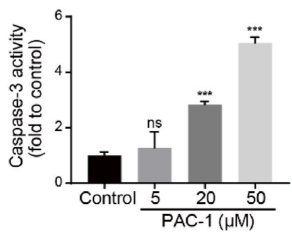
Caspase-3 activity was measured in HepaRG cells treated with PAC-1 (5-50 μM; 12 h). PAC-1 increased caspase-3 activity in a dose-dependent manner.
-
Mol Cancer Ther
Discovery of New Targets to Control Metastasis in Pancreatic Cancer by Single-cell Transcriptomics Analysis of Circulating Tumor Cells. [Abstract]2020 Aug;19(8):1751-1760. PMID: 32499301 -
Cell Mol Life Sci
Suppression of Skp2 contributes to sepsis-induced acute lung injury by enhancing ferroptosis through the ubiquitination of SLC3A2. [Abstract]2024 Jul 30;81(1):325. PMID: 39079969 -
Cancers (Basel)
High-Dosage NMN Promotes Ferroptosis to Suppress Lung Adenocarcinoma Growth through the NAM-Mediated SIRT1-AMPK-ACC Pathway. [Abstract]2023 Apr 23;15(9):2427. PMID: 37173894 -
Poult Sci
Protective effects of hypericin against infectious bronchitis virus induced apoptosis and reactive oxygen species in chicken embryo kidney cells. [Abstract]2019 Dec 1;98(12):6367-6377. PMID: 31399732
PAC-1 purchased from MedChemExpress. Usage Cited in: Poult Sci. 2019 Dec 1;98(12):6367-6377. [Abstract]
The immunofluorescence was performed in chicken embryo kidney cells treated with PAC-1 (20 μM; 30 h) as control.
-
Vet Microbiol
Lipid metabolism is a novel and practical source of potential targets for antiviral discovery against porcine parvovirus. [Abstract]2021 Oct:261:109177. PMID: 34391196
PAC-1 purchased from MedChemExpress. Usage Cited in: Vet Microbiol. 2021 Oct:261:109177. [Abstract]
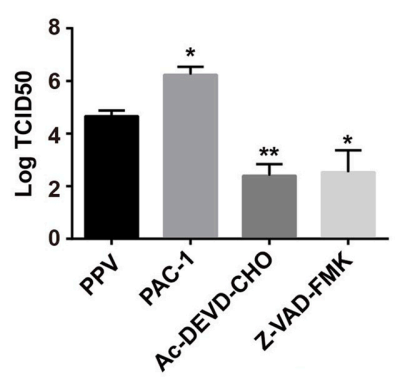
PK15 cells were infected with porcine parvovirus (MOI = 1) and simultaneously incubated with PAC-1 (10 μM; 24 h), followed by determination Log TCID50. PAC-1 increased porcine parvovirus yield.
-
Oncotarget
RXRα ligand Z-10 induces PML-RARα cleavage and APL cell apoptosis through disrupting PML-RARα/RXRα complex in a cAMP-independent manner. [Abstract]2017 Feb 14;8(7):12311-12322. PMID: 28129653
PAC-1 purchased from MedChemExpress. Usage Cited in: Oncotarget. 2017 Feb 14;8(7):12311-12322. [Abstract]
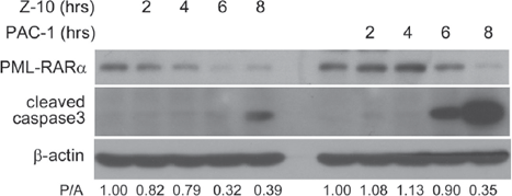
NB4 cells are treated with 5 μM Z-10 or 50 μM PAC-1 for the indicated time, and the expression of PML-RARα and cleaved caspase3 is analyzed by western blot.


Solvent & Solubility
In Vitro:
DMSO : 50 mg/mL (127.39 mM; Need ultrasonic; Hygroscopic DMSO has a significant impact on the solubility of product, please use newly opened DMSO)
H2O : < 0.1 mg/mL (insoluble)
Please refer to the solubility information to select the appropriate solvent. Once prepared, please aliquot and store the solution to prevent product inactivation from repeated freeze-thaw cycles.
Storage method and period of stock solution: -80°C, 2 years; -20°C, 1 year. When stored at -80°C, please use it within 2 years. When stored at -20°C, please use it within 1 year.
Please refer to the solubility information to select the appropriate solvent. Once prepared, please aliquot and store the solution to prevent product inactivation from repeated freeze-thaw cycles.
Storage method and period of stock solution: -80°C, 2 years; -20°C, 1 year. When stored at -80°C, please use it within 2 years. When stored at -20°C, please use it within 1 year.
Concentration (start) × Volume (start) = Concentration (final) × Volume (final)
In Vivo:
Select the appropriate dissolution method based on your experimental animal and administration route.
- For the following dissolution methods, please ensure to first prepare a clear stock solution using an In Vitro approach and then sequentially add co-solvents:
- To ensure reliable experimental results, the clarified stock solution can be appropriately stored based on storage conditions. As for the working solution for In Vivo experiments, it is recommended to prepare freshly and use it on the same day.
- The percentages shown for the solvents indicate their volumetric ratio in the final prepared solution. If precipitation or phase separation occurs during preparation, heat and/or sonication can be used to aid dissolution.
Add each solvent one by one: 10% DMSO 40% PEG300 5% Tween-80 45% Saline
Solubility: ≥ 2.5 mg/mL (6.37 mM); Clear solution
This protocol yields a clear solution of ≥ 2.5 mg/mL (saturation unknown).
Taking 1 mL working solution as an example, add 100 μL DMSO stock solution (25.0 mg/mL) to 400 μL PEG300, and mix evenly; then add 50 μL Tween-80 and mix evenly; then add 450 μL Saline to adjust the volume to 1 mL.
Preparation of Saline: Dissolve 0.9 g sodium chloride in ddH₂O and dilute to 100 mL to obtain a clear Saline solution.
Add each solvent one by one: 10% DMSO 90% (20% SBE-β-CD in Saline)
Solubility: 2.5 mg/mL (6.37 mM); Suspended solution; Need ultrasonic
This protocol yields a suspended solution of 2.5 mg/mL. Suspended solution can be used for oral and intraperitoneal injection.
Taking 1 mL working solution as an example, add 100 μL DMSO stock solution (25.0 mg/mL) to 900 μL 20% SBE-β-CD in Saline, and mix evenly.
Preparation of 20% SBE-β-CD in Saline (4°C, storage for one week): 2 g SBE-β-CD powder is dissolved in 10 mL Saline, completely dissolve until clear.
In Vivo Dissolution Calculator
Please enter the basic information of animal experiments:
-
-
-
-
Recommended: Prepare an additional quantity of animals to account for potential losses during experiments.
Please enter your animal formula composition:
-
%DMSO +
Recommended: Keep the proportion of DMSO in working solution below 2% if your animal is weak.
-
%+
-
+%Tween-80 + +
-
%Saline +
The co-solvents required include: DMSO, . All of co-solvents are available by MedChemExpress (MCE). , Tween 80. All of co-solvents are available by MedChemExpress (MCE).
Working solution concentration: 0.22 mg/mL
Method for preparing stock solution: mg drug dissolved in μL DMSO. Stock solution concentration: mg/mL.
1. Take μL DMSO stock solution;
2. Add μL .
μL , mix evenly;
3. Then add μL Tween 80, mix evenly;
4. Then add μL
Please ensure that the stock solution in the first step is dissolved to a clear state, and add co-solvents in sequence. You can use ultrasonic heating (ultrasonic cleaner, recommended frequency 20-40 kHz), vortexing, etc. to assist dissolution.

Protocols
-
Apoptosis
Apoptosis, also called programmed cell death, is generally characterized by distinct morphological characteristics.
-
TUNEL staining for apoptotic DNA fragmentation
TUNEL staining detects DNA strand breaks by using terminal deoxynucleotidyl transferase to add labeled nucleotides to exposed 3′-OH DNA termini, generating either microscopic staining in fixed cells or tissue sections, or fluorescence/cytometric signal in cell suspensions. TUNEL positivity reflects DNA fragmentation but should not be interpreted alone as definitive apoptosis, because TUNEL can also label necrotic, autolytic, mechanically damaged, or DNA-repair-associated DNA breaks.
-
Annexin V plus membrane-impermeant dye apoptosis staining
Annexin V-based apoptosis assays rely on the detection of phosphatidylserine (PS) externalization from the inner leaflet of the plasma membrane to the outer leaflet, an early biochemical hallmark of apoptosis. Fluorescently labeled Annexin V binds PS in a calcium-dependent manner, enabling identification of early apoptotic cells by flow cytometry or fluorescence microscopy. When combined with a membrane-impermeant DNA-binding dye (e. g. , propidium iodide), this approach allows discrimination between viable (Annexin V−/dye−), early apoptotic (Annexin V+/dye−), and late apoptotic or necrotic (Annexin V+/dye+) cell populations by assessing membrane integrity and PS exposure.
-
Apoptosis Solutions
Apoptosis is a regulated, generally non-lytic cell-death pathway that removes unwanted, damaged, infected, or abnormal cells through coordinated morphological changes, caspase activation, DNA fragmentation, and membrane remodeling. The intrinsic apoptosis pathway is controlled mainly by mitochondrial outer membrane permeabilization, BCL-2 family proteins, cytochrome c release, apoptosome formation, caspase-9 activation, and downstream executioner caspase-3/7 activation. The extrinsic apoptosis pathway is initiated by death receptors such as Fas, TNFR, and TRAIL receptors, which recruit adaptor proteins and activate caspase-8 before engaging executioner caspases or mitochondrial amplification through BID cleavage. Apoptosis is linked to many phenotypes, including cancer cell killing, tissue homeostasis, immune regulation, neurodegeneration, infection response, and treatment-induced cytotoxicity; unresolved questions include how apoptosis interacts with necroptosis, pyroptosis, ferroptos


Purity & Documentation
-
Data Sheet (277 KB)
-
SDS (597 KB)
- English - EN (597 KB)
- Français - FR (597 KB)
- Deutsch - DE (597 KB)
- Norwegian - NO (597 KB)
- Español - ES (597 KB)
- Swedish - SV (597 KB)
- Italian - IT (597 KB)
- Korean - KR (597 KB)
- Portuguese - PT (597 KB)
-
Handling Instructions (2659 KB)
References
[1]. Wang F, et al. A novel small-molecule activator of procaspase-3 induces apoptosis in cancer cells and reduces tumor growth in human breast, liver and gallbladder cancer xenografts. Mol Oncol. 2014 Dec;8(8):1640-52. [Content Brief]
[2]. Seervi M, et al. ERO1α-dependent endoplasmic reticulum-mitochondrial calcium flux contributes to ER stress and mitochondrial permeabilization by procaspase-activating compound-1 (PAC-1). Cell Death Dis. 2013 Dec 19;4:e968. [Content Brief]
[3]. Putt KS, et al. Small-molecule activation of procaspase-3 to caspase-3 as a personalized anticancer strategy. Nat Chem Biol. 2006 Oct;2(10):543-50. [Content Brief]


Complete Stock Solution Preparation Table
Please refer to the solubility information to select the appropriate solvent. Once prepared, please aliquot and store the solution to prevent product inactivation from repeated freeze-thaw cycles.
Storage method and period of stock solution: -80°C, 2 years; -20°C, 1 year. When stored at -80°C, please use it within 2 years. When stored at -20°C, please use it within 1 year.
| Optional Solvent | Concentration Solvent Mass | 1 mg | 5 mg | 10 mg | 25 mg |
|---|---|---|---|---|---|
| DMSO | 1 mM | 2.5478 mL | 12.7392 mL | 25.4784 mL | 63.6959 mL |
| 5 mM | 0.5096 mL | 2.5478 mL | 5.0957 mL | 12.7392 mL | |
| 10 mM | 0.2548 mL | 1.2739 mL | 2.5478 mL | 6.3696 mL | |
| 15 mM | 0.1699 mL | 0.8493 mL | 1.6986 mL | 4.2464 mL | |
| 20 mM | 0.1274 mL | 0.6370 mL | 1.2739 mL | 3.1848 mL | |
| 25 mM | 0.1019 mL | 0.5096 mL | 1.0191 mL | 2.5478 mL | |
| 30 mM | 0.0849 mL | 0.4246 mL | 0.8493 mL | 2.1232 mL | |
| 40 mM | 0.0637 mL | 0.3185 mL | 0.6370 mL | 1.5924 mL | |
| 50 mM | 0.0510 mL | 0.2548 mL | 0.5096 mL | 1.2739 mL | |
| 60 mM | 0.0425 mL | 0.2123 mL | 0.4246 mL | 1.0616 mL | |
| 80 mM | 0.0318 mL | 0.1592 mL | 0.3185 mL | 0.7962 mL | |
| 100 mM | 0.0255 mL | 0.1274 mL | 0.2548 mL | 0.6370 mL |


PAC-1 Related Classifications
HY-13523 Related Classifications
 Powered by Bioz