



Protosappanin A

Based on 1 Customer Validation
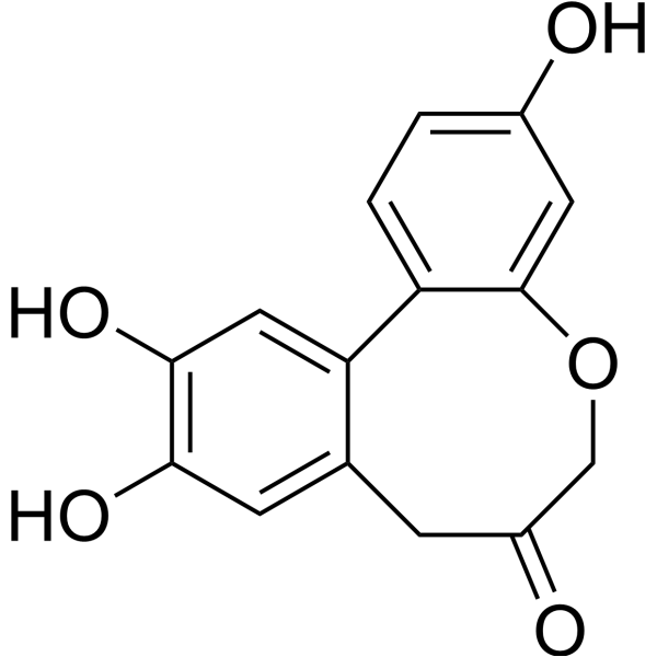
Protosappanin A (PTA), an immunosuppressive ingredient and major biphenyl compound isolated from Caesalpinia sappan L, suppresses JAK2/STAT3-dependent inflammation pathway through down-regulating the phosphorylation of JAK2 and STAT3.
For research use only. We do not sell to patients.
- Purity : 99.95%
- CAS No.: 102036-28-2
- Formula: C15H12O5
- Molecular Weight:272.25
-
Storage:
4°C, sealed storage, away from moisture and light
* In solvent : -80°C, 6 months; -20°C, 1 month (sealed storage, away from moisture and light)

To place orders, for customer services and technical support, please contact: MedChemExpress USA
Tel: 609-228-6898 E-mail: [email protected] [email protected]


-
 Biological Activity
-
Chemical Information
-
Solvent & Solubility
- Protocols
- Purity & Documentation
- References
-
 Help & FAQs


-
HY-15312
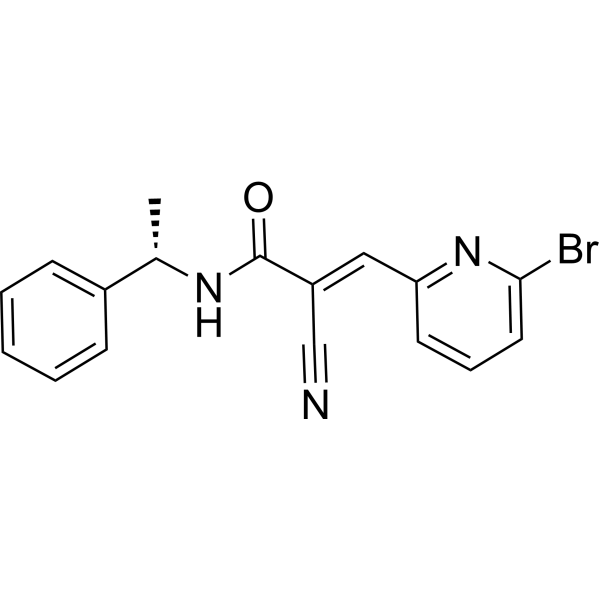
WP1066JAK2/STAT3 Inhibitor -
HY-115452
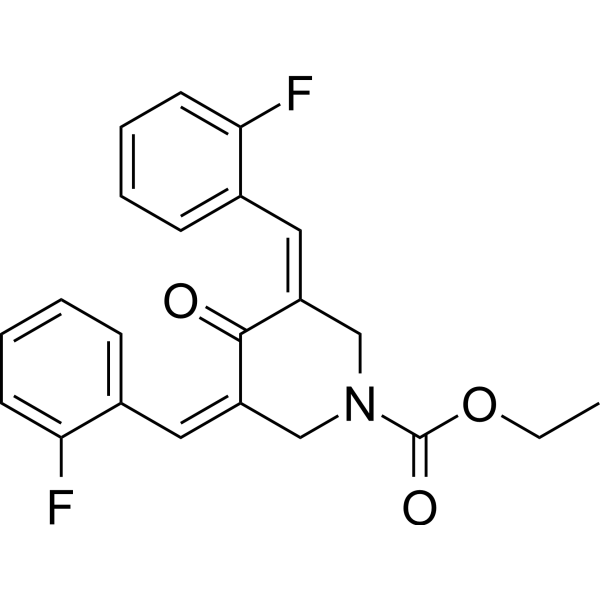
G5-7JAK2 Inhibitor -
HY-107429
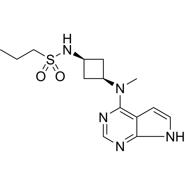
AbrocitinibJAK1 Inhibitor -
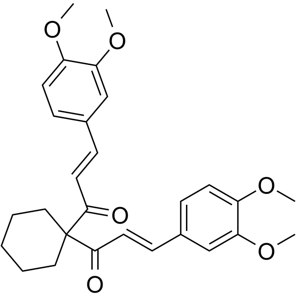
HY-100544
FLLL32JAK2/STAT3 Inhibitor -
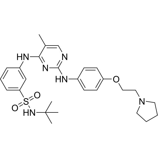
HY-10409
FedratinibJAK2 Inhibitor -
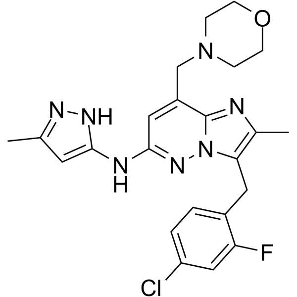
HY-13034
GandotinibJAK2 Inhibitor -
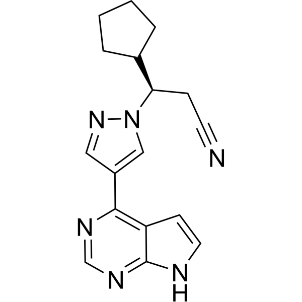
HY-50856
RuxolitinibJAK1/2 Inhibitor -
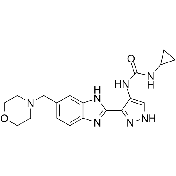
HY-50514
AT9283Aurora/JAK Inhibitor -
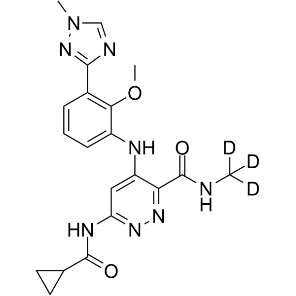
HY-117287
DeucravacitinibTYK2 Inhibitor -
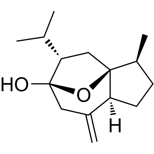
HY-N0104
CurcumolApoptosis Inducer -
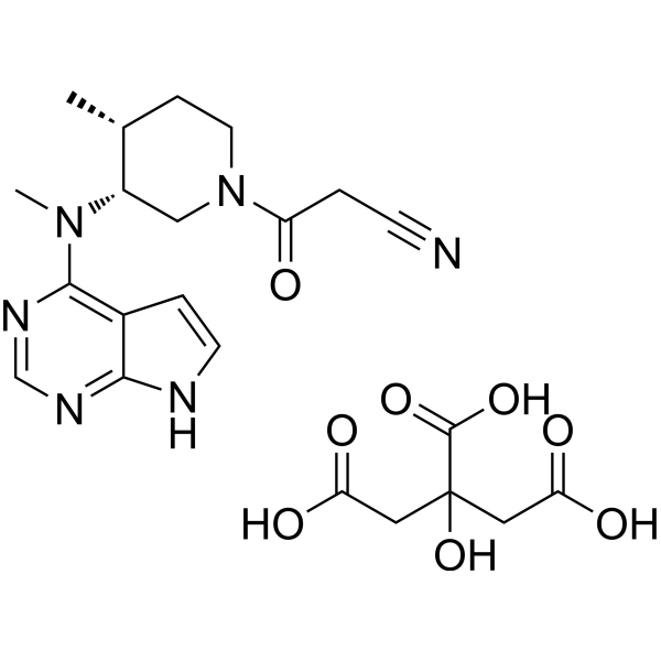
HY-40354A
Tofacitinib citrateJAK1/JAK3 Inhibitor -
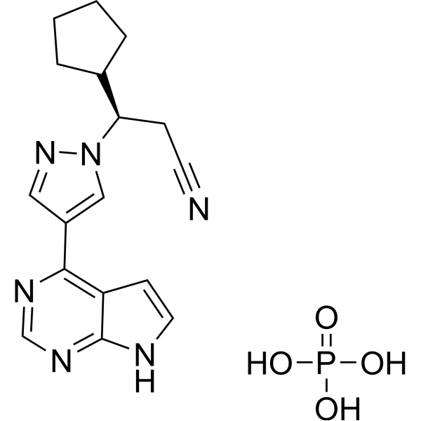
HY-50858
Ruxolitinib phosphateJAK1/2 Inhibitor


Biological Activity
Description
IC50 & Target
[1]|
JAK2 |
STAT3 |
Cellular Effect
|
Cell Line

|
Type | Value | Description | References |
|---|---|---|---|---|
| RAW264.7 | IC50 |
12.5 μM
Compound: 10
|
Antiinflammatory activity in mouse RAW264.7 cells assessed as inhibition of LPS-induced NO production after 24 hrs by Griess method
Antiinflammatory activity in mouse RAW264.7 cells assessed as inhibition of LPS-induced NO production after 24 hrs by Griess method
|
[PMID: 23127886] |
In Vitro
Protosappanin A (PTA: 12.5, 25, 50 μM, 24 hours) significantly inhibits the production of TNF-α and IL-1β in LPS-activated BV2 microglia. And the mRNA expressions of IL-6, IL-1β, and MCP-1 are reduced by PTA in a dose-dependent manner in BV2 microglial cell line[1].
Protosappanin A (PTA: 12.5, 25, 50 μM, 24 hours) suppresses JAK2/STAT3-dependent inflammation pathway through down-regulating the phosphorylation of JAK2 and STAT3, as well as STAT3 nuclear translocation against LPS treatment[1].
Protosappanin A (PTA: 12.5, 25, 50 μM, 24 hours) shows obvious effect on disturbing the interaction of transmembrane protein CD14 with Toll-like receptor-4, resulting in the inhibition of NF-κB-dependent oxidative and nitrative stress in LPS-induced BV2 microglia[2].
MedChemExpress (MCE) has not independently confirmed the accuracy of these methods. They are for reference only. Further protocols information, click here.
-
Cell Line:Murine BV2 microglial cell line.
-
Concentration:12.5, 25, 50 μM.
-
Incubation Time:24 hours.
-
Result:Inhibits the releases of NO, TNF-α and IL-1β in LPS-induced BV2 cells.
Attenuated IL-6, IL-1β and MCP-1 gene expressions in the LPS-induced BV2 cells.
Suppressed JAK2/STAT3 pathway activation in the LPS-induced BV2 cells.


Chemical Information
-
CAS No. 102036-28-2
-
Appearance Solid
-
Molecular Weight 272.25
-
Formula C15H12O5
-
Color White to off-white
-
SMILES
O=C1CC2=CC(O)=C(O)C=C2C3=CC=C(O)C=C3OC1
-
Synonyms
PTA
-
Structure Classification
-
Initial Source
-
Shipping
Room temperature in continental US; may vary elsewhere.
-
Storage
4°C, sealed storage, away from moisture and light
* In solvent : -80°C, 6 months; -20°C, 1 month (sealed storage, away from moisture and light)


Solvent & Solubility
In Vitro:
DMSO : 125 mg/mL (459.14 mM; Need ultrasonic; Hygroscopic DMSO has a significant impact on the solubility of product, please use newly opened DMSO)
Please refer to the solubility information to select the appropriate solvent. Once prepared, please aliquot and store the solution to prevent product inactivation from repeated freeze-thaw cycles.
Storage method and period of stock solution: -80°C, 6 months; -20°C, 1 month (sealed storage, away from moisture and light). When stored at -80°C, please use it within 6 months. When stored at -20°C, please use it within 1 month.
Please refer to the solubility information to select the appropriate solvent. Once prepared, please aliquot and store the solution to prevent product inactivation from repeated freeze-thaw cycles.
Storage method and period of stock solution: -80°C, 6 months; -20°C, 1 month (sealed storage, away from moisture and light). When stored at -80°C, please use it within 6 months. When stored at -20°C, please use it within 1 month.
Concentration (start) × Volume (start) = Concentration (final) × Volume (final)
In Vivo:
Select the appropriate dissolution method based on your experimental animal and administration route.
- For the following dissolution methods, please ensure to first prepare a clear stock solution using an In Vitro approach and then sequentially add co-solvents:
- To ensure reliable experimental results, the clarified stock solution can be appropriately stored based on storage conditions. As for the working solution for In Vivo experiments, it is recommended to prepare freshly and use it on the same day.
- The percentages shown for the solvents indicate their volumetric ratio in the final prepared solution. If precipitation or phase separation occurs during preparation, heat and/or sonication can be used to aid dissolution.
Add each solvent one by one: 10% DMSO 40% PEG300 5% Tween-80 45% Saline
Solubility: ≥ 2.08 mg/mL (7.64 mM); Clear solution
This protocol yields a clear solution of ≥ 2.08 mg/mL (saturation unknown).
Taking 1 mL working solution as an example, add 100 μL DMSO stock solution (20.8 mg/mL) to 400 μL PEG300, and mix evenly; then add 50 μL Tween-80 and mix evenly; then add 450 μL Saline to adjust the volume to 1 mL.
Preparation of Saline: Dissolve 0.9 g sodium chloride in ddH₂O and dilute to 100 mL to obtain a clear Saline solution.
Add each solvent one by one: 10% DMSO 90% (20% SBE-β-CD in Saline)
Solubility: ≥ 2.08 mg/mL (7.64 mM); Clear solution
This protocol yields a clear solution of ≥ 2.08 mg/mL (saturation unknown).
Taking 1 mL working solution as an example, add 100 μL DMSO stock solution (20.8 mg/mL) to 900 μL 20% SBE-β-CD in Saline, and mix evenly.
Preparation of 20% SBE-β-CD in Saline (4°C, storage for one week): 2 g SBE-β-CD powder is dissolved in 10 mL Saline, completely dissolve until clear.
In Vivo Dissolution Calculator
Please enter the basic information of animal experiments:
-
-
-
-
Recommended: Prepare an additional quantity of animals to account for potential losses during experiments.
Please enter your animal formula composition:
-
%DMSO +
Recommended: Keep the proportion of DMSO in working solution below 2% if your animal is weak.
-
%+
-
+%Tween-80 + +
-
%Saline +
The co-solvents required include: DMSO, . All of co-solvents are available by MedChemExpress (MCE). , Tween 80. All of co-solvents are available by MedChemExpress (MCE).
Working solution concentration: 0.22 mg/mL
Method for preparing stock solution: mg drug dissolved in μL DMSO. Stock solution concentration: mg/mL. * In solvent : -80°C, 6 months; -20°C, 1 month (sealed storage, away from moisture and light)
1. Take μL DMSO stock solution;
2. Add μL .
μL , mix evenly;
3. Then add μL Tween 80, mix evenly;
4. Then add μL
Please ensure that the stock solution in the first step is dissolved to a clear state, and add co-solvents in sequence. You can use ultrasonic heating (ultrasonic cleaner, recommended frequency 20-40 kHz), vortexing, etc. to assist dissolution.

Protocols
-
Kinase activity and phosphorylation assays
Kinase activity assays measure the ability of kinases to transfer phosphate groups from ATP to specific substrates, while phosphorylation assays detect the presence and levels of phosphorylated proteins. Common methods include radiolabeled ATP incorporation (e. g. ,), ADP release detection via bioluminescence (e. g. ,[3]), enzyme-linked immunosorbent assays (ELISA) for phospho-specific epitopes (e. g. ,[6]), and microtiter-based formats for high-throughput screening (e. g. ,[8]). The ADP-Glo assay quantifies kinase activity by measuring ADP produced during phosphorylation using a luciferase-based system. Radiometric assays involve autoradiography or scintillation counting after incorporation of 32P-labeled ATP into substrate proteins. ELISA-based approaches rely on phospho-specific antibodies to detect activated kinases in cell lysates or purified samples.
-
Western Blot
Western blotting (WB) is a commonly used experimental method in molecular biology, biochemistry, and immunogenetics for identifying and quantifying target proteins. It combines gel electrophoresis with immunoassay, enabling researchers to analyze protein expression, post-translational modifications, and molecular weight.
-
Research Protocol for Inflammation-related Diseases
The NLRP3 inflammasome is a cytosolic innate immune signaling platform that integrates priming signals and danger-signal activation to promote caspase-1 activation, maturation of IL-1β and IL-18, and gasdermin D-mediated pyroptotic cell death. The core experimental logic is to determine whether inflammatory disease phenotypes are driven by increased NLRP3 expression, ASC-containing inflammasome assembly, caspase-1 cleavage, GSDMD cleavage, and extracellular release of IL-1β/IL-18 rather than by nonspecific cell injury alone. The pathway is strongly linked to inflammation-related disease phenotypes because monosodium urate crystals activate NALP3/NLRP3 inflammasome signaling in gout-like crystal inflammation, cholesterol crystals activate NLRP3 inflammasomes in atherogenesis models, and DSS-induced intestinal inflammation has been reported to involve NLRP3 inflammasome activity. However, experimental colitis studies also show context-dependent protective effects of NLRP3 inflammasome co
-
Protocol for Kinase activity and phosphorylation assays
Kinase activity assays measure transfer of phosphate from ATP to a protein or peptide substrate, generating phosphorylated substrate, ADP, or incorporated radiolabeled phosphate as the readout; phosphorylation assays measure site-specific phosphorylation in cells or tissues as a proxy for kinase-pathway activation, inhibition, or substrate regulation. Phosphorylation can be detected by phospho-specific Western blot, immunoprecipitation kinase assay, phospho-immunofluorescence, phospho-flow cytometry, luminescent ADP detection, radiolabeled ATP incorporation, or reporter-based pathway assays, and these readouts can be applied to cancer cells, primary neurons, mouse tumors, organoids, inflammatory macrophages, ferroptosis studies, and mitophagy studies when the kinase target is biologically relevant.


Purity & Documentation
-
Data Sheet (280 KB)
-
SDS (393 KB)
- English - EN (393 KB)
- Français - FR (393 KB)
- Deutsch - DE (393 KB)
- Norwegian - NO (393 KB)
- Español - ES (393 KB)
- Swedish - SV (393 KB)
- Italian - IT (393 KB)
- Korean - KR (393 KB)
- Portuguese - PT (393 KB)
-
Handling Instructions (2659 KB)
References
[1]. Wang LC, et al. Protosappanin A exerts anti-neuroinflammatory effect by inhibiting JAK2-STAT3 pathway in lipopolysaccharide-induced BV2 microglia. Chin J Nat Med. 2017 Sep;15(9):674-679. [Content Brief]
[2]. Zeng KW, et al. Protosappanin A inhibits oxidative and nitrative stress via interfering the interaction of transmembrane protein CD14 with Toll-like receptor-4 in lipopolysaccharide-induced BV-2 microglia. Int Immunopharmacol, 2012, 14(4): 558- 569. [Content Brief]


Complete Stock Solution Preparation Table
Please refer to the solubility information to select the appropriate solvent. Once prepared, please aliquot and store the solution to prevent product inactivation from repeated freeze-thaw cycles.
Storage method and period of stock solution: -80°C, 6 months; -20°C, 1 month (sealed storage, away from moisture and light). When stored at -80°C, please use it within 6 months. When stored at -20°C, please use it within 1 month.
| Optional Solvent | Concentration Solvent Mass | 1 mg | 5 mg | 10 mg | 25 mg |
|---|---|---|---|---|---|
| DMSO | 1 mM | 3.6731 mL | 18.3655 mL | 36.7309 mL | 91.8274 mL |
| 5 mM | 0.7346 mL | 3.6731 mL | 7.3462 mL | 18.3655 mL | |
| 10 mM | 0.3673 mL | 1.8365 mL | 3.6731 mL | 9.1827 mL | |
| 15 mM | 0.2449 mL | 1.2244 mL | 2.4487 mL | 6.1218 mL | |
| 20 mM | 0.1837 mL | 0.9183 mL | 1.8365 mL | 4.5914 mL | |
| 25 mM | 0.1469 mL | 0.7346 mL | 1.4692 mL | 3.6731 mL | |
| 30 mM | 0.1224 mL | 0.6122 mL | 1.2244 mL | 3.0609 mL | |
| 40 mM | 0.0918 mL | 0.4591 mL | 0.9183 mL | 2.2957 mL | |
| 50 mM | 0.0735 mL | 0.3673 mL | 0.7346 mL | 1.8365 mL | |
| 60 mM | 0.0612 mL | 0.3061 mL | 0.6122 mL | 1.5305 mL | |
| 80 mM | 0.0459 mL | 0.2296 mL | 0.4591 mL | 1.1478 mL | |
| 100 mM | 0.0367 mL | 0.1837 mL | 0.3673 mL | 0.9183 mL |


Protosappanin A Related Classifications
HY-113573 Related Classifications
 Powered by Bioz