



Solenopsin
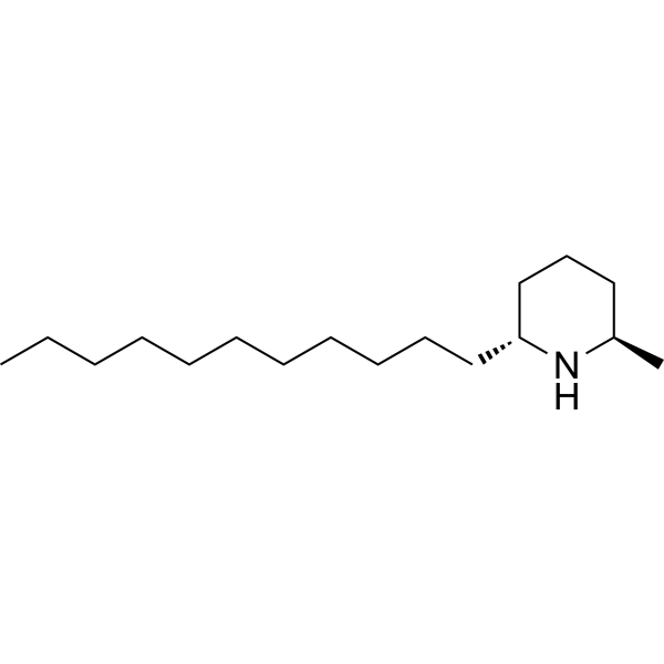
Solenopsin ((-)-Solenopsin A) is an ATP-competitive and selective Akt-1 inhibitor with an IC50 of 5-10 μM, and also acts as an RSK1 inhibitor. Solenopsin inhibits the activities of PDK1 in lipid rafts, downregulates PI3K, blocks PI3K-dependent generation of 3-phosphoinositides, and suppresses the phosphorylation of FOXO1a. Solenopsin induces Mitophagy and ROS production, reduces mitochondrial oxygen consumption, and exhibits antiproliferative and antiangiogenic activities. Solenopsin can be used in research related to hyperproliferative skin diseases and malignant diseases.
연구목적의 판매만을 진행합니다. 환자를 대상으로 한 판매는 하지 않습니다.
- CAS No.: 137038-57-4
- 화학식: C17H35N
- 분자량:253.47
-
보관:
Please store the product under the recommended conditions in the Certificate of Analysis.

To place orders, for customer services and technical support, please contact: MedChemExpress USA
Tel: 609-228-6898 E-mail: [email protected] [email protected]


-
 Biological Activity
-
Chemical Information
- Protocol
- 순도&문서
- References
-
 Help & FAQs


-
HY-18749
SC79Akt Activator -
HY-15186
IpatasertibPan-Akt Inhibitor -
HY-10249A
AKT Kinase InhibitorAkt Inhibitor -
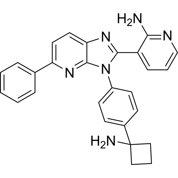
HY-19719
MiransertibAKT Inhibitor -
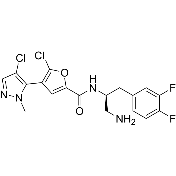
HY-15965
UprosertibAkt Inhibitor -
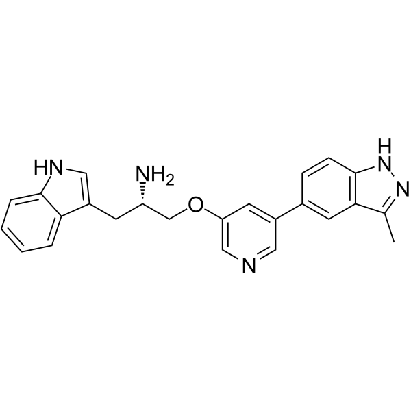
HY-10425
Rizavasertibpan-Akt Inhibitor -
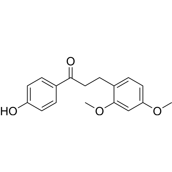
HY-N1505
Loureirin AAkt Inhibitor -
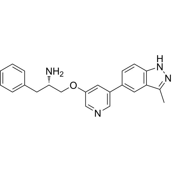
HY-13254
A-674563Akt1 Inhibitor -
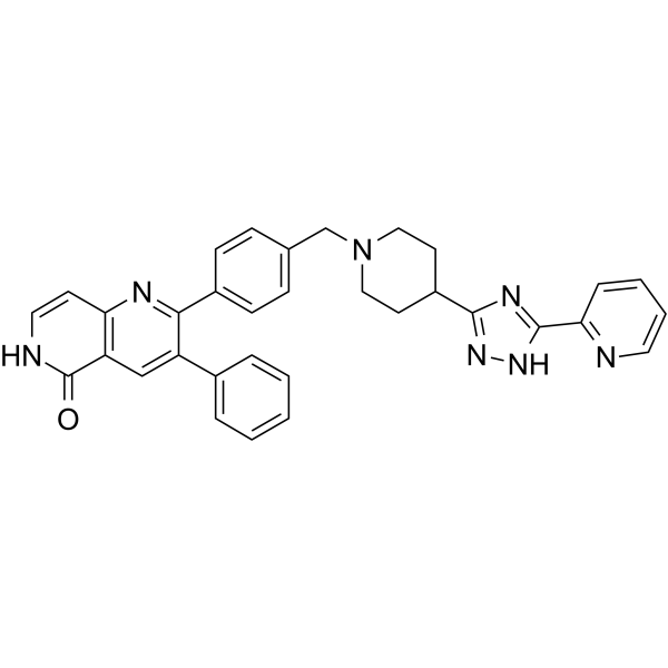
HY-50862
Akt1/Akt2-IN-1Akt Inhibitor -
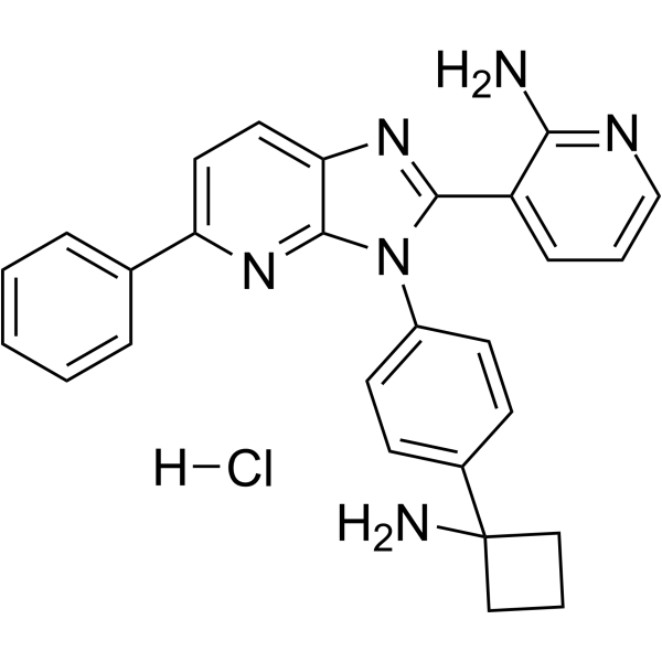
HY-19719A
Miransertib hydrochlorideAkt Inhibitor


Biological Activity
제품 설명
IC50 & Target
[2]|
Akt1 5-10 μM (IC50) |
RSK1 |
FOXO1a |
In Vitro
Solenopsin (10 μM; 24 h) potently inhibits proliferation of human A375 melanoma, human A2058 melanoma, and murine SVR angiosarcoma cells, with activity equivalent to its enantiomer (+)-solenopsin A and greater than cis isomer or long side-chain analogs[1].
Solenopsin (10-20 μM; 1 h) inhibits PDGF-induced Akt activity and PDK1 activation in membrane rafts of murine embryonic NIH3T3 fibroblast cells, with full inhibition at 20 μM for 1 hour and partial inhibition at 10 μM for 1 hour[1].
Solenopsin (10 μM; 24 h) upregulates p-Akt S473 and p-MAPK 44/42 in human A375 and A2058 melanoma cells, while downregulating these phosphorylated proteins in murine SVR angiosarcoma cells, demonstrating a context-dependent effect based on p53 status[1].
Solenopsin (10 μM; 24 h) reduces mitochondrial oxygen consumption rate in human UM-SCC1A squamous carcinoma cells[1].
Solenopsin (10 μM; 18 h) induces mitophagy in human UM-SCC1A squamous carcinoma cells[1].
Solenopsin (10 μM; 24 h) elevates reactive oxygen species levels by 1.7-2.3 fold in human A375 melanoma and murine SVR angiosarcoma cells[1].
Solenopsin (10 μM) potently and selectively inhibits purified recombinant Akt1 in an ATP-competitive manner with an IC50 of 5 to 10 μM at 0.1 mM ATP, while only significantly inhibiting one additional kinase (RSK1) out of 28 tested enzymes, and not inhibiting purified PI3K or PDK1[2].
MedChemExpress (MCE) has not independently confirmed the accuracy of these methods. They are for reference only.
-
Cell Line:human A375 melanoma cells, human A2058 melanoma cells, murine SVR angiosarcoma cells
-
Concentration:10 μM
-
Incubation Time:24 h
-
Result:Reduced cell counts significantly compared to DMSO control in all three tumor cell lines.
Exhibited equivalent anti-proliferative potency to its enantiomer (+)-solenopsin A.
Was more potent than cis isomer analog S12, and more potent than long side-chain analogs S11 and S13.
Was less potent than analog S14 in A375 and SVR cells, and less potent than analog S15 in A2058 cells.
-
Cell Line:human UM-SCC1A squamous carcinoma cells
-
Concentration:10 μM
-
Incubation Time:18 h
-
Result:Caused a significant increase in autophagosome-specific fluorescence compared to DMSO control, verifying induction of autophagy/mitophagy.
In Vivo
MedChemExpress (MCE) has not independently confirmed the accuracy of these methods. They are for reference only.
-
Animal Model:transgenic TG(fli1:EGFP)y1[2]
-
Dosage:3.75 μg/mL; 5 μg/mL; 6 μg/mL
-
Administration:immersion; continuous; 6 hours after fertilization until 32 hours after fertilization
-
Result:Delayed intersomitic vessel sprouts arising from the dorsal aorta.
Prevented primary sprouts from reaching the dorsolateral surface of the neural tube at 32 hours after fertilization.
Inhibited formation of paired longitudinal anastomotic vessels.
Allowed appropriate formation of vasculogenic vessels (dorsal aorta, posterior cardinal vein).


Chemical Information
-
CAS No. 137038-57-4
-
분자량 253.47
-
화학식 C17H35N
-
SMILES
CCCCCCCCCCC[C@@H]1CCC[C@@H](C)N1
-
Synonyms
(-)-Solenopsin A
-
Structure Classification
-
Initial Source
Pseudomonas aeruginosa
-
선적
Room temperature in continental US; may vary elsewhere.
-
보관
Please store the product under the recommended conditions in the Certificate of Analysis.


순도&문서
References
[1]. Karlsson I, et al. Solenopsin A and analogs exhibit ceramide-like biological activity. Vasc Cell. 2015;7:5. Published 2015 May 8. [Content Brief]
[2]. Arbiser JL, et al. Solenopsin, the alkaloidal component of the fire ant (Solenopsis invicta), is a naturally occurring inhibitor of phosphatidylinositol-3-kinase signaling and angiogenesis. Blood. 2007;109(2):560-565. [Content Brief]

Calculators
Concentration (start) × Volume (start) = Concentration (final) × Volume (final)


Solenopsin Related Classifications
HY-16461 Related Classifications
 Powered by Bioz