



Acetyl-L-carnitine-d3 hydrochloride

Based on 1 Customer Validation
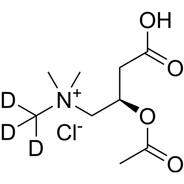
Acetyl-L-carnitine-d3 (O-Acetyl-L-carnitine-d3) hydrochloride is the deuterium labeled Acetyl-L-carnitine hydrochloride (HY-B0762). Acetyl-L-carnitine (O-Acetyl-L-carnitine; ALCAR) hydrochloride is an orally active mitochondrial energy metabolism regulator and neuroprotectant that can penetrate the blood-brain barrier. Acetyl-L-carnitine hydrochloride selectively enters cells and the brain through the organic cation transporter OCTN2. Acetyl-L-carnitine hydrochloride can participate in fatty acid β-oxidation, promote acetylcholine synthesis, regulate mitochondrial function and inhibit oxidative stress as an acetyl donor. Acetyl-L-carnitine hydrochloride exerts its activity by enhancing energy metabolism, protecting neurons and improving synaptic plasticity. Acetyl-L-carnitine hydrochloride is mainly used in the study of neurodegenerative diseases and metabolic disorder-related diseases such as neonatal hypoxic-ischemic brain damage, Alzheimer's disease, and depression.
For research use only. We do not sell to patients.
- Purity : 99.79%
- CAS No.: 1334532-17-0
- Formula: C9H15D3ClNO4
- Molecular Weight:242.72
-
Storage:
4°C, sealed storage, away from moisture
* In solvent : -80°C, 6 months; -20°C, 1 month (sealed storage, away from moisture)

To place orders, for customer services and technical support, please contact: MedChemExpress USA
Tel: 609-228-6898 E-mail: [email protected] [email protected]


-
 Biological Activity
-
Chemical Information
-
Solvent & Solubility
- Protocols
- Purity & Documentation
- References
-
 Help & FAQs


-
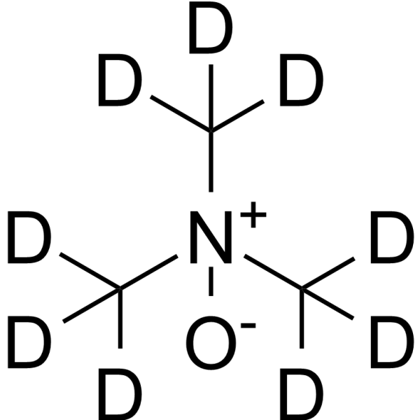
HY-116084S
Trimethylamine N-oxide-d9Stable Isotope -
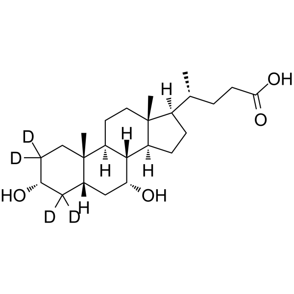
HY-76847S
Chenodeoxycholic Acid-d4Stable Isotope -
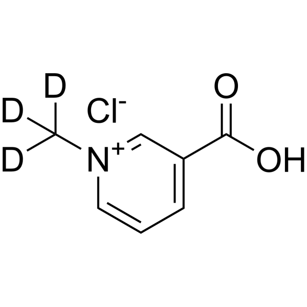
HY-N0415S
Trigonelline-d3 chlorideStable Isotope -
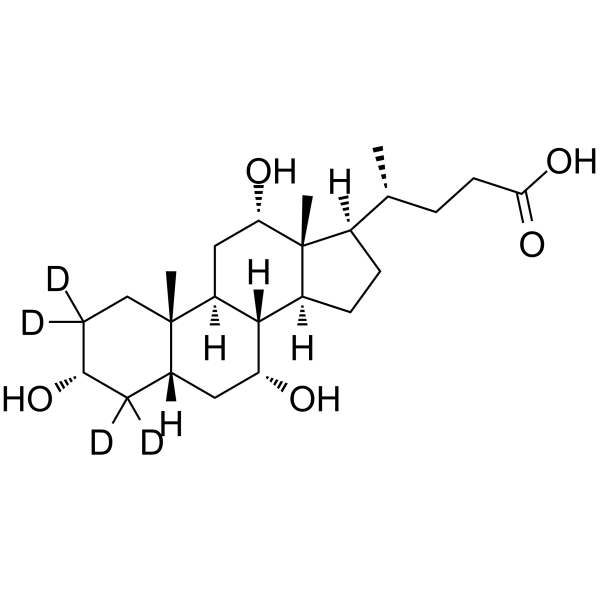
HY-N0324S
Cholic acid-d4Stable Isotope -
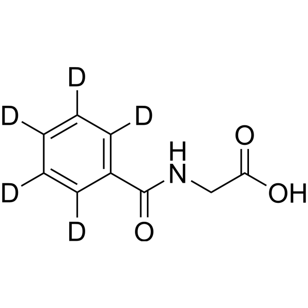
HY-W016562S
Hippuric acid-d5Stable Isotope -
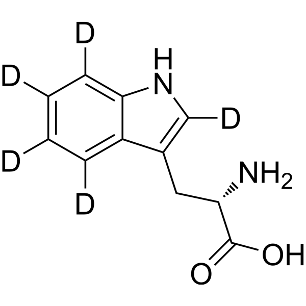
HY-N0623S
L-Tryptophan-d5Stable Isotope -
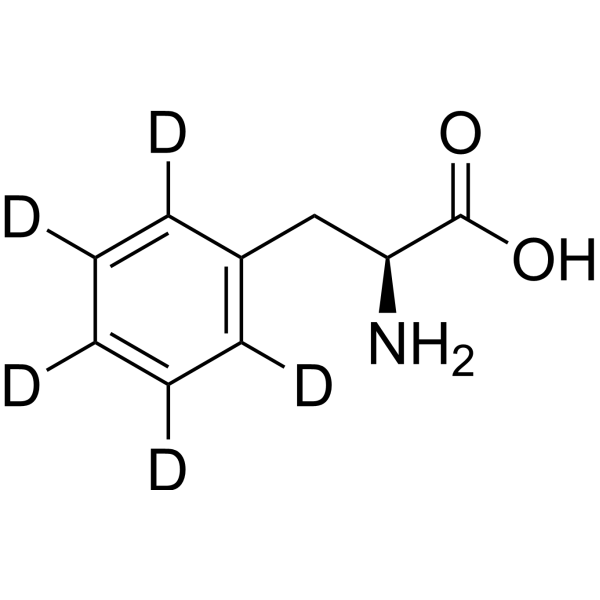
HY-N0215S12
L-Phenylalanine-d5Stable Isotope -
HY-B0617S
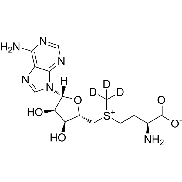
S-Adenosyl-L-methionine-d3Stable Isotope -
HY-B1337S1
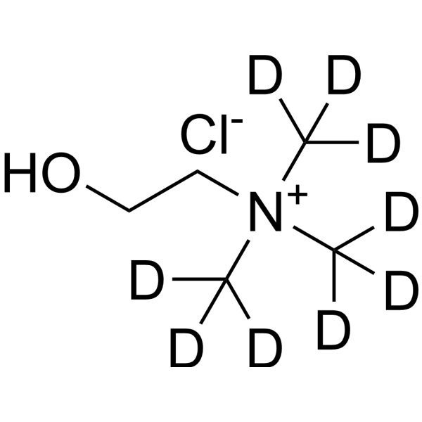
Choline-d9 chlorideStable Isotope -
HY-W010347S
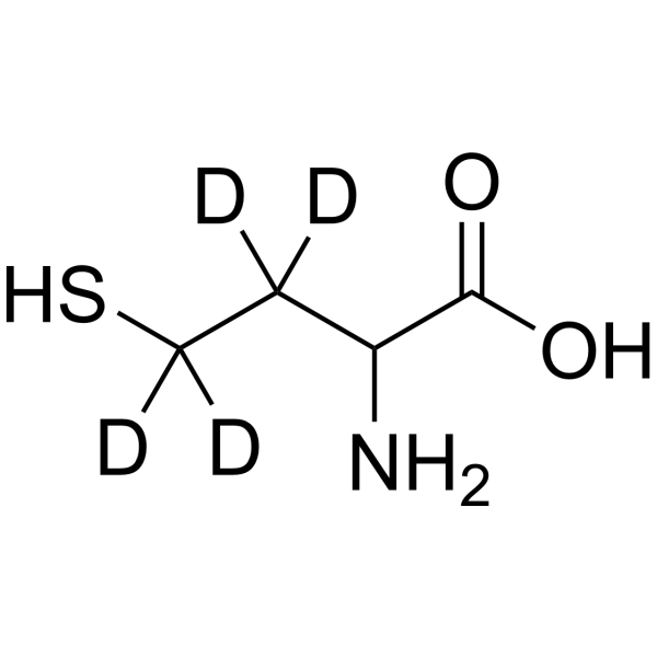
DL-Homocysteine-d4Stable Isotope -
HY-B0351S
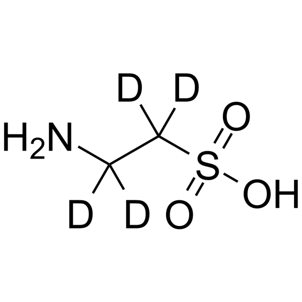
Taurine-d4Stable Isotope -
HY-N0391S4
L-Citrulline-d4Stable Isotope
All Caspase Isoforms
More

Biological Activity
Description
In Vitro
Stable heavy isotopes of hydrogen, carbon, and other elements have been incorporated into drug molecules, largely as tracers for quantitation during the drug development process. Deuteration has gained attention because of its potential to affect the pharmacokinetic and metabolic profiles of drugs[1].
MedChemExpress (MCE) has not independently confirmed the accuracy of these methods. They are for reference only. Further protocols information, click here.
Application
1. This compound can be used as a tracer
2. This compound can be used as an internal standard for quantitative analysis by NMR, GC-MS, or LC-MS.


Chemical Information
-
CAS No. 1334532-17-0
-
Unlabeled CAS 5080-50-2
-
Appearance Solid
-
Molecular Weight 242.72
-
Formula C9H15D3ClNO4
-
Color White to off-white
-
SMILES
OC(C[C@@H](OC(C)=O)C[N+](C)(C)C([2H])([2H])[2H])=O.[Cl-]
-
Synonyms
O-Acetyl-L-carnitine-d3 hydrochloride
-
Shipping
Room temperature in continental US; may vary elsewhere.
-
Storage
4°C, sealed storage, away from moisture
* In solvent : -80°C, 6 months; -20°C, 1 month (sealed storage, away from moisture)


Solvent & Solubility
In Vitro:
H2O : 100 mg/mL (412.00 mM; Need ultrasonic)
DMSO : 31.25 mg/mL (128.75 mM; Need ultrasonic; Hygroscopic DMSO has a significant impact on the solubility of product, please use newly opened DMSO)
Please refer to the solubility information to select the appropriate solvent. Once prepared, please aliquot and store the solution to prevent product inactivation from repeated freeze-thaw cycles.
Storage method and period of stock solution: -80°C, 6 months; -20°C, 1 month (sealed storage, away from moisture). When stored at -80°C, please use it within 6 months. When stored at -20°C, please use it within 1 month.
* Note: If you choose water as the stock solution, please dilute it to the working solution, then filter and sterilize it with a 0.22 μm filter before use.
Please refer to the solubility information to select the appropriate solvent. Once prepared, please aliquot and store the solution to prevent product inactivation from repeated freeze-thaw cycles.
Storage method and period of stock solution: -80°C, 6 months; -20°C, 1 month (sealed storage, away from moisture). When stored at -80°C, please use it within 6 months. When stored at -20°C, please use it within 1 month.
* Note: If you choose water as the stock solution, please dilute it to the working solution, then filter and sterilize it with a 0.22 μm filter before use.
Concentration (start) × Volume (start) = Concentration (final) × Volume (final)

Protocols
-
Mitochondrial membrane-potential fluorescent assay
Mitochondrial membrane potential fluorescent assays estimate ΔΨm in living cells using lipophilic cationic dyes such as TMRM, TMRE, rhodamine 123, and JC-1, which accumulate in mitochondria according to membrane polarization; loss of signal after FCCP or CCCP treatment is interpreted as mitochondrial depolarization. TMRM/TMRE and rhodamine 123 are commonly used for semi-quantitative live-cell microscopy or flow cytometry, while JC-1 can report a shift from red aggregate fluorescence to green monomer fluorescence during depolarization; interpretation requires controls because dye concentration, quenching mode, cell type, dye efflux, and mitochondrial mass can affect fluorescence independently of ΔΨm.
-
ROS/oxidative-stress fluorescent staining
ROS/oxidative-stress fluorescent staining uses cell-permeant fluorogenic probes that become fluorescent after oxidation inside cells or tissues; commonly used examples include DCFH-DA/DCFDA for broad cellular oxidant detection, DHE for superoxide-related signal detection, MitoSOX for mitochondrial superoxide-related signal detection, and CellROX probes for oxidative-stress-associated fluorescence readouts. The assay detects probe oxidation rather than a single ROS species unless the probe and analysis method have been chemically validated for that species. DCFH-DA enters cells, is deacetylated by intracellular esterases to DCFH, and produces fluorescent DCF after oxidation, so the readout is used as an operational measure of total cellular oxidative stress rather than a species-specific ROS measurement. DHE and MitoSOX can report superoxide-related oxidation, but red fluorescence alone can include non-specific ethidium-like oxidation products; HPLC or optimized spectral approaches are
-
Mitochondrial membrane-potential and mitochondrial mass staining
Mitochondrial membrane potential staining measures the electrochemical polarization across the mitochondrial inner membrane in live cells using lipophilic cationic fluorescent probes; early rhodamine-based work showed that selective mitochondrial dye accumulation is lost when the mitochondrial transmembrane potential is dissipated. JC-1 reports mitochondrial polarization by shifting from green monomer fluorescence to red J-aggregate fluorescence as dye concentration increases within energized mitochondria; therefore, the red/green fluorescence ratio is used as a relative readout of mitochondrial membrane potential. TMRE or TMRM staining provides a single-channel relative readout because these cationic rhodamine esters accumulate in polarized mitochondria, and lower fluorescence indicates reduced mitochondrial polarization when acquisition and dye-loading conditions are controlled. Mitochondrial mass staining is commonly performed with MitoTracker Green FM or related MitoTracker dyes as
-
Research Protocol for Metabolic Diseases
AMP-activated protein kinase, AMPK, is a conserved cellular energy sensor that responds to reduced cellular energy status and coordinates metabolism by increasing ATP-generating catabolic pathways while suppressing ATP-consuming anabolic processes. In metabolic disease research, the AMPK pathway is experimentally relevant because it regulates hepatic lipid synthesis, fatty acid oxidation, glucose production, skeletal-muscle glucose disposal, mTORC1-linked biosynthesis, autophagy, mitochondrial homeostasis, and whole-body energy balance. The central pathway logic is that energy stress, metformin, exercise-like stimulation, or direct AMPK activators increase AMPKα Thr172 phosphorylation and downstream substrate phosphorylation, including ACC and RAPTOR. Phosphorylation of ACC suppresses lipogenesis and supports fatty acid oxidation, whereas phosphorylation of RAPTOR suppresses mTORC1 signaling and links cellular energy status to growth and protein synthesis control. The pathway is linked
-
Alzheimer’s Disease Modeling
Alzheimer’s Disease (AD) is a neurodegenerative disorder characterized by a progressive decline in cognitive functions and loss of specific types of neurons and synapses. Alzheimer's symptoms can be simulated in mice by injecting drugs (such as Aβ) or genetically modified.


Purity & Documentation
-
Data Sheet (278 KB)
-
SDS (252 KB)
- English - EN (252 KB)
- Français - FR (252 KB)
- Deutsch - DE (252 KB)
- Norwegian - NO (252 KB)
- Español - ES (252 KB)
- Swedish - SV (252 KB)
- Italian - IT (252 KB)
- Korean - KR (252 KB)
- Portuguese - PT (252 KB)
-
Handling Instructions (2659 KB)
References
[1]. Russak EM, et al. Impact of Deuterium Substitution on the Pharmacokinetics of Pharmaceuticals. Ann Pharmacother. 2019;53(2):211-216. [Content Brief]
[2]. Zhu X, et al. Acetyl-L-carnitine suppresses apoptosis of thioredoxin 2-deficient DT40 cells. Arch Biochem Biophys. 2008 Oct 15;478(2):154-60. [Content Brief]
[3]. Wang SM, et al. A review of current evidence for acetyl-l-carnitine in the treatment of depression. J Psychiatr Res. 2014 Jun;53:30-7. [Content Brief]
[4]. Ferreira GC, et al. L-Carnitine and Acetyl-L-carnitine Roles and Neuroprotection in Developing Brain. Neurochem Res. 2017 Jun;42(6):1661-1675. [Content Brief]


Complete Stock Solution Preparation Table
Please refer to the solubility information to select the appropriate solvent. Once prepared, please aliquot and store the solution to prevent product inactivation from repeated freeze-thaw cycles.
Storage method and period of stock solution: -80°C, 6 months; -20°C, 1 month (sealed storage, away from moisture). When stored at -80°C, please use it within 6 months. When stored at -20°C, please use it within 1 month.
| Optional Solvent | Concentration Solvent Mass | 1 mg | 5 mg | 10 mg | 25 mg |
|---|---|---|---|---|---|
| DMSO / H2O | 1 mM | 4.1200 mL | 20.5999 mL | 41.1997 mL | 102.9993 mL |
| 5 mM | 0.8240 mL | 4.1200 mL | 8.2399 mL | 20.5999 mL | |
| 10 mM | 0.4120 mL | 2.0600 mL | 4.1200 mL | 10.2999 mL | |
| 15 mM | 0.2747 mL | 1.3733 mL | 2.7466 mL | 6.8666 mL | |
| 20 mM | 0.2060 mL | 1.0300 mL | 2.0600 mL | 5.1500 mL | |
| 25 mM | 0.1648 mL | 0.8240 mL | 1.6480 mL | 4.1200 mL | |
| 30 mM | 0.1373 mL | 0.6867 mL | 1.3733 mL | 3.4333 mL | |
| 40 mM | 0.1030 mL | 0.5150 mL | 1.0300 mL | 2.5750 mL | |
| 50 mM | 0.0824 mL | 0.4120 mL | 0.8240 mL | 2.0600 mL | |
| 60 mM | 0.0687 mL | 0.3433 mL | 0.6867 mL | 1.7167 mL | |
| 80 mM | 0.0515 mL | 0.2575 mL | 0.5150 mL | 1.2875 mL | |
| 100 mM | 0.0412 mL | 0.2060 mL | 0.4120 mL | 1.0300 mL |
* Note: If you choose water as the stock solution, please dilute it to the working solution, then filter and sterilize it with a 0.22 μm filter before use.


Acetyl-L-carnitine-d3 hydrochloride Related Classifications
HY-B0762S Related Classifications
 Powered by Bioz