



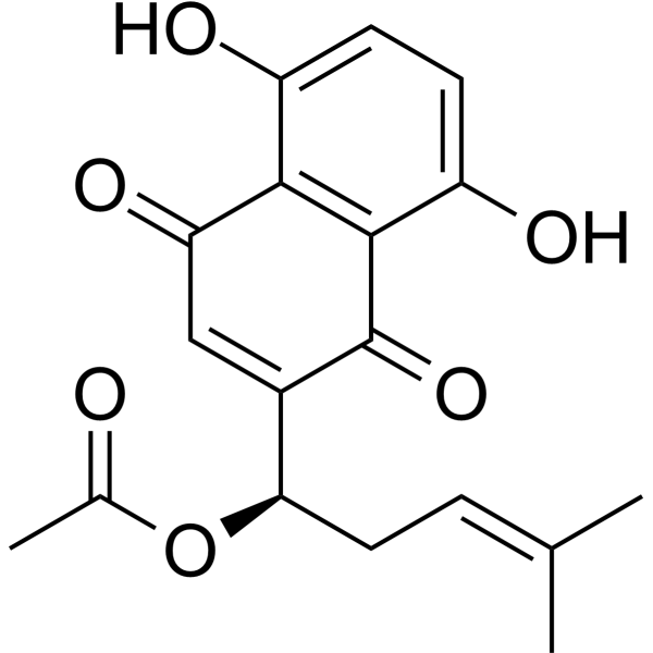
Acetylshikonin
Based on 3 publication(s) in Google Scholar
Acetylshikonin is an oral active anti-cancer, anti-inflammatory, antioxidant, anti-fertility, antibacterial, and neuroprotective agent. Acetylshikonin is a inhibitor of acetylcholinase (AChE) (IC50=34.6 μM) and nonselective cytochrome P450. Acetylshikonin can induce Apoptosis and Autophagy in cancer cells. Acetylshikonin regulates blood glucose, liver fat metabolism, and renal fibrosis, and is used in the study of diabetes, diabetic nephropathy (DN), obesity, and nonalcoholic fatty liver disease (NAFLD).
For research use only. We do not sell to patients.
- Purity : 99.89%
- CAS No.: 24502-78-1
- Formula: C18H18O6
- Molecular Weight:330.33
-
Storage:
4°C, protect from light
* In solvent : -80°C, 6 months; -20°C, 1 month (protect from light)

To place orders, for customer services and technical support, please contact: MedChemExpress USA
Tel: 609-228-6898 E-mail: [email protected] [email protected]


-
 Biological Activity
-
Chemical Information
-
Solvent & Solubility
- Protocols
- Purity & Documentation
- References
-
 Help & FAQs


-
HY-N2187
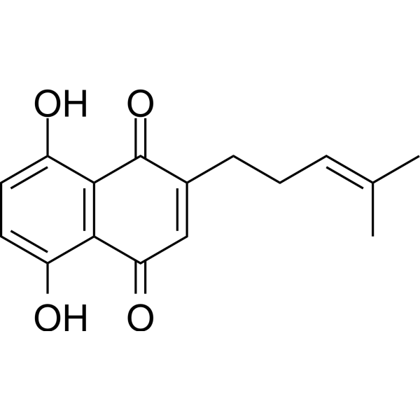
DeoxyshikoninAntibacterial Agent -
HY-N5112A
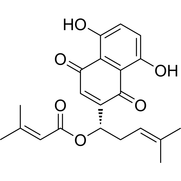
β,β-DimethylacrylalkanninFGFR1 Inhibitor -
HY-N0822
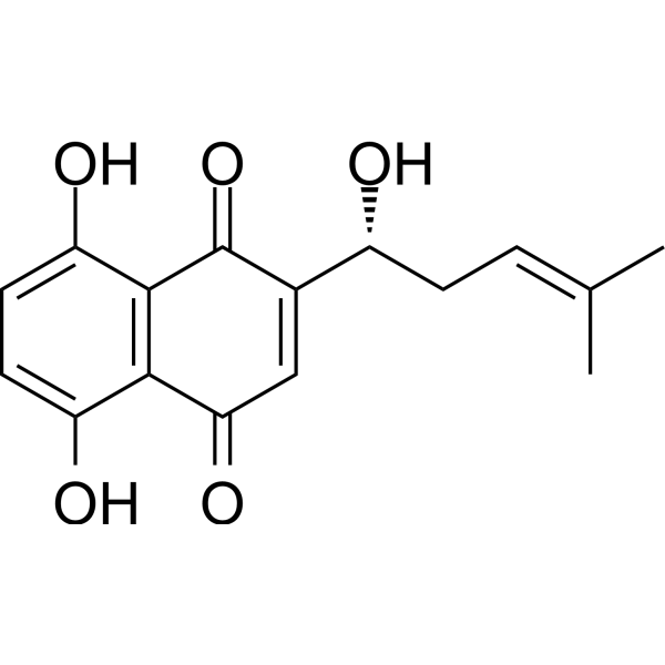
ShikoninChloride Channel Inhibitor -
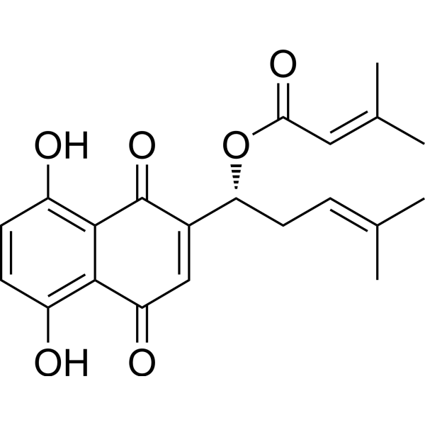
HY-N5112B
β,β-DimethylacrylshikoninAnticancer Compound -
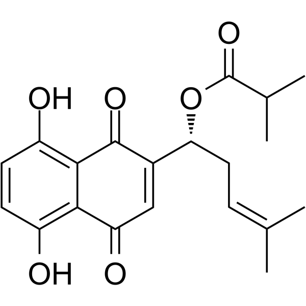
HY-N2592
IsobutylshikoninShikonin Pigment -
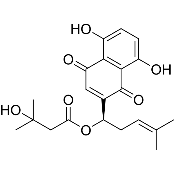
HY-N4201
β-HydroxyisovalerylshikoninSrc Inhibitor -
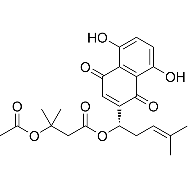
HY-N2188
Beta-AcetoxyisovalerylshikoninHIV Inhibitor -
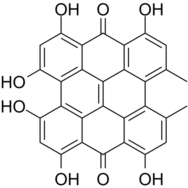
HY-N0453
HypericinPKC Inhibitor -

HY-Y0320C
Dimethyl sulfoxideAprotic Solvent -
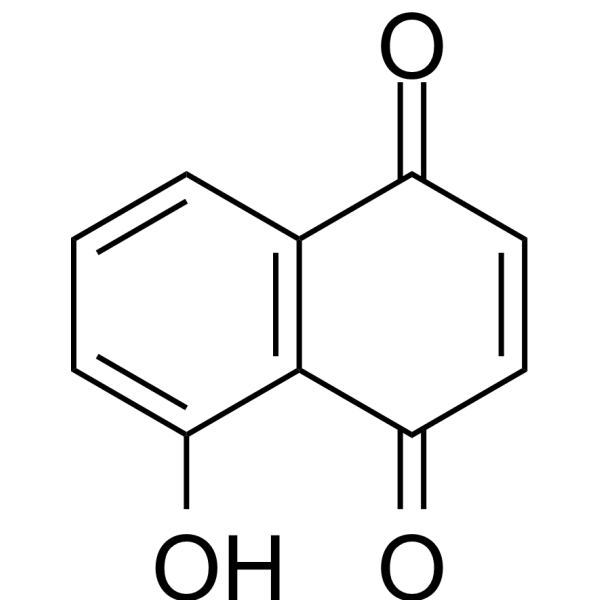
HY-N6949
JugloneAntimicrobial Drug -
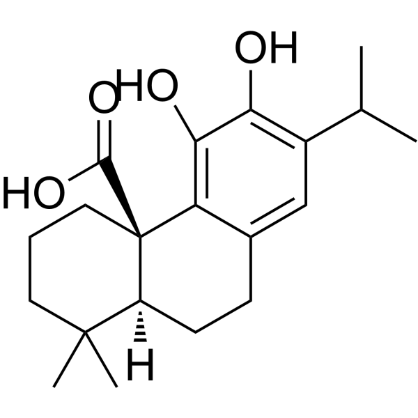
HY-N0644
Carnosic acidLipid Absorption Inhibitor -
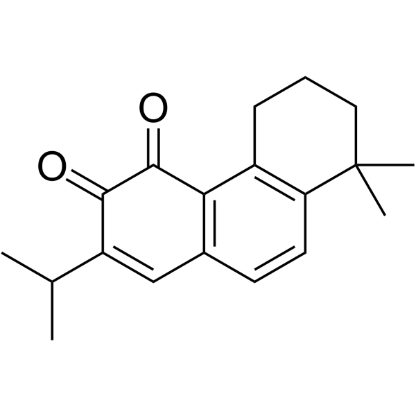
HY-N1951
MiltironeBenzodiazepine Receptor Partial Agonist
Publications Citing Use of MedChemExpress (MCE) Acetylshikonin
More

Biological Activity
Description
IC50 & Target
|
AChE |
Cellular Effect
|
Cell Line

|
Type | Value | Description | References |
|---|---|---|---|---|
| A-375 | IC50 |
1.91 μM
Compound: SK-4
|
Antiproliferative activity against human A-375 cells assessed as inhibition of cell proliferation measured after 3 days by MTT assay
Antiproliferative activity against human A-375 cells assessed as inhibition of cell proliferation measured after 3 days by MTT assay
|
[PMID: 39129245] |
| A549 | IC50 |
39 μM
Compound: 12
|
Tested in vitro for inhibitory effect on the growth of human A-549 cancer cell line
Tested in vitro for inhibitory effect on the growth of human A-549 cancer cell line
|
[PMID: 11992780] |
| Bel-7402 | IC50 |
0.068 μM
Compound: 12
|
Tested in vitro for inhibitory effect on the growth of BEL -7402 cell line
Tested in vitro for inhibitory effect on the growth of BEL -7402 cell line
|
[PMID: 11992780] |
| BTI-TN-5B1-4 | IC50 |
112.2 μM
Compound: 1
|
Inhibition of human ACAT2 expressed in Hi5 cells
Inhibition of human ACAT2 expressed in Hi5 cells
|
[PMID: 17157006] |
| BTI-TN-5B1-4 | IC50 |
128.9 μM
Compound: 1
|
Inhibition of human ACAT1 expressed in Hi5 cells
Inhibition of human ACAT1 expressed in Hi5 cells
|
[PMID: 17157006] |
| Calu-3 | CC50 |
>100 μM
Compound: 30
|
Cytotoxicity against human Calu-3 cells assessed as reduction in cell viability
Cytotoxicity against human Calu-3 cells assessed as reduction in cell viability
|
[PMID: 36651644] |
| CCRF-CEM | IC50 |
1 μM
Compound: 2
|
Cytotoxicity against human CCRF-CEM cells after 72 hrs by XTT assay
Cytotoxicity against human CCRF-CEM cells after 72 hrs by XTT assay
|
[PMID: 22530779] |
| HCT-116 | IC50 |
0.45 μM
Compound: 16
|
Cytotoxicity against MDR1 Pgp under-expressing human HCT116 cells after 24 hrs by MTT assay
Cytotoxicity against MDR1 Pgp under-expressing human HCT116 cells after 24 hrs by MTT assay
|
[PMID: 17949858] |
| HCT-116 | IC50 |
3.56 μM
Compound: SK-4
|
Antiproliferative activity against human HCT-116 cells assessed as inhibition of cell proliferation measured after 3 days by MTT assay
Antiproliferative activity against human HCT-116 cells assessed as inhibition of cell proliferation measured after 3 days by MTT assay
|
[PMID: 39129245] |
| HCT-116 | IC50 |
9 μM
Compound: 2
|
Cytotoxicity against human HCT116 cells after 72 hrs by XTT assay
Cytotoxicity against human HCT116 cells after 72 hrs by XTT assay
|
[PMID: 22530779] |
| HeLa | IC50 |
45 μM
Compound: 1
|
Inhibitory activity against DNA topoisomerase-1 obtained from Hela cells
Inhibitory activity against DNA topoisomerase-1 obtained from Hela cells
|
[PMID: 7699697] |
| HepG2 | IC50 |
0.38 μM
Compound: 16
|
Cytotoxicity against MDR1 Pgp overexpressing human HepG2 cells after 24 hrs by MTT assay
Cytotoxicity against MDR1 Pgp overexpressing human HepG2 cells after 24 hrs by MTT assay
|
[PMID: 17949858] |
| HepG2 | IC50 |
6.48 μM
Compound: SK-4
|
Antiproliferative activity against human HepG2 cells assessed as inhibition of cell proliferation measured after 3 days by MTT assay
Antiproliferative activity against human HepG2 cells assessed as inhibition of cell proliferation measured after 3 days by MTT assay
|
[PMID: 39129245] |
| K562 | IC50 |
29 μM
Compound: 12
|
Tested in vitro for inhibitory effect on the growth of K-562 cell line
Tested in vitro for inhibitory effect on the growth of K-562 cell line
|
[PMID: 11992780] |
| MCF7 | IC50 |
31 μM
Compound: 12
|
Tested in vitro for inhibitory effect on the growth of MCF-7 cell line
Tested in vitro for inhibitory effect on the growth of MCF-7 cell line
|
[PMID: 11992780] |
| MDA-MB-231 | IC50 |
11.3 μM
Compound: 2
|
Cytotoxicity against human MDA-MB-231 cells after 72 hrs by XTT assay
Cytotoxicity against human MDA-MB-231 cells after 72 hrs by XTT assay
|
[PMID: 22530779] |
| MRC5 | IC50 |
2.6 μM
Compound: 2
|
Cytotoxicity against human MRC5 cells after 72 hrs by XTT assay
Cytotoxicity against human MRC5 cells after 72 hrs by XTT assay
|
[PMID: 22530779] |
| RD | CC50 |
9.4 μM
Compound: AS
|
Cytotoxicity against human RD cells assessed as reduction in cell viability after 12 hrs by CCK8 assay
Cytotoxicity against human RD cells assessed as reduction in cell viability after 12 hrs by CCK8 assay
|
[PMID: 31063370] |
| SBcl2 | IC50 |
13 μM
Compound: 2
|
Cytotoxicity against human SBcl2 cells after 72 hrs by XTT assay
Cytotoxicity against human SBcl2 cells after 72 hrs by XTT assay
|
[PMID: 22530779] |
| SGC-7901 | IC50 |
0.29 μM
Compound: 12
|
Tested in vitro for inhibitory effect on the growth of human gastric carcinoma SGC-7901 cell line
Tested in vitro for inhibitory effect on the growth of human gastric carcinoma SGC-7901 cell line
|
[PMID: 11992780] |
| U-251 | IC50 |
15.9 μM
Compound: 2
|
Cytotoxicity against human U251 cells after 72 hrs by XTT assay
Cytotoxicity against human U251 cells after 72 hrs by XTT assay
|
[PMID: 22530779] |
| WM164 | IC50 |
71.5 μM
Compound: 2
|
Cytotoxicity against human WM164 cells after 72 hrs by XTT assay
Cytotoxicity against human WM164 cells after 72 hrs by XTT assay
|
[PMID: 22530779] |
In Vitro
Acetylshikonin(1.6-100 μM) inhibits the proliferation of oral cancer cells KB-R5 with IC50 40 μM[6].
Acetylshikonin (20-80 μM; 24 h) can induce Apoptosis and (20-80 μM) Autophagy of KB-R5 cells, and block mTOR/PI3K/AKT signaling pathway in KB-R5 cells[6].
Acetylshikonin (1-10 μM; 12 h) inhibits H2O2 (500 μM; 4 h) H2O2- (500 μM; 4 h ) induced Apoptosis of neuroblastoma SH-SY5Y and pc12 cells[8].
Acetylshikonin (0.01-5 μM/L; 2 h) has anti-CoxSackievirus A16 (CVA16) activity in the adsorption/invasion stage with EC50 was 0.04 μmol/L. However, has no effective in the pre-infection, replication and release stages[2].
Acetylshikonin (0.01-1 μM; 30 min) promotes glucose uptake by skeletal muscle cells L6 through PLC-β3/PKCδ mediation, thereby reducing blood glucose level[3].
Acetylshikonin (1-5 μg/mL; 48 h) inhibits TGF-β1 (5 ng/mL) induced renal fibrosis in HK2 cells[5].
MedChemExpress (MCE) has not independently confirmed the accuracy of these methods. They are for reference only. Further protocols information, click here.
-
Cell Line:KB-R5 (oral cancer cell line)
-
Concentration:20 μM, 40 μM, 80 μM
-
Incubation Time:24 h
-
Result:Changed the morphology of the nucleus.
Increased apoptosis ratio.
-
Cell Line:KB-R5 (oral cancer cell line)
-
Concentration:20 μM, 40 μM, 80 μM
-
Incubation Time:
-
Result:Increased the expression of Beclin-1 and LC3-II and inhibited the expression of p62. However, had no effect on the expression of LC3-I and Vps34.
Decreased the expression of p-mTOR, p-PI3K and p-AKT in a concentration-dependent manner.
-
Cell Line:CVA16-induced human rhabdomyosarcoma (RD) cells
-
Concentration:0.01-5 μM/L (1:1 mix with CVA16 strain TA271)
-
Incubation Time:2 h
-
Result:Reduced CVA16-induced cytopathic effect with inhibition rates of 80% at the concentration of 0.08 μmol/L.
-
Cell Line:L6 (rat skeletal muscle cells)
-
Concentration:0.01 μM, 0.1 μM, 1 μM
-
Incubation Time:2 h
-
Result:Significantly up-regulated phosphorylation of PKCδ.
Up-regulates the expression of glucose transporter 4 (GLUT4).and PLC-β3.
In Vivo
Acetylshikonin (270-1080 mg/kg; Intragastric administration; Once a day for 30 days) reduces D-galactose-induced (150 mg/kg) cognitive impairment and hippohippoal aging by reducing oxidative stress and neuroinflammation and inhibiting the activation of the p53/p21 signaling pathway in mice[1].
Acetylshikonin (2 mg/kg; Intramuscular injection; Single dose) inhibits Coxsackievirus A16 (CVA16) replication in mice[2].
Acetylshikonin (100 mg/kg; Intragastric administration; Once a day for eight weeks) inhibits renal fibrosis by inhibiting TGF-β1/Smad pathway without affecting blood glucose, thus reducing the damage of renal function in streptozotocin (STZ) -induced diabetic C57BL/6 mice[5].
Acetylshikonin (10 mk/kg; Intraperitoneal injection; Once a day for three days) reduces alloxouracil- (180 mg/kg; Intraperitoneal injection; Single dose) induced blood glucose level in diabetic mice[3].
Acetylshikonin (540 mg/kg; Oral administration; Once a day for eight weeks) reduces liver fat accumulation by regulating fat metabolism and liver inflammation in obese C57BL/6J mice, thereby improving obesity and nonalcoholic fatty liver disease (NAFLD)[4].
Acetylshikonin (120-1080 mg/kg; Intragastric administration;) doesn’t affect the ability of pregnancy in Sprague-Dawley rats at low doses (120 mg/kg and 360 mg/kg), but inhibits the ability of pregnancy in Sprague-Dawley rats at high doses (1080 mg/kg) by affecting the secretion of gonadotropin (GTH) and reducing the levels of serum follicle-stimulating hormone (FSH) and luteinizing hormone (LH)[7].
MedChemExpress (MCE) has not independently confirmed the accuracy of these methods. They are for reference only.
-
Animal Model:D-galactose (D-gal)-induced sub-acuteaging mouse model of Alzheimer’s disease (AD)[1]
-
Dosage:270 mg/kg, 540 mg/kg, 1080 mg/kg
-
Administration:Intragastrical administration (i.g.); Once daily for 30 days. After D-gal treatment (150 mg/kg; Subcutaneous injection (s.c.); Once daily for 30 days)
-
Result:Decreased levels of the pro-inflammatory cytokines IL-1β and TNF-α.
Decreased the content of MDA and increased the activity of SOD.
Significantly mitigated D-Gal-induced downregulation of SIRT1 in hippocampal neurons.
Significantly inhibited the expression of p53, acetyl-p53, and p21 in mice (all proteins associated with hippocampal aging).
-
Animal Model:CAV16-indeced ICR suckling mice model[2]
-
Dosage:2 mg/kg
-
Administration:Intramuscular injection (i.m.); Single dose. After CVA16 treatment (10[5.5] TCID50/g; Intramuscular injection (i.m.); Single dose )
-
Result:Delayed death of the mice (6 days post-infection and 7 dpi), and eventually resulted in a survival rate of 50% and 70% for the mice in the treatment and prevention groups, respectively (the death of the control mice began at 4 days after infection and all died at 6 days after infection).
-
Animal Model:Obese male C57BL/6J Mice model[4]
-
Dosage:540 mg/kg
-
Administration:Oral gavage (P.O.); Once daily for 8 weeks
-
Result:Reduced body mass index (BMI) and food efficiency in obese mice by 17.1% and 48.2%, respectively.
Decreased plasma glucose, CHE, AST and ALT levels by 34.1%, 45.5% and 27.2%, respectively.
Significantly inhibited the levels of serum proinflammatory cytokines TNF-α, IL-6 and IL-1β by 49.1%, 41.1% and 45.6%, respectively.


Chemical Information
-
CAS No. 24502-78-1
-
Appearance Solid
-
Molecular Weight 330.33
-
Formula C18H18O6
-
Color Brown to khaki
-
SMILES
O=C1C([C@H](OC(C)=O)C/C=C(C)\C)=CC(C2=C1C(O)=CC=C2O)=O
-
Structure Classification
-
Initial Source
-
Shipping
Room temperature in continental US; may vary elsewhere.
-
Storage
4°C, protect from light
* In solvent : -80°C, 6 months; -20°C, 1 month (protect from light)


Publications (3)
-
Journal Impact Factor
-
Most Recent
-
Mol Med Rep
Acetylshikonin induces ferroptosis via the lipid peroxidation pathway in osteosarcoma cells. [Abstract]2026 Feb;33(2):55. PMID: 41312749 -
Vet Microbiol
The Chinese medicine monomer Schisandrin C inhibits PRRSV infection by regulating the OGT-PI3K/AKT/mTOR signaling pathway. [Abstract]2026 May:316:110992. PMID: 41865607 -


Solvent & Solubility
In Vitro:
DMSO : 50 mg/mL (151.36 mM; Need ultrasonic; Hygroscopic DMSO has a significant impact on the solubility of product, please use newly opened DMSO)
Please refer to the solubility information to select the appropriate solvent. Once prepared, please aliquot and store the solution to prevent product inactivation from repeated freeze-thaw cycles.
Storage method and period of stock solution: -80°C, 6 months; -20°C, 1 month (protect from light). When stored at -80°C, please use it within 6 months. When stored at -20°C, please use it within 1 month.
Please refer to the solubility information to select the appropriate solvent. Once prepared, please aliquot and store the solution to prevent product inactivation from repeated freeze-thaw cycles.
Storage method and period of stock solution: -80°C, 6 months; -20°C, 1 month (protect from light). When stored at -80°C, please use it within 6 months. When stored at -20°C, please use it within 1 month.
Concentration (start) × Volume (start) = Concentration (final) × Volume (final)
In Vivo:
Select the appropriate dissolution method based on your experimental animal and administration route.
- For the following dissolution methods, please ensure to first prepare a clear stock solution using an In Vitro approach and then sequentially add co-solvents:
- To ensure reliable experimental results, the clarified stock solution can be appropriately stored based on storage conditions. As for the working solution for In Vivo experiments, it is recommended to prepare freshly and use it on the same day.
- The percentages shown for the solvents indicate their volumetric ratio in the final prepared solution. If precipitation or phase separation occurs during preparation, heat and/or sonication can be used to aid dissolution.
Add each solvent one by one: 10% DMSO 90% (20% SBE-β-CD in Saline)
Solubility: ≥ 2.5 mg/mL (7.57 mM); Clear solution
This protocol yields a clear solution of ≥ 2.5 mg/mL (saturation unknown).
Taking 1 mL working solution as an example, add 100 μL DMSO stock solution (25.0 mg/mL) to 900 μL 20% SBE-β-CD in Saline, and mix evenly.
Preparation of 20% SBE-β-CD in Saline (4°C, storage for one week): 2 g SBE-β-CD powder is dissolved in 10 mL Saline, completely dissolve until clear.
In Vivo Dissolution Calculator
Please enter the basic information of animal experiments:
-
-
-
-
Recommended: Prepare an additional quantity of animals to account for potential losses during experiments.
Please enter your animal formula composition:
-
%DMSO +
Recommended: Keep the proportion of DMSO in working solution below 2% if your animal is weak.
-
%+
-
+%Tween-80 + +
-
%Saline +
The co-solvents required include: DMSO, . All of co-solvents are available by MedChemExpress (MCE). , Tween 80. All of co-solvents are available by MedChemExpress (MCE).
Working solution concentration: 0.22 mg/mL
Method for preparing stock solution: mg drug dissolved in μL DMSO. Stock solution concentration: mg/mL. * In solvent : -80°C, 6 months; -20°C, 1 month (protect from light)
1. Take μL DMSO stock solution;
2. Add μL .
μL , mix evenly;
3. Then add μL Tween 80, mix evenly;
4. Then add μL
Please ensure that the stock solution in the first step is dissolved to a clear state, and add co-solvents in sequence. You can use ultrasonic heating (ultrasonic cleaner, recommended frequency 20-40 kHz), vortexing, etc. to assist dissolution.

Protocols
-
Reproductive and Developmental Toxicity Study
Reproductive and developmental toxicity studies detect adverse effects of prenatal or peri/postnatal exposure on maternal condition, pregnancy maintenance, embryo-fetal survival, fetal growth, structural development, and offspring reproductive or developmental endpoints; classic rat protocols generate readouts by comparing treated groups with vehicle, pair-fed, or untreated controls for implantation, resorption, fetal weight, crown-rump length, external morphology, visceral morphology, skeletal ossification, anogenital distance, nipple/areola retention, and postnatal cohort outcomes.
-
Research Protocol for Infectious Diseases
Infectious-disease experiments test how pathogens interact with host barriers, innate immune receptors, inflammatory signaling, pathogen replication, and tissue injury; pattern-recognition receptors such as TLRs, RIG-I-like receptors, NOD-like receptors, and inflammasomes detect microbial molecules and activate NF-κB, interferon, and cytokine responses. The central hypothesis is that infection severity reflects the balance between pathogen burden and host response: protective inflammation restricts pathogen growth, whereas excessive or mislocalized inflammation contributes to tissue damage and disease phenotype. Unresolved questions include which host pathways are protective versus pathogenic, why some infection models fail to translate to human disease, and which combined readouts best predict clinically relevant infection outcomes.
-
Collagen: Sirius Red Staining
Sirius Red or picrosirius red staining is a histochemical method for visualizing collagen-rich extracellular matrix in tissue sections, and collagen fibers are detected as red-stained structures under bright-field microscopy with enhanced birefringence under polarized light. Picrosirius red is useful for assessing total collagen organization, distribution, and fibrosis burden, but polarized color should not be interpreted as a definitive collagen type I versus type III readout because color is affected by fiber orientation, thickness, and packing.
-
Annexin V plus membrane-impermeant dye apoptosis staining
Annexin V-based apoptosis assays rely on the detection of phosphatidylserine (PS) externalization from the inner leaflet of the plasma membrane to the outer leaflet, an early biochemical hallmark of apoptosis. Fluorescently labeled Annexin V binds PS in a calcium-dependent manner, enabling identification of early apoptotic cells by flow cytometry or fluorescence microscopy. When combined with a membrane-impermeant DNA-binding dye (e. g. , propidium iodide), this approach allows discrimination between viable (Annexin V−/dye−), early apoptotic (Annexin V+/dye−), and late apoptotic or necrotic (Annexin V+/dye+) cell populations by assessing membrane integrity and PS exposure.
-
Connective Tissue: Masson's Trichrome/Collagen Trichrome Staining
Masson’s Trichrome (collagen/trichrome staining) is a histological technique that differentially stains tissue compartments using sequential acidic dyes to distinguish collagen from muscle and cytoplasmic components based on dye affinity and tissue permeability differences, enabling visualization of fibrosis and connective tissue architecture in histological sections. The classical formulation typically uses Weigert's iron hematoxylin for nuclear staining, Biebrich scarlet-acid fuchsin for cytoplasm and muscle, and aniline blue (or light green variants) for collagen, producing a characteristic blue/green collagen signal contrasted against red cytoplasm and dark nuclei. The staining principle relies on selective displacement of smaller dye molecules by larger anionic dyes in collagen-rich regions under controlled acidified conditions, which enhances collagen-specific dye retention. This property makes the method widely used for fibrosis assessment in organs such as heart, liver, lung, a
-
Research Protocol for Inflammation-related Diseases
The NLRP3 inflammasome is a cytosolic innate immune signaling platform that integrates priming signals and danger-signal activation to promote caspase-1 activation, maturation of IL-1β and IL-18, and gasdermin D-mediated pyroptotic cell death. The core experimental logic is to determine whether inflammatory disease phenotypes are driven by increased NLRP3 expression, ASC-containing inflammasome assembly, caspase-1 cleavage, GSDMD cleavage, and extracellular release of IL-1β/IL-18 rather than by nonspecific cell injury alone. The pathway is strongly linked to inflammation-related disease phenotypes because monosodium urate crystals activate NALP3/NLRP3 inflammasome signaling in gout-like crystal inflammation, cholesterol crystals activate NLRP3 inflammasomes in atherogenesis models, and DSS-induced intestinal inflammation has been reported to involve NLRP3 inflammasome activity. However, experimental colitis studies also show context-dependent protective effects of NLRP3 inflammasome co
-
Research Protocol for Metabolic Diseases
AMP-activated protein kinase, AMPK, is a conserved cellular energy sensor that responds to reduced cellular energy status and coordinates metabolism by increasing ATP-generating catabolic pathways while suppressing ATP-consuming anabolic processes. In metabolic disease research, the AMPK pathway is experimentally relevant because it regulates hepatic lipid synthesis, fatty acid oxidation, glucose production, skeletal-muscle glucose disposal, mTORC1-linked biosynthesis, autophagy, mitochondrial homeostasis, and whole-body energy balance. The central pathway logic is that energy stress, metformin, exercise-like stimulation, or direct AMPK activators increase AMPKα Thr172 phosphorylation and downstream substrate phosphorylation, including ACC and RAPTOR. Phosphorylation of ACC suppresses lipogenesis and supports fatty acid oxidation, whereas phosphorylation of RAPTOR suppresses mTORC1 signaling and links cellular energy status to growth and protein synthesis control. The pathway is linked
-
Fibrosis/Collagen Morphometry
Fibrosis and collagen morphometry is based on the quantitative visualization of fibrillar collagen deposition in tissue sections using histochemical stains such as Sirius Red (Picrosirius Red) or Masson's trichrome, followed by image-based or polarization-enhanced analysis to estimate collagen proportional area as a surrogate of extracellular matrix accumulation during fibrotic remodeling. Sirius Red combined with polarized light microscopy enhances detection of collagen fibers due to birefringence properties, enabling more specific visualization of collagen type I and III fibrils compared to conventional bright-field histology, while whole-section or region-restricted digital morphometry reduces field-selection bias in fibrosis assessment. Alternative quantitative approaches include second harmonic generation (SHG) and two-photon excited fluorescence microscopy, which enable label-free detection of fibrillar collagen and have been validated against histological staining and biochemica
-
Apoptosis
Apoptosis, also called programmed cell death, is generally characterized by distinct morphological characteristics.
-
TUNEL staining for apoptotic DNA fragmentation
TUNEL staining detects DNA strand breaks by using terminal deoxynucleotidyl transferase to add labeled nucleotides to exposed 3′-OH DNA termini, generating either microscopic staining in fixed cells or tissue sections, or fluorescence/cytometric signal in cell suspensions. TUNEL positivity reflects DNA fragmentation but should not be interpreted alone as definitive apoptosis, because TUNEL can also label necrotic, autolytic, mechanically damaged, or DNA-repair-associated DNA breaks.
-
Cell Cytotoxicity Assay
Cytotoxicity assays are usually based on the assessment of cell membrane damage, which can also be indirectly detected by measuring cell viability. Detection methods include MTT assay, CKK-8 assay, LDH assay and ATP assay, etc.
-
Autophagy
Autophagy is a process in which eukaryotic cells use lysosomes to degrade their own cytoplasmic proteins and damaged organelles under the regulation of autophagy related gene (Atg). Microtubule-associated proteins light chain 3 (LC3) is recognized as autophagy marker, which transfers from cytoplasmic LC3 (LC3-I) to membrane type (LC3-II). LC3-II/I ratio could be detected by Western Blot and fluorescence microscopy.
-
Apoptosis Solutions
Apoptosis is a regulated, generally non-lytic cell-death pathway that removes unwanted, damaged, infected, or abnormal cells through coordinated morphological changes, caspase activation, DNA fragmentation, and membrane remodeling. The intrinsic apoptosis pathway is controlled mainly by mitochondrial outer membrane permeabilization, BCL-2 family proteins, cytochrome c release, apoptosome formation, caspase-9 activation, and downstream executioner caspase-3/7 activation. The extrinsic apoptosis pathway is initiated by death receptors such as Fas, TNFR, and TRAIL receptors, which recruit adaptor proteins and activate caspase-8 before engaging executioner caspases or mitochondrial amplification through BID cleavage. Apoptosis is linked to many phenotypes, including cancer cell killing, tissue homeostasis, immune regulation, neurodegeneration, infection response, and treatment-induced cytotoxicity; unresolved questions include how apoptosis interacts with necroptosis, pyroptosis, ferroptos
-
Lysosome and acidic-vesicle live-cell staining
Lysosome and acidic-vesicle live-cell staining detects acidic intracellular compartments by using membrane-permeant acidotropic probes that accumulate in low-pH vesicles, including lysosomes, late endosomes, autolysosomes, and acidic phagosomes. LysoTracker staining is commonly used as an intensity-based readout of acidic lysosomal compartment abundance or enlargement, while acridine orange produces green fluorescence in less concentrated compartments and red fluorescence after concentration-dependent accumulation in acidic vesicular organelles. Loss or reduction of acridine-orange red signal can be used as a readout of lysosomal membrane permeabilization or reduced acidic-vesicle integrity. This protocol is designed for live cultured cells and can be adapted for fluorescence microscopy, high-content imaging, plate-reader readout, or flow cytometry when the selected literature supports the readout. Because these dyes report acidotropic accumulation rather than lysosome identity alone,
-
Macroautophagy Solutions
Macroautophagy is a conserved lysosome-dependent degradation pathway in which cytoplasmic material is sequestered into double-membrane autophagosomes and delivered to lysosomes for degradation and recycling. The pathway supports cellular homeostasis during nutrient limitation, organelle stress, protein-aggregate accumulation, infection, differentiation, and tissue remodeling by coupling cargo sequestration, autophagosome maturation, lysosomal fusion, and degradation of cargo-derived macromolecules. The core molecular sequence includes initiation by nutrient- and stress-regulated autophagy machinery, autophagosome nucleation, LC3/ATG8-family conjugation to autophagosomal membranes, cargo selection through receptors such as SQSTM1/p62, autophagosome-lysosome fusion, and lysosomal degradation. LC3 was identified as a mammalian homolog of yeast Atg8 that localizes to autophagosomal membranes after processing, and p62/SQSTM1 was shown to connect ubiquitinated cargo with autophagic degradati


Purity & Documentation
-
Data Sheet (295 KB)
-
SDS (252 KB)
- English - EN (252 KB)
- Français - FR (252 KB)
- Deutsch - DE (252 KB)
- Norwegian - NO (252 KB)
- Español - ES (252 KB)
- Swedish - SV (252 KB)
- Italian - IT (252 KB)
- Korean - KR (252 KB)
- Portuguese - PT (252 KB)
-
Handling Instructions (2659 KB)
References
[1]. Li Q, et al. Acetylshikonin from Zicao attenuates cognitive impairment and hippocampus senescence in d-galactose-induced aging mouse model via upregulating the expression of SIRT1. Brain Res Bull. 2018 Mar;137:311-318. [Content Brief]
[2]. Liu X, et al. Effects of Acetylshikonin on the Infection and Replication of Coxsackievirus A16 in Vitro and in Vivo. J Nat Prod. 2019 May 24;82(5):1089-1097. [Content Brief]
[3]. Huang W, et al. Acetylshikonin stimulates glucose uptake in L6 myotubes via a PLC-β3/PKCδ-dependent pathway. Biomed Pharmacother. 2019 Apr;112:108588. [Content Brief]
[4]. Su ML, et al. Efficacy of Acetylshikonin in Preventing Obesity and Hepatic Steatosis in db/db Mice. Molecules. 2016 Jul 28;21(8):976. [Content Brief]
[5]. Li Z, et al. Acetylshikonin from Zicao ameliorates renal dysfunction and fibrosis in diabetic mice by inhibiting TGF-β1/Smad pathway. Hum Cell. 2018 Jul;31(3):199-209. [Content Brief]
[6]. Wang P, et al. Acetylshikonin inhibits in vitro and in vivo tumorigenesis in cisplatin-resistant oral cancer cells by inducing autophagy, programmed cell death and targeting m-TOR/PI3K/Akt signalling pathway. J BUON. 2019 Sep-Oct;24(5):2062-2067. Retraction in: J BUON. 2021 Jul-Aug;26(4):1691. PMID: 31786876. [Content Brief]
[7]. He Y, et al. Acetylshikonin from Zicao exerts antifertility effects at high dose in rats by suppressing the secretion of GTH. Biochem Biophys Res Commun. 2016 Aug 5;476(4):560-565. [Content Brief]
[8]. Wang Y, et al. Acetylshikonin, a Novel AChE Inhibitor, Inhibits Apoptosis via Upregulation of Heme Oxygenase-1 Expression in SH-SY5Y Cells. Evid Based Complement Alternat Med. 2013;2013:937370. [Content Brief]
[9]. Shon JC, et al. Acetylshikonin is a novel non-selective cytochrome P450 inhibitor. Biopharm Drug Dispos. 2017 Dec;38(9):553-556. [Content Brief]


Complete Stock Solution Preparation Table
Please refer to the solubility information to select the appropriate solvent. Once prepared, please aliquot and store the solution to prevent product inactivation from repeated freeze-thaw cycles.
Storage method and period of stock solution: -80°C, 6 months; -20°C, 1 month (protect from light). When stored at -80°C, please use it within 6 months. When stored at -20°C, please use it within 1 month.
| Optional Solvent | Concentration Solvent Mass | 1 mg | 5 mg | 10 mg | 25 mg |
|---|---|---|---|---|---|
| DMSO | 1 mM | 3.0273 mL | 15.1364 mL | 30.2728 mL | 75.6819 mL |
| 5 mM | 0.6055 mL | 3.0273 mL | 6.0546 mL | 15.1364 mL | |
| 10 mM | 0.3027 mL | 1.5136 mL | 3.0273 mL | 7.5682 mL | |
| 15 mM | 0.2018 mL | 1.0091 mL | 2.0182 mL | 5.0455 mL | |
| 20 mM | 0.1514 mL | 0.7568 mL | 1.5136 mL | 3.7841 mL | |
| 25 mM | 0.1211 mL | 0.6055 mL | 1.2109 mL | 3.0273 mL | |
| 30 mM | 0.1009 mL | 0.5045 mL | 1.0091 mL | 2.5227 mL | |
| 40 mM | 0.0757 mL | 0.3784 mL | 0.7568 mL | 1.8920 mL | |
| 50 mM | 0.0605 mL | 0.3027 mL | 0.6055 mL | 1.5136 mL | |
| 60 mM | 0.0505 mL | 0.2523 mL | 0.5045 mL | 1.2614 mL | |
| 80 mM | 0.0378 mL | 0.1892 mL | 0.3784 mL | 0.9460 mL | |
| 100 mM | 0.0303 mL | 0.1514 mL | 0.3027 mL | 0.7568 mL |


Acetylshikonin Related Classifications
HY-N2181 Related Classifications
 Powered by Bioz