



Anti-Mouse/Rat/Human CCL2/MCP-1 Antibody (2H5)
Based on 1 publication(s) in Google Scholar
Anti-Mouse/Rat/Human CCL2/MCP-1 Antibody (2H5) is an anti-mouse/rat/human CCL2/MCP-1 IgG monoclonal antibody. Anti-Mouse/Rat/Human CCL2/MCP-1 Antibody (2H5) can reverse the immunosuppressive microenvironment by blocking the CCL2 signaling pathway. Anti-Mouse/Rat/Human CCL2/MCP-1 Antibody (2H5) can reduce the secretion of IFN-γ and the infiltration of macrophages. CCL2/MCP-1 Antibody (2H5) can reduce the amount of HIV virus by increasing the proportion of T cells. Anti-Mouse/Rat/Human CCL2/MCP-1 Antibody (2H5) can be used for researches on inflammation conditions, virus infection and cancer such as inflammatory bowel disease (IBD), hepatocellular carcinoma (HCC) and HIV.
For research use only. We do not sell to patients.
- Purity : 99.5%
- Molecular Weight:150 kDa
-
Storage:
Please store the product under the recommended conditions in the Certificate of Analysis.

To place orders, for customer services and technical support, please contact: MedChemExpress USA
Tel: 609-228-6898 E-mail: [email protected] [email protected]


-
 Biological Activity
-
Chemical Information
- Protocols
- Purity & Documentation
- References
-
 Help & FAQs


-

HY-18739
Phorbol 12-myristate 13-acetatePKC Activator -

HY-P7237
MCP-1/CCL2 Protein, Human -
HY-P990305
Polyclonal Armenian hamster IgG, Isotype ControlInhibitory Antibody -
HY-13818
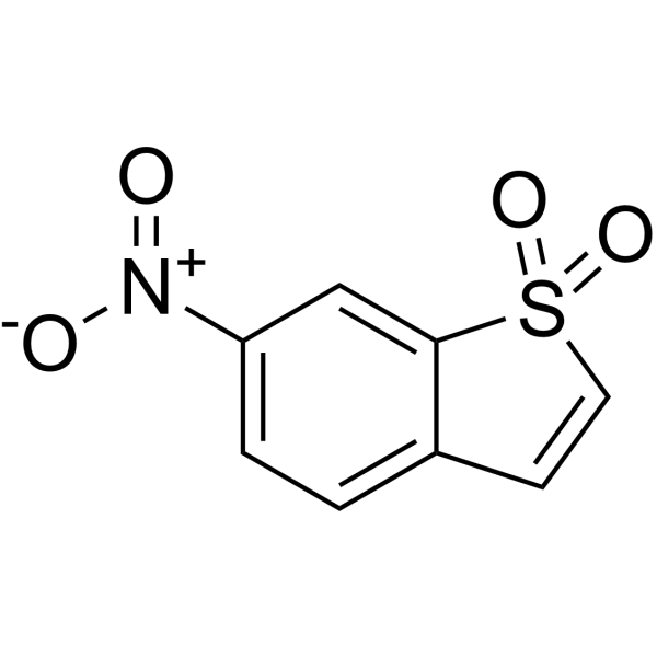
StatticSTAT3 Inhibitor -

HY-P99001
Human IgG1 kappa, Isotype ControlIgG1κ Isotype Control -
HY-P7764
MCP-1/CCL2 Protein, Mouse
Publications Citing Use of MedChemExpress (MCE) Anti-Mouse/Rat/Human CCL2/MCP-1 Antibody (2H5)
More

Biological Activity
Description
Isotype
Armenian hamster IgG
Recommend Isotype Controls
Species Reactivity
Human/Mouse/Rat
IC50 & Target
CCL2/MCP-1
In Vitro
Anti-Mouse/Rat/Human CCL2/MCP-1 Antibody (2H5) (5 μg/mL, 16 h) significantly reduces the migration of THP-1 cells to infected enteroid-derived monolayers (EDMs)[1].
Anti-Mouse/Rat/Human CCL2/MCP-1 Antibody (2H5) (10 μg/mL, 24 h) significantly reduces the enhancing effect of mesenchymal stromal/stem cells (MSCs) on natural killer (NK) cell IFN-γ secretion[2].
Anti-Mouse/Rat/Human CCL2/MCP-1 Antibody (2H5) can inhibit the recruitment of CD8+ T cells by LILRB4 knockdown in BV2 cells[3].
MedChemExpress (MCE) has not independently confirmed the accuracy of these methods. They are for reference only. Further protocols information, click here.
-
Cell Line:THP-1 cells
-
Concentration:5 μg/mL
-
Incubation Time:16 h
-
Result:Significantly inhibited the migration of THP-1 cells.
-
Cell Line:NK cells, co-culture with MSCs
-
Concentration:10 μg/mL
-
Incubation Time:24 h
-
Result:Significantly reduced the level of IFN-γ.
In Vivo
Anti-Mouse/Rat/Human CCL2/MCP-1 Antibody (2H5) (100-200 μg, i.p., once every 48 h, for 12 days) significantly reduces the amount of HIV virus in TKO-BLT mice infected with HIV virus[5].
MedChemExpress (MCE) has not independently confirmed the accuracy of these methods. They are for reference only.
-
Animal Model:DEN (20 mg/kg) and CCl4 (2 μL/g) induced C57BL/6J mice[4]
-
Dosage:8 mg/kg, combined with BLZ945 (200 mg/kg)
-
Administration:Intraperitoneal injection (i.p.), 4 times weekly from week 18 to week 24
-
Result:Significantly reduced the number of tumors.
Reduced M2 macrophage infiltration (CD68+ and CD206+).
Had no significant change in weight.
-
Animal Model:5×10⁴ tissue culture infectious units (TCIU) HIV virus injected TKO-BLT mice[5]
-
Dosage:100 μg for first-dose, 200 μg for subsequent doses
-
Administration:Intraperitoneal injection (i.p.), once every 48 h for 12 days
-
Result:Caused only 14 % of mice to detect HIV DNA, compared to 41 % in the control group.
Did not cause virus rebound but 5 mice showed virus rebound in the control group.
Significantly increased the proportion of CCR2/5+ CD4 T cells.
Gene ID
Accession
Conjugated
Unconjugated
Reconsititution
The product can be reconstituted/diluted with sterile PBS or saline.
Format
-
Product Image
Application
in vivo CCL2 neutralization; Immunohistochemistry (frozen)


Chemical Information
-
Appearance Liquid
-
Molecular Weight 150 kDa
-
Color Colorless to light yellow
-
SMILES
[Anti-Mouse/Rat/Human CCL2/MCP-1 Antibody (2H5)]
-
Formulation
Please refer to the lot-specific COA for specific buffer information.
-
Storage
Please store the product under the recommended conditions in the Certificate of Analysis.


Publications (1)
-
Journal Impact Factor
-
Most Recent
-
Proc Natl Acad Sci U S A
Unraveling the neuroimmune mechanisms in cancer-induced bone pain: New horizons for therapeutic intervention of the two-phase paradigm. [Abstract]2025 Aug 26;122(34):e2503779122. PMID: 40828016

Protocols
-
Research Protocol for Infectious Diseases
Infectious-disease experiments test how pathogens interact with host barriers, innate immune receptors, inflammatory signaling, pathogen replication, and tissue injury; pattern-recognition receptors such as TLRs, RIG-I-like receptors, NOD-like receptors, and inflammasomes detect microbial molecules and activate NF-κB, interferon, and cytokine responses. The central hypothesis is that infection severity reflects the balance between pathogen burden and host response: protective inflammation restricts pathogen growth, whereas excessive or mislocalized inflammation contributes to tissue damage and disease phenotype. Unresolved questions include which host pathways are protective versus pathogenic, why some infection models fail to translate to human disease, and which combined readouts best predict clinically relevant infection outcomes.
-
DSS-Induced Colitis
Dextran sulfate sodium (DSS)-induced colitis is generated by administering DSS in mouse drinking water, producing epithelial injury, barrier disruption, weight loss, diarrhea, fecal blood, colon shortening, histologic mucosal damage, and inflammatory mediator changes; the model is mainly used to study acute or chronic intestinal inflammation resembling selected features of ulcerative colitis. DSS injury is interpreted through clinical and tissue readouts rather than a single molecular endpoint: daily body weight, stool consistency, and bleeding are combined into a disease activity index, while colon length, histology, cytokines, myeloperoxidase activity, intestinal permeability, and tight-junction markers provide complementary measures of inflammation and barrier damage.
-
TNBS-Induced Colitis
TNBS-induced colitis is produced by intrarectal delivery of 2,4,6-trinitrobenzene sulfonic acid in ethanol, where ethanol disrupts the mucosal barrier and TNBS haptenates colonic proteins, generating immune-mediated colonic inflammation with weight loss, diarrhea, ulceration, transmural injury, inflammatory-cell infiltration, and cytokine responses. The model is used as an experimental intestinal inflammation model with Crohn’s disease–like features, especially when Th1-type responses, IL-12–dependent inflammation, chronic relapsing inflammation, or fibrosis-related endpoints are studied.
-
Research Protocol for Inflammation-related Diseases
The NLRP3 inflammasome is a cytosolic innate immune signaling platform that integrates priming signals and danger-signal activation to promote caspase-1 activation, maturation of IL-1β and IL-18, and gasdermin D-mediated pyroptotic cell death. The core experimental logic is to determine whether inflammatory disease phenotypes are driven by increased NLRP3 expression, ASC-containing inflammasome assembly, caspase-1 cleavage, GSDMD cleavage, and extracellular release of IL-1β/IL-18 rather than by nonspecific cell injury alone. The pathway is strongly linked to inflammation-related disease phenotypes because monosodium urate crystals activate NALP3/NLRP3 inflammasome signaling in gout-like crystal inflammation, cholesterol crystals activate NLRP3 inflammasomes in atherogenesis models, and DSS-induced intestinal inflammation has been reported to involve NLRP3 inflammasome activity. However, experimental colitis studies also show context-dependent protective effects of NLRP3 inflammasome co
-
Liver Cancer Modeling
Liver cancer can be classified into primary liver cancer and secondary liver cancer. Secondary liver cancer is the metastatic liver cancer. Primary liver cancer includes hepatocellular carcinoma (HCC), intrahepatic cholangiocarcinoma (ICC) and fibrolamellar HCC, of which HCC is the most common form, accounting for approximately 90% of primary liver cancers[1]. HCC mouse models include chemical agent-induced models, transplanted tumor models, and genetic engineered models.


Purity & Documentation
-
Data Sheet (267 KB)
-
SDS (251 KB)
- English - EN (251 KB)
- Français - FR (251 KB)
- Deutsch - DE (251 KB)
- Norwegian - NO (251 KB)
- Español - ES (251 KB)
- Swedish - SV (251 KB)
- Italian - IT (251 KB)
- Korean - KR (251 KB)
- Portuguese - PT (251 KB)
-
Inhibitory Antibodies User Guide (603 KB)
References
[1]. Sayed IM, et al. Host engulfment pathway controls inflammation in inflammatory bowel disease. FEBS J. 2020 Sep;287(18):3967-3988. [Content Brief]
[2]. Cui R, et al. Human mesenchymal stromal/stem cells acquire immunostimulatory capacity upon cross-talk with natural killer cells and might improve the NK cell function of immunocompromised patients. Stem Cell Res Ther. 2016 Jul 7;7(1):88. [Content Brief]
[3]. Ma Y, et al. Microglia LILRB4 upregulation reduces brain damage after acute ischemic stroke by limiting CD8+ T cell recruitment. J Neuroinflammation. 2024 Aug 31;21(1):214. [Content Brief]
[4]. Du Y, et al. ASH1L in Hepatoma Cells and Hepatic Stellate Cells Promotes Fibrosis-Associated Hepatocellular Carcinoma by Modulating Tumor-Associated Macrophages. Adv Sci (Weinh). 2024 Dec;11(45):e2404756. [Content Brief]
[5]. Packard TA, et al. CCL2: a Chemokine Potentially Promoting Early Seeding of the Latent HIV Reservoir. mBio. 2022 Oct 26;13(5):e0189122. [Content Brief]

Calculators
Concentration (start) × Volume (start) = Concentration (final) × Volume (final)


Anti-Mouse/Rat/Human CCL2/MCP-1 Antibody (2H5) Related Classifications
HY-P990297 Related Classifications
 Powered by Bioz