



ATN-161
Based on 12 publication(s) in Google Scholar
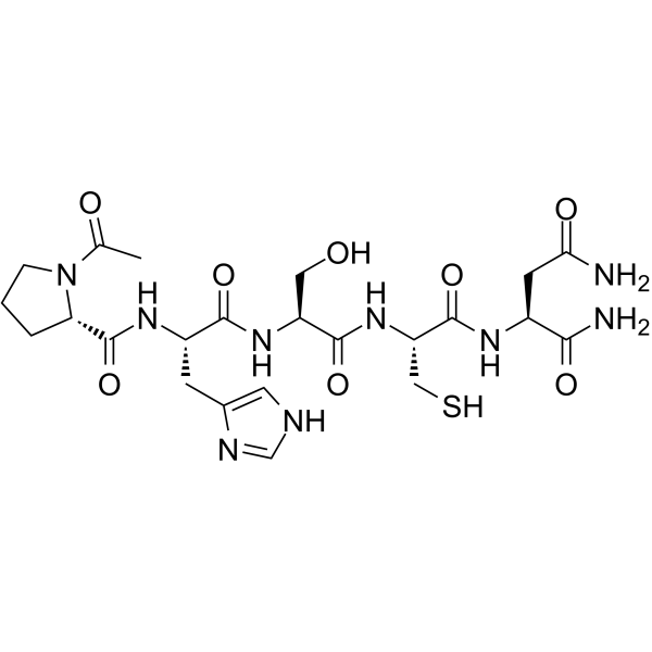
ATN-161 is an antagonist of α5β1 integrin and αvβ3 integrin, as well as an inhibitor of fibrosis, an inhibitor of oxidative stress, a tight junction stabilizer, a mitochondrial integrity protector, an angiogenesis inhibitor and an antiviral agent. ATN-161 inhibits NLRP3, MAPK, and ROS. ATN-161 protects mice from SARS-CoV-2 infection. ATN-161 reduces infarct volume, alleviates edema, decreases immune cell infiltration, reduces the area of choroidal neovascularization lesions, lowers tumor microvessel density and viral load. ATN-161 can be used in research related to ischemic stroke, choroidal neovascularization, breast cancer, coronavirus disease 2019 (COVID-19), and liver metastasis of colorectal cancer.
For research use only. We do not sell to patients.
- Purity : 99.50%
- CAS No.: 262438-43-7
- Formula: C23H35N9O8S
- Molecular Weight:597.64
-
Storage:
Sealed storage, away from moisture.
Powder -80°C, 2 years , -20°C, 1 year* In solvent : -80°C, 6 months; -20°C, 1 month (sealed storage, away from moisture)

To place orders, for customer services and technical support, please contact: MedChemExpress USA
Tel: 609-228-6898 E-mail: [email protected] [email protected]


-
 Biological Activity
-
Chemical Information
-
Solvent & Solubility
- Protocols
- Purity & Documentation
- References
-
 Help & FAQs


-
HY-16141
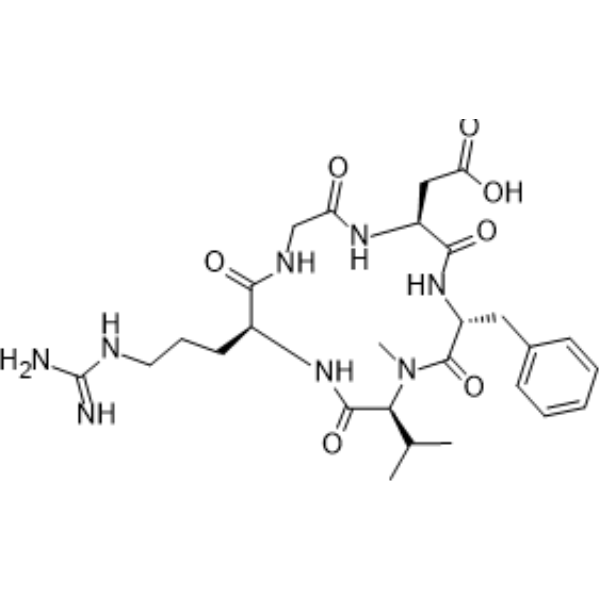
CilengitideIntegrin Inhibitor -
HY-P0023
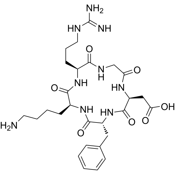
Cyclo(-RGDfK)αvβ3 Integrin Inhibitor -
HY-15102
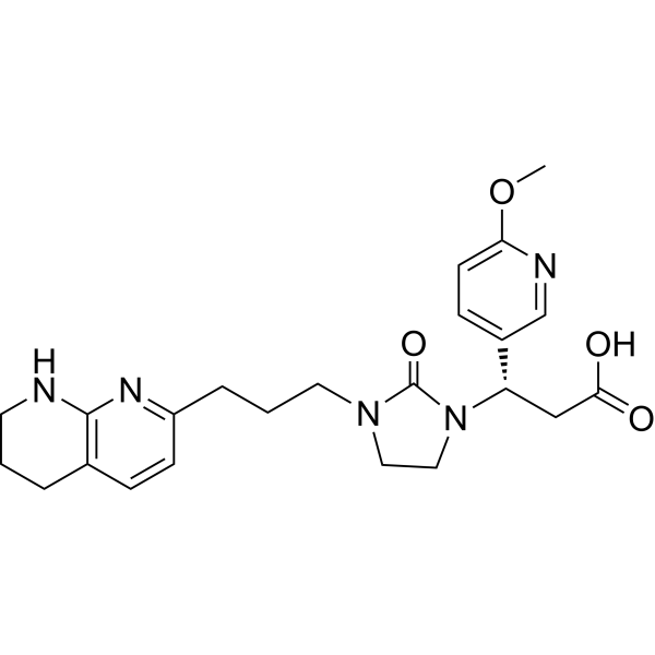
MK-0429αvβ3 Integrin Inhibitor -
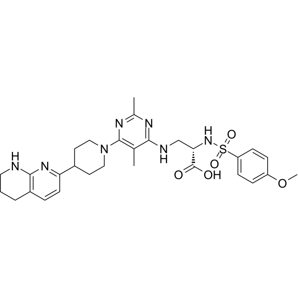
HY-100506
GLPG0187Integrin Receptor Antagonist -
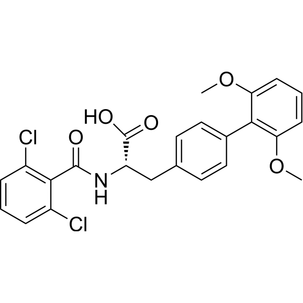
HY-15770
TR-14035α4β7/α4β1 Antagonist -
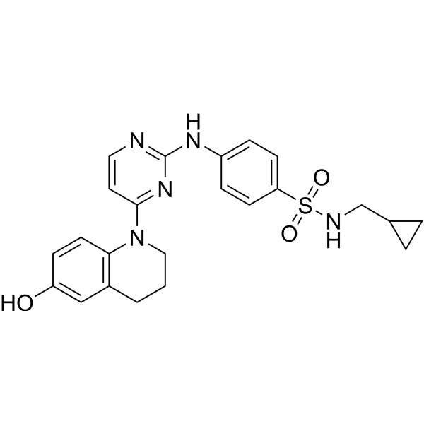
HY-13306
Pyrintegrinβ1-integrin Agonist -
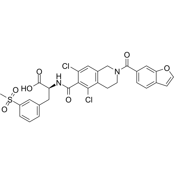
HY-19344
LifitegrastLFA-1/ICAM-1 Antagonist -
HY-17369
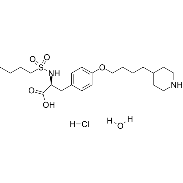
Tirofiban hydrochloride monohydrateGp IIb/IIIa Receptor Antagonist -
HY-14857
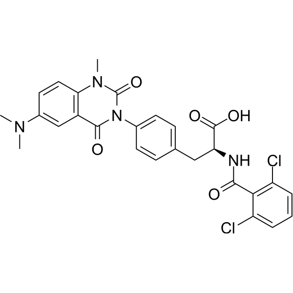
CarotegrastIntegrin Inhibitor -
HY-18644
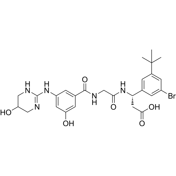
CWHM-12αV Integrins Inhibitor -
HY-13535A
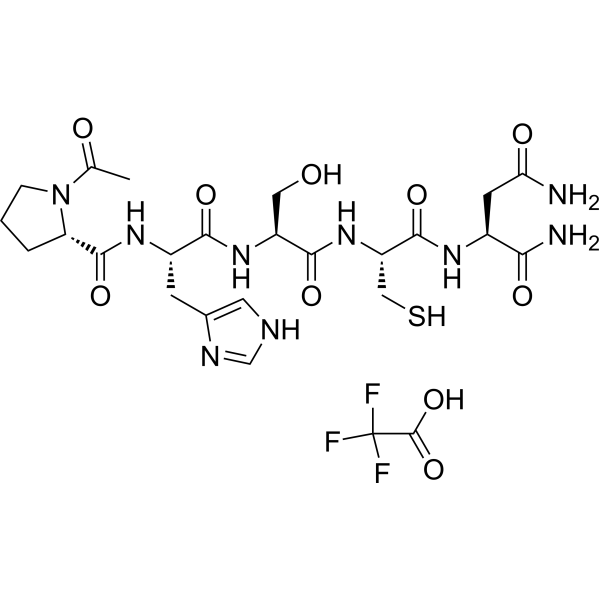
ATN-161 trifluoroacetate saltIntegrin Antagonist -
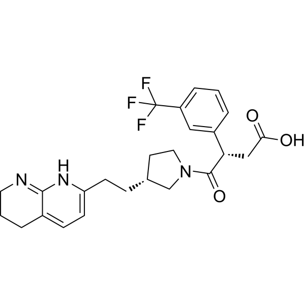
HY-145363
αvβ5 integrin-IN-1Integrin Inhibitor
Publications Citing Use of MedChemExpress (MCE) ATN-161
More- Cancer Cell. 2019 Jan 14;35(1):64-80.e7. [Abstract]
- Nat Nanotechnol. 2026 Jun;21(6):880-891. [Abstract]
- Int J Oral Sci. 2026 Jul 9;18(1):50.
- ACS Nano. 2025 Jun 10;19(22):20564-20577. [Abstract]
- J Adv Res. 2025 May 25:S2090-1232(25)00370-4. [Abstract]
- Stem Cell Res Ther. 2022 Jul 18;13(1):327. [Abstract]
- Cell Prolif. 2021 Apr;54(4):e13012. [Abstract]
- Sci Signal. 2022 Dec 6;15(763):eabn2743. [Abstract]
- ACS Biomater Sci Eng. 2023 May 8;9(5):2524-2533. [Abstract]
- Colloids Surf B Biointerfaces. 2022 Feb:210:112227. [Abstract]
- J Cell Sci. 2025 Dec 1;138(23):jcs264412. [Abstract]
- Res Sq. 2025 Dec 17.
 Customer Validation & Images

-
Cell Imaging/Staining

-
RT-PCR

-
Flow Cytometry

-
Flow Cytometry


Biological Activity
Description
IC50 & Target
[1]|
α5β1 |
αvβ3 |
NLRP3 |
In Vitro
ATN-161 (5-25 μM; 1 h) significantly inhibits the upregulation of NLRP3 inflammasome induced by OGD/R in bEnd.3 cells[1].
ATN-161 (pretreatment for 1 h, oxygen-glucose deprivation for 6 h + reoxygenation for 24 h, 10 μM) significantly inhibits oxidative stress in bEnd.3 cells induced by oxygen-glucose deprivation/reoxygenation (OGD/R), including mitochondrial superoxide anion production and intracellular ROS generation[1].
ATN-161 (10 μM) inhibits the VEGF-induced upregulation of integrin α5-β1 expression in human choroidal endothelial cells (hCEC)[2].
ATN-161 (1 nM-100 μM; 30 min) inhibits VEGF-induced migration of human choroidal endothelial cells (hCEC) in a dose-dependent manner, with a significant effect observed starting at the concentration of 100 nM[2].
ATN-161 (1 nM-100 μM; 24 h) does not inhibit VEGF-induced proliferation of human choroidal endothelial cells (hCEC)[2].
ATN-161 competes with the biotinylated analog ATN-453 for binding to purified human integrin α5β1 and αvβ3, which is confirmed by the specific high-affinity binding of ATN-453 to these integrins[3].
ATN-161 (1-1000 μM) increases the survival rate of VeroE6 cells infected with SARS-CoV-2, with a minimum effective concentration of only 1 μM, and the survival rate peaks at 10 μM[4].
ATN-161 (1.0 μM; 48 h) significantly reduces the viability and proliferation capacity of human umbilical vein endothelial cells (HUVEC) by 21% after 48 hours of incubation on fibronectin-coated culture plates under low-serum conditions, while exerting no effect on CT26 mouse colon cancer cells[5].
ATN-161 (1-100 μM; up to 5 days) exerts no significant effect on the proliferation of MDA-MB-231 breast cancer cells during 5 days of in vitro culture[3].
ATN-161 (20 μM; 30 min) significantly inhibits the level of phosphorylated MAPK in MDA-MB-231 breast cancer cells in vitro, but has no effect on the levels of FAK or phosphorylated FAK[3].
MedChemExpress (MCE) has not independently confirmed the accuracy of these methods. They are for reference only. Further protocols information, click here.
-
Cell Line:human choroidal endothelial cells (hCECs)
-
Concentration:1 nM, 10 nM, 100 nM, 1 μM, 10 μM, 100 μM
-
Incubation Time:24 h
-
Result:Did not alter VEGF-induced proliferation at any tested concentration.
-
Cell Line:human choroidal endothelial cells (hCECs)
-
Concentration:1 nM, 10 nM, 100 nM, 1 μM, 10 μM, 100 μM
-
Incubation Time:30-minute preincubation, followed by 8-hour migration
-
Result:Reduced VEGF-induced migration in a dose-dependent manner starting at 100 nM, with significant inhibition observed at 100 nM, 1 μM, 10 μM, and 100 μM (P < 0.001 vs.
VEGF-only group).
-
Cell Line:human MDA-MB-231 breast cancer cells
-
Concentration:1-100 μmol/L
-
Incubation Time:up to 5 days
-
Result:Showed no significant effect on MDA-MB-231 cell proliferation compared to vehicle-treated controls over 5 days.
-
Cell Line:human umbilical vein endothelial cells (HUVEC), CT26 murine colon cancer cells
-
Concentration:1.0 μM
-
Incubation Time:48 h
-
Result:Reduced HUVEC number by 21% compared to control (p < 0.03).
Exhibited no significant effect on CT26 murine colon cancer cell number.
In Vivo
ATN-161 (0.1-10 μg/mL; intravitreal injection; single dose administered immediately after laser photocoagulation) inhibits laser-induced choroidal neovascularization (CNV) in Brown-Norway rats in a dose-dependent manner. At the dose of 10 μg/mL, it reduces the average CNV leakage score to 0.7, the average CNV area to 12186 μm2, and the average CNV thickness to 72.3 μm (all *P* < 0.001 compared with the sham-operated group), while also decreasing VEGF expression levels on postoperative days 7, 10, and 14[2].
ATN-161 protects k18-hACE2 transgenic mice from SARS-CoV-2 infection[1].
ATN-161 inhibits VEGF-induced retinal neovascularization in mice by targeting integrin α5β1 and the NLRP3 inflammasome[1].
ATN-161 (0.05-1 mg/kg; intravenous injection; three times per week; for 6 consecutive weeks) induces dose-dependent, statistically significant inhibition of subcutaneous breast cancer xenograft growth in female BALB/c nu/nu mice, with maximal efficacy observed at doses of 0.1 mg/kg and 1 mg/kg, while reducing tumor angiogenesis, cell proliferation levels, and the expression of P-MAPK[3].
ATN-161 (100 mg/kg; i.p.; once every 3 days; administered starting on day 4 after tumor cell injection) alone reduces tumor microvessel density by 48% in a mouse model of colorectal cancer liver metastasis. When combined with continuously infused 5-FU (HY-90006), it significantly reduces liver tumor burden, number of metastatic lesions, and tumor cell proliferation, increases the apoptosis rate, and improves mouse survival (p < 0.03)[5].
MedChemExpress (MCE) has not independently confirmed the accuracy of these methods. They are for reference only.
-
Animal Model:Brown-Norway (BN) (12-week-old male, 200 to 250 g, laser-induced choroidal neovascularization)[2]
-
Dosage:0.1 μg/mL; 10 μg/mL
-
Administration:intravitreal injection; single dose immediately after laser photocoagulation
-
Result:Reduced mean CNV leakage score to 1.1, mean CNV area to 16,890 μm2, and mean CNV thickness to 81.3 μm at 0.1 μg/mL dose on day 14 (P < 0.001 vs. sham for leakage score and area, P < 0.05 vs. sham for thickness).
Reduced mean CNV leakage score to 0.7, mean CNV area to 12,186 μm2, and mean CNV thickness to 72.3 μm at 10 μg/mL dose on day 14 (all P < 0.001 vs. sham).
Lowered VEGF levels in RPE-choroid complexes on days 7, 10, and 14 compared to sham group (P < 0.001).
Showed remarkable reduction of CNV thickness on days 7 and 14, with less evident fluorescein leakage at day 14 relative to sham-injected eyes at 10 μg/mL dose.
-
Animal Model:BALB/c nu/nu (5-week-old, 15-20 g, female, subcutaneous xenograft of MDA-MB-231 human breast cancer cells)[3]
-
Dosage:0.05 mg/kg; 0.1 mg/kg; 1 mg/kg
-
Administration:i.v.; thrice a week; 6 weeks
-
Result:Caused a dose-dependent reduction in tumor volume compared to vehicle controls.
Produced marked, statistically significant tumor volume reduction at 0.1 and 1 mg/kg doses with no significant difference in efficacy between these two doses.
Reduced tumor microvessel density by 43% at 1 mg/kg dose.
Significantly decreased tumor cell mitotic index at 1 mg/kg dose.
Significantly reduced Ki-67 proliferation index at 1 mg/kg dose.
Decreased expression of phosphorylated mitogen-activated protein kinase (P-MAPK) in tumor tissue at 1 mg/kg dose.
Left levels of focal adhesion kinase (FAK) and phosphorylated FAK (P-FAK) unchanged at 1 mg/kg dose.
Caused no significant weight loss in treated animals, indicating good tolerability.
-
Animal Model:BALB/c (8-week-old male; induced by 10,000 CT26 murine colon carcinoma cells injected into spleens)[5]
-
Dosage:100 mg/kg (monotherapy); 100 mg/kg + 100 mg/kg/2 weeks 5-FU (combination)
-
Administration:i.p.; every third day; starting day 4 after tumor cell injection; continuous infusion (5-FU, 2 weeks)
-
Result:Reduced microvessel density in liver metastases by 48% compared to controls (p < 0.03).
Did not significantly reduce liver weight, number of liver metastases, tumor cell proliferation, increase tumor cell apoptosis, or improve survival when administered alone.
Significantly reduced liver weight (p < 0.02), number of liver metastases (p < 0.05), tumor cell proliferation (p < 0.01), reduced microvessel density by 44% compared to controls (p < 0.03), increased tumor cell apoptosis (p < 0.03), and improved overall survival (p < 0.03, log-rank test) relative to single-agent treatments when combined with continuous-infusion 5-FU.
Left six mice alive and healthy on day 21 when the experiment was terminated.
Clinical Trial
| NCT Number | Sponsor | Condition | Start Date |
Phase

|
|---|---|---|---|---|
| NCT01329991 | Plexxikon| | 2011-05 | PHASE1 |


Chemical Information
-
CAS No. 262438-43-7
-
Appearance Solid
-
Molecular Weight 597.64
-
Formula C23H35N9O8S
-
Color White to off-white
-
SMILES
O=C(N)C[C@@H](C(N)=O)NC([C@H](CS)NC([C@H](CO)NC([C@H](CC1=CNC=N1)NC([C@H]2N(C(C)=O)CCC2)=O)=O)=O)=O
-
Shipping
Room temperature in continental US; may vary elsewhere.
-
Storage
Sealed storage, away from moisture
Powder -80°C 2 years -20°C 1 year * In solvent : -80°C, 6 months; -20°C, 1 month (sealed storage, away from moisture)


Publications (12)
-
Journal Impact Factor
-
Most Recent
-
Cancer Cell
Tinagl1 Suppresses Triple-Negative Breast Cancer Progression and Metastasis by Simultaneously Inhibiting Integrin/FAK and EGFR Signaling. [Abstract]2019 Jan 14;35(1):64-80.e7. PMID: 30612941 -
Nat Nanotechnol
2026 Jun;21(6):880-891. PMID: 42286217 -
-
ACS Nano
Deciphering the Distinct Roles of Molecular and Supramolecular Chirality in Osteogenic Differentiation of Mesenchymal Stem Cells in 3D Hydrogels. [Abstract]2025 Jun 10;19(22):20564-20577. PMID: 40425514 -
J Adv Res
Fibrinogen exacerbates α-synuclein aggregation and mitochondrial dysfunction via alpha5beta3 integrin in Parkinson's disease. [Abstract]2025 May 25:S2090-1232(25)00370-4. PMID: 40425084 -
Stem Cell Res Ther
Extracellular matrix derived from Wharton's Jelly-derived mesenchymal stem cells promotes angiogenesis via integrin αVβ3/c-Myc/P300/VEGF. [Abstract]2022 Jul 18;13(1):327. PMID: 35851415
ATN-161 purchased from MedChemExpress. Usage Cited in: Stem Cell Res Ther. 2022 Jul 18;13(1):327. [Abstract]
ATN-161 trifluoroacetate salt (8 h) and ECM significantly inhibited the tube formation ability but did not change the tube formation ability of HUVECs.
-
Cell Prolif
Vitronectin-activated αvβ3 and αvβ5 integrin signalling specifies haematopoietic fate in human pluripotent stem cells. [Abstract]2021 Apr;54(4):e13012. PMID: 33656760
ATN-161 purchased from MedChemExpress. Usage Cited in: Cell Prolif. 2021 Apr;54(4):e13012. [Abstract]
ATN-161 trifluoroacetate salt (10 μM; from day 2 to day 6) did not affect the mRNA expression of RUNX1, GATA2 and MYB in hPSCs.
ATN-161 purchased from MedChemExpress. Usage Cited in: Cell Prolif. 2021 Apr;54(4):e13012. [Abstract]
ATN-161 trifluoroacetate salt (10 μM; from day 2 to day 6) did not affect the production of CD43+ HPCs in VTN‐coated hPSCs.
ATN-161 purchased from MedChemExpress. Usage Cited in: Cell Prolif. 2021 Apr;54(4):e13012. [Abstract]
ATN-161 trifluoroacetate salt (10 μM; from day 2 to day 6) did not affect the production of CD43+ HPCs in VTN‐coated hPSCs.
-
Sci Signal
Cross-talk between TSC2 and the extracellular matrix controls pulmonary vascular proliferation and pulmonary hypertension. [Abstract]2022 Dec 6;15(763):eabn2743. PMID: 36473049 -
ACS Biomater Sci Eng
Regulation of Macrophage Polarization on Chiral Potential Distribution of CFO/P(VDF-TrFE) Films. [Abstract]2023 May 8;9(5):2524-2533. PMID: 37092816 -
Colloids Surf B Biointerfaces
Anisotropic magneto-mechanical stimulation on collagen coatings to accelerate osteogenesis. [Abstract]2022 Feb:210:112227. PMID: 34838419 -
J Cell Sci
Bile canaliculi formation in primary hepatocytes requires α1β1 integrin-dependent adherens junction re-organization. [Abstract]2025 Dec 1;138(23):jcs264412. PMID: 41347643 -


Solvent & Solubility
In Vitro:
DMSO : 25 mg/mL (41.83 mM; Need ultrasonic; Hygroscopic DMSO has a significant impact on the solubility of product, please use newly opened DMSO)
H2O : 5 mg/mL (8.37 mM; ultrasonic and warming and heat to 60°C)
Please refer to the solubility information to select the appropriate solvent. Once prepared, please aliquot and store the solution to prevent product inactivation from repeated freeze-thaw cycles.
Storage method and period of stock solution: -80°C, 6 months; -20°C, 1 month (sealed storage, away from moisture). When stored at -80°C, please use it within 6 months. When stored at -20°C, please use it within 1 month.
* Note: If you choose water as the stock solution, please dilute it to the working solution, then filter and sterilize it with a 0.22 μm filter before use.
Please refer to the solubility information to select the appropriate solvent. Once prepared, please aliquot and store the solution to prevent product inactivation from repeated freeze-thaw cycles.
Storage method and period of stock solution: -80°C, 6 months; -20°C, 1 month (sealed storage, away from moisture). When stored at -80°C, please use it within 6 months. When stored at -20°C, please use it within 1 month.
* Note: If you choose water as the stock solution, please dilute it to the working solution, then filter and sterilize it with a 0.22 μm filter before use.
Concentration (start) × Volume (start) = Concentration (final) × Volume (final)

Protocols
-
Collagen: Sirius Red Staining
Sirius Red or picrosirius red staining is a histochemical method for visualizing collagen-rich extracellular matrix in tissue sections, and collagen fibers are detected as red-stained structures under bright-field microscopy with enhanced birefringence under polarized light. Picrosirius red is useful for assessing total collagen organization, distribution, and fibrosis burden, but polarized color should not be interpreted as a definitive collagen type I versus type III readout because color is affected by fiber orientation, thickness, and packing.
-
Research Protocol for Infectious Diseases
Infectious-disease experiments test how pathogens interact with host barriers, innate immune receptors, inflammatory signaling, pathogen replication, and tissue injury; pattern-recognition receptors such as TLRs, RIG-I-like receptors, NOD-like receptors, and inflammasomes detect microbial molecules and activate NF-κB, interferon, and cytokine responses. The central hypothesis is that infection severity reflects the balance between pathogen burden and host response: protective inflammation restricts pathogen growth, whereas excessive or mislocalized inflammation contributes to tissue damage and disease phenotype. Unresolved questions include which host pathways are protective versus pathogenic, why some infection models fail to translate to human disease, and which combined readouts best predict clinically relevant infection outcomes.
-
Multiplex immunofluorescence IHC
Multiplex immunofluorescence IHC detects multiple protein biomarkers in one tissue section by sequential antibody staining, HRP-mediated tyramide fluorophore deposition, heat-mediated antibody stripping, nuclear counterstaining, multispectral imaging, spectral unmixing, and digital cell phenotyping; TSA deposits fluorophore near the antigen so the fluorescence signal remains after primary and secondary antibodies are removed, enabling repeated staining cycles, including with antibodies from the same host species. Classic FFPE tumor immune-profiling applications use panels such as CD3, CD8, CD68/CD163, FOXP3, PD-1, PD-L1, pancytokeratin, Ki67, and DAPI to identify tumor cells, immune-cell subsets, checkpoint-marker expression, co-expression phenotypes, cell density, and spatial relationships in the tumor microenvironment.
-
Human pluripotent stem cell endothelial-cell differentiation
Human pluripotent stem cell endothelial differentiation is based on stepwise developmental patterning: early activation of WNT/GSK3β inhibition promotes mesodermal or vascular progenitor entry, followed by endothelial specification using VEGF-related signaling, BMP4, FGF2, Notch modulation, or cAMP depending on the published protocol. Endothelial differentiation is read out by acquisition of CD31, CD34, VE-cadherin/CD144, KDR/VEGFR2, vWF, Tie2, NOS3, acetylated LDL uptake, tube/network formation, barrier function, and in vivo vessel-forming capacity where tested.
-
Vascular/Branching Fractal Analysis
Vascular/branching fractal analysis quantifies the geometric complexity of vessel trees or vascular networks from segmented 2D images, commonly by converting vessels into binary and/or skeletonized maps and estimating fractal dimension using box-counting or related approaches. Fractal dimension is interpreted as an image-derived readout of vascular branching complexity, space filling, or density, and has been applied to retinal photographs, fluorescein angiography, OCT angiography, capillary perfusion maps, and in vitro Matrigel angiogenesis networks. The assay readout is generated from vessel-positive pixels after image preprocessing, vessel segmentation, binarization, and optional skeletonization; reported outputs include fractal dimension, vessel density, branchpoint density, endpoint density, vessel length density, tortuosity, and generation-based branching metrics when VESGEN-style analysis is used. The biological interpretation is limited to quantitative vascular patterning and s
-
ROS/oxidative-stress fluorescent staining
ROS/oxidative-stress fluorescent staining uses cell-permeant fluorogenic probes that become fluorescent after oxidation inside cells or tissues; commonly used examples include DCFH-DA/DCFDA for broad cellular oxidant detection, DHE for superoxide-related signal detection, MitoSOX for mitochondrial superoxide-related signal detection, and CellROX probes for oxidative-stress-associated fluorescence readouts. The assay detects probe oxidation rather than a single ROS species unless the probe and analysis method have been chemically validated for that species. DCFH-DA enters cells, is deacetylated by intracellular esterases to DCFH, and produces fluorescent DCF after oxidation, so the readout is used as an operational measure of total cellular oxidative stress rather than a species-specific ROS measurement. DHE and MitoSOX can report superoxide-related oxidation, but red fluorescence alone can include non-specific ethidium-like oxidation products; HPLC or optimized spectral approaches are
-
Connective Tissue: Masson's Trichrome/Collagen Trichrome Staining
Masson’s Trichrome (collagen/trichrome staining) is a histological technique that differentially stains tissue compartments using sequential acidic dyes to distinguish collagen from muscle and cytoplasmic components based on dye affinity and tissue permeability differences, enabling visualization of fibrosis and connective tissue architecture in histological sections. The classical formulation typically uses Weigert's iron hematoxylin for nuclear staining, Biebrich scarlet-acid fuchsin for cytoplasm and muscle, and aniline blue (or light green variants) for collagen, producing a characteristic blue/green collagen signal contrasted against red cytoplasm and dark nuclei. The staining principle relies on selective displacement of smaller dye molecules by larger anionic dyes in collagen-rich regions under controlled acidified conditions, which enhances collagen-specific dye retention. This property makes the method widely used for fibrosis assessment in organs such as heart, liver, lung, a
-
Multiplex immunohistochemistry
Multiplex immunohistochemistry (mIHC), also known as tyramide dignal amplification (TSA), is an enzymatic detection method that uses horseradish peroxidase (HRP) to perform high-density in-situ labeling of target proteins or nucleic acids.
-
Breast Cancer Modeling
Breast cancer is a heterogeneous cancer, and it has been distinguished into four subtypes: luminal A, luminal B, HER2-positive and basal-like. Molecular mutations, epigenetic alterations, hormone exposure and immune microenvironment are related to the progression of breast cancer.
-
Research Protocol for Cancer Immunology
Cancer immunology studies how the immune system recognizes, suppresses, edits, or fails to eliminate malignant cells through tumor antigen release, antigen presentation, T-cell priming, immune trafficking, tumor-cell killing, and feedback inhibition in the tumor microenvironment. The cancer-immunity cycle links tumor antigenicity, dendritic-cell priming, CD8+ T-cell infiltration, cytotoxic function, and immune-checkpoint regulation to tumor rejection or immune escape. Immune-checkpoint pathways such as PD-1/PD-L1 and CTLA-4 suppress antitumor T-cell activity and can be therapeutically blocked, but many tumors remain resistant because of poor antigen presentation, weak T-cell infiltration, suppressive myeloid cells, regulatory T cells, and tumor-intrinsic immune-exclusion programs. Unresolved questions include which immune-cell states predict response, how tumor-intrinsic pathways exclude immune cells, how myeloid suppression limits checkpoint blockade, and which combination strategies
-
Fibrosis/Collagen Morphometry
Fibrosis and collagen morphometry is based on the quantitative visualization of fibrillar collagen deposition in tissue sections using histochemical stains such as Sirius Red (Picrosirius Red) or Masson's trichrome, followed by image-based or polarization-enhanced analysis to estimate collagen proportional area as a surrogate of extracellular matrix accumulation during fibrotic remodeling. Sirius Red combined with polarized light microscopy enhances detection of collagen fibers due to birefringence properties, enabling more specific visualization of collagen type I and III fibrils compared to conventional bright-field histology, while whole-section or region-restricted digital morphometry reduces field-selection bias in fibrosis assessment. Alternative quantitative approaches include second harmonic generation (SHG) and two-photon excited fluorescence microscopy, which enable label-free detection of fibrillar collagen and have been validated against histological staining and biochemica
-
Endothelial Tube Formation Assay
Endothelial tube formation assay evaluates the ability of endothelial cells to attach, migrate, align, and organize into capillary-like networks when cultured on gelled basement membrane extract or Matrigel; the readout is the morphology and quantity of tube-like networks, which reflects an in vitro endothelial morphogenesis step related to angiogenesis. Basement membrane extract/Matrigel provides laminin-rich extracellular matrix cues that support endothelial differentiation into capillary-like structures, but it can contain biologically active growth factors, so growth-factor-reduced matrix is preferred when testing defined angiogenic stimulators or inhibitors.
-
Splenic/Portal-Vein Liver Metastasis Xenograft
Splenic and portal-vein liver metastasis xenograft models deliver tumor cells into the portal circulation so that cells reach the liver first and form hepatic metastatic lesions; splenic injection uses the spleen as an access route to the portal system, while direct portal-vein injection introduces cells into the portal vein without requiring splenectomy. The assay detects liver colonization, intrahepatic tumor growth, tumor distribution, treatment response, survival, and liver-metastasis microenvironment changes; readouts include bioluminescence or fluorescence imaging, gross liver nodule counts, liver weight or tumor burden, histology, and survival.
-
Endothelial Cell Migration/Angiogenic Sprouting Assay
Endothelial cell migration and angiogenic sprouting assays are in vitro (and partially ex vivo-adapted) functional models that quantify the ability of endothelial cells to undergo coordinated migration, extracellular matrix invasion, and multicellular organization into capillary-like sprouts in response to pro-angiogenic stimuli such as VEGF, bFGF, or conditioned microenvironments. These assays are used to model early angiogenic events including tip-cell formation, directional migration, and lumen-like sprout extension, which collectively reflect angiogenic activation and vascular morphogenesis processes observed in vivo.
-
Orthotopic Cell-Line Xenograft
Orthotopic cell-line xenograft models involve implantation of human cancer cell lines into the anatomically corresponding organ of immunodeficient mice to reproduce tumor growth within a native microenvironment, enabling more clinically relevant tumor behavior compared with subcutaneous models. These models are widely used because orthotopic placement better recapitulates tumor progression, including invasion and metastatic spread, which are often underrepresented in heterotopic implantation systems. Compared with conventional xenografts, orthotopic implantation is described as more technically complex but provides improved simulation of tumor-microenvironment interactions and metastatic behavior, making it particularly valuable for translational oncology research. Surgical orthotopic implantation approaches have been emphasized as enabling faithful reproduction of clinical cancer features, including metastasis and disease progression patterns that align with the tumor’s organ of origi


Purity & Documentation
-
Data Sheet (302 KB)
-
SDS (396 KB)
- English - EN (396 KB)
- Français - FR (396 KB)
- Deutsch - DE (396 KB)
- Norwegian - NO (396 KB)
- Español - ES (396 KB)
- Swedish - SV (396 KB)
- Italian - IT (396 KB)
- Korean - KR (396 KB)
- Portuguese - PT (396 KB)
-
Handling Instructions (2659 KB)
References
[1]. Amruta N, et al. ATN-161 Ameliorates Ischemia/Reperfusion-induced Oxidative Stress, Fibro-inflammation, Mitochondrial damage, and Apoptosis-mediated Tight Junction Disruption in bEnd.3 Cells. Inflammation. 2021 Dec;44(6):2377-2394. [Content Brief]
[2]. Wang W, et al. The antiangiogenic effects of integrin alpha5beta1 inhibitor (ATN-161) in vitro and in vivo. Invest Ophthalmol Vis Sci. 2011 Sep 14;52(10):7213-20. [Content Brief]
[3]. Khalili P, et al. A non-RGD-based integrin binding peptide (ATN-161) blocks breast cancer growth and metastasis in vivo. Molecular cancer therapeutics. 2006 Sep;5(9):2271-80. [Content Brief]
[4]. Beddingfield BJ, et al. The Integrin Binding Peptide, ATN-161, as a Novel Therapy for SARS-CoV-2 Infection. JACC Basic Transl Sci. 2021 Jan;6(1):1-8. [Content Brief]
[5]. Stoeltzing O, et al. Inhibition of integrin alpha5beta1 function with a small peptide (ATN-161) plus continuous 5-FU infusion reduces colorectal liver metastases and improves survival in mice. International journal of cancer. 2003 Apr 20;104(4):496-503. [Content Brief]


Complete Stock Solution Preparation Table
Please refer to the solubility information to select the appropriate solvent. Once prepared, please aliquot and store the solution to prevent product inactivation from repeated freeze-thaw cycles.
Storage method and period of stock solution: -80°C, 6 months; -20°C, 1 month (sealed storage, away from moisture). When stored at -80°C, please use it within 6 months. When stored at -20°C, please use it within 1 month.
| Optional Solvent | Concentration Solvent Mass | 1 mg | 5 mg | 10 mg | 25 mg |
|---|---|---|---|---|---|
| H2O / DMSO | 1 mM | 1.6732 mL | 8.3662 mL | 16.7325 mL | 41.8312 mL |
| 5 mM | 0.3346 mL | 1.6732 mL | 3.3465 mL | 8.3662 mL | |
| DMSO | 10 mM | 0.1673 mL | 0.8366 mL | 1.6732 mL | 4.1831 mL |
| 15 mM | 0.1115 mL | 0.5577 mL | 1.1155 mL | 2.7887 mL | |
| 20 mM | 0.0837 mL | 0.4183 mL | 0.8366 mL | 2.0916 mL | |
| 25 mM | 0.0669 mL | 0.3346 mL | 0.6693 mL | 1.6732 mL | |
| 30 mM | 0.0558 mL | 0.2789 mL | 0.5577 mL | 1.3944 mL | |
| 40 mM | 0.0418 mL | 0.2092 mL | 0.4183 mL | 1.0458 mL |
* Note: If you choose water as the stock solution, please dilute it to the working solution, then filter and sterilize it with a 0.22 μm filter before use.


ATN-161 Related Classifications
HY-13535 Related Classifications
 Powered by Bioz
Keywords
- ATN-161
- 262438-43-7
- ATN161
- ATN 161
- Integrin
- Apoptosis
- NOD-like Receptor (NLR)
- p38 MAPK
- Reactive Oxygen Species (ROS)
- SARS-CoV
- α5β1 integrin
- bEnd.3 cells
- ischemic stroke
- SARS-CoV-2
- colorectal liver metastases
- αvβ3 integrin
- choroidal neovascularization
- human choroidal endothelial cells
- VeroE6 cells
- MDA-MB-231 breast cancer cells
- Inhibitor
- inhibitor
- inhibit